- Goals
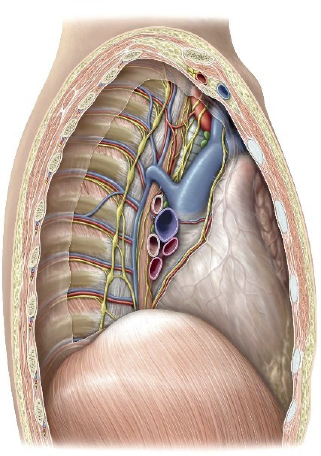
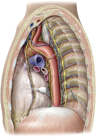
- Define the extent and boundaries of the mediastinum as a whole; describe the boundaries and contents of the subdivisions of the mediastinum.
- Clean and demonstrate the structures in the superior mediastinum.
- Open the pericardium, study its parts, and demonstrate the “great” vessels of the heart.
- Remove the heart.
- Clean and demonstrate the contents of the posterior mediastinum.
- Identify the sympathetic trunk.
Muscles and structure of the thoracic wall
The Mediastinum
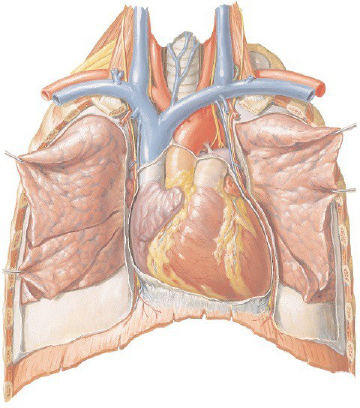
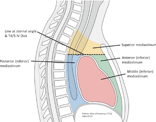
The mediastinum (“median septum”) is the part of the thoracic cavity in the midline, between the two pleural sacs. The mediastinum contains all the thoracic organs, except for the lungs and pleura.
Boundaries of the mediastinum:
■Superior to inferior: From the superior thoracic aperture to the diaphragm
■Anterior to posterior: from the sternum/ costal cartilages to the bodies of the thoracic vertebrae
A horizontal plane drawn from the sternal angle to the intervertebral disc between T-4/T-5 subdivides the mediastinum into superior and inferior parts.
The pericardium and heart further subdivides the inferior mediastinum into posterior, middle, and anterior parts.
Superior Mediastinum
Superior mediastinum
Boundaries:
■Above: Superior thoracic aperture = continuous with the neck above.
■Below: Horizontal plane through T-4/T-5 intervertebral disc and through the sternal angle.
Main contents (shortened):
■Thymus
■Superior vena cava; Right and Left Brachiocephalic veins
■Arch of the aorta and its branches
■Vagus and phrenic nerves; Left recurrent laryngeal nerve
■Trachea, Esophagus, Thoracic duct
Big Picture of the Superior Mediastinum
Rules of thumb:
1The derivatives of the embryonic foregut are placed in the CENTER of the superior mediastinum—verify this by locating the trachea and esophagus with blunt dissection.
2Major arteries predominate on the LEFT side: verify this by locating the course of the aorta.
3Major veins predominate on the RIGHT side: verify this by locating the superior vena cava and azygos vein.
Clean and identify major veins in the superior mediastinum.
1Identify and remove the fatty thymus gland. (If visible – this involutes with age)
2Clean and follow the courses of the left and right brachiocephalic veins. Which one is longer? Why? At the base of the neck, each brachiocephalic vein (left and right) is formed by the union of the internal jugular and subclavian veins. The junctions of these veins on each side of the neck are called the left and right “venous angles.”
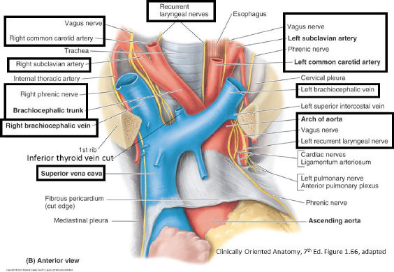
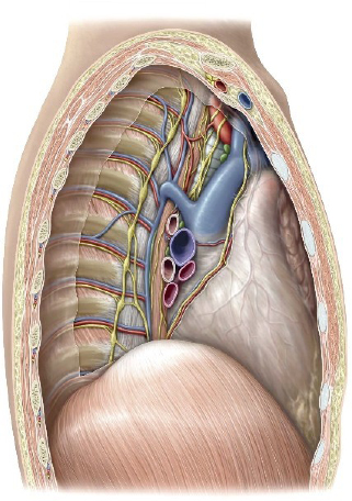
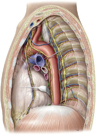
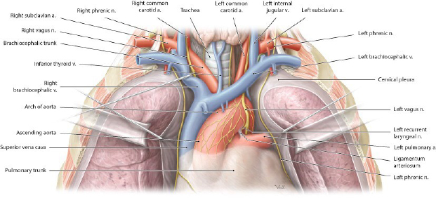
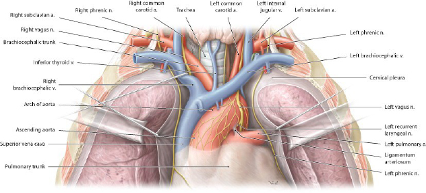
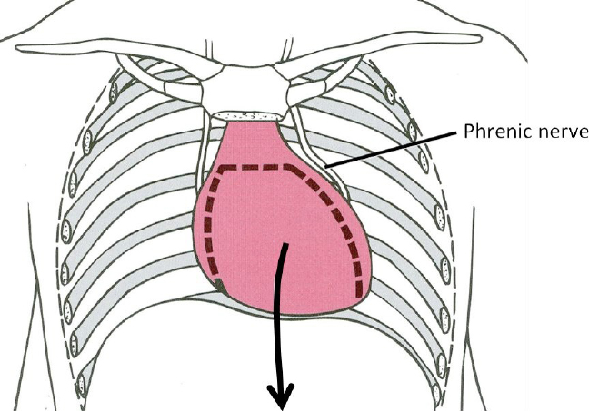
3On the left and right sides of the mediastinum, use forceps and scissors to remove any remaining mediastinal (parietal) pleura. Before you cut, see if you can visualize the courses of the phrenic nerves as they pass through the mediastinum on their way to the diaphragm, so you don’t damage them. On the left, the phrenic nerve crosses the arch of the aorta and the left side of the pericardium. On the right, it is on the lateral side of the superior vena cava before it reaches the right side of the pericardium. Remember that on BOTH sides, the phrenic nerves pass anterior to the lung roots. See Figures 9.3 and 9.4.
4Clean off the superior vena cava.
5Clean off the arch of the azygos vein as it passes over the root of the right lung to enter the SVC.
Clean and identify the major arteries in the superior mediastinum.
1Follow the course of the aorta as it emerges from the heart and arches to the left.
2Clean and identify the three large arteries that branch from the arch of the aorta from right to left:
■Brachiocephalic artery
■Left common carotid artery
■Left subclavian artery
3Remove any mediastinal pleura covering the arch of the aorta. Take care not to damage the phrenic nerve or vagus nerve as they cross the aorta.
Clean and identify major nerves passing through the superior mediastinum.
1Start at the superior thoracic aperture and locate the phrenic and vagus nerves. Both pass from the neck into the thorax, sandwiched between the subclavian vein and artery.
■The vagi are near the common carotid arteries.
■The phrenic nerves are lateral to the vagi.
2Carefully clean both sets of nerves with forceps and follow them into the superior mediastinum.
Phrenic nerves pass around the periphery of the pericardium on their way to the left and right domes of the diaphragm. You will find them tethered to the fibrous pericardium—they are located ANTERIOR to the root structures of the lungs. Carefully free them up and clean them with scissors and forceps.
Question
Can you identify the phrenic and vagus nerves on the right side? How about on the left?
Vagus Nerves target the esophagus, so they pass POSTERIOR to the root structures of the lungs. Carefully free them up and clean them with scissors and forceps.
■The right vagus descends from the neck across the right subclavian artery. As it does so, it gives off the right recurrent laryngeal nerve, which loops under the subclavian artery and ascends in the neck to the larynx. In the mediastinum, the right vagus squeezes between the arch of the azygos vein and the trachea before reaching the esophagus. Clean the right side of the trachea to find the right vagus.
■The left vagus passes onto the lateral surface of the arch of the aorta and is flattened and wide. The left recurrent laryngeal nerve leaves the vagus here and passes under the arch of the aorta (you will revisit the recurrent laryngeal nerves during your head and neck and respiratory labs).
Open the pericardium.
Opening the pericardium will allow you to view the heart and the roots of the great vessels that leave/enter the heart.
Pericardium
EXAMINE the Layers of the Pericardium and the Pericardial Cavity.
The pericardium is a bag around the heart—the bottom of the bag is fused to the superior surface of the diaphragm, while the mouth of the bag is sealed around the great vessels that enter and exit the heart from above.
The outer part of the pericardium (the part we made a flap in) is formed from two fused layers = the external layer is the fibrous pericardium; the internal layer is the parietal layer of the serous pericardium (a serous membrane).
Grasp the pericardial flap between your fingers and feel the textures of the two layers. Both come from somatic mesoderm. Think of the fibrous pericardium as a detached part of the body wall (it peeled off the body wall in the embryo).
The heart is surrounded by a serous sac: the serous pericardium.
■The parietal layer of the serous pericardium is fused to the inside of the fibrous pericardium.
■The visceral layer of the serous pericardium (aka = epicardium) is the outer layer of the heart itself. This layer is laden with fat in most hearts.
The epicardium continues superiorly from the heart’s surface onto the roots of the great vessels that are entering and leaving the heart. Thus the pericardial sac surrounds not only the heart, but the roots of the great vessels as well.
The diagram below is a schematic coronal section of the heart and pericardium. The layers of the serous pericardium (parietal and visceral) are indicated with the dashed line. Between the two layers is the pericardial cavity, a potential space that is normally filled with a small amount of lubricating serous fluid.
interactive 9.11
In the diagram, label:
-
- Fibrous pericardium
- Parietal layer of serous pericardium
- Visceral layer of serous pericardium (epicardium)
- Pericardial cavity
- Superior vena cava
- Aorta
- Pulmonary trunk
Now, put your fingers in the open pericardium and feel all of the above structures.
HAND-DRAWN CONLEY- GRAM.
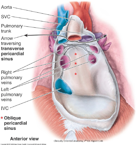
Place a finger in the pericardial cavity, between the parietal and visceral layers of serous pericardium. A snug fit, eh? The pericardial cavity has two specialized regions:
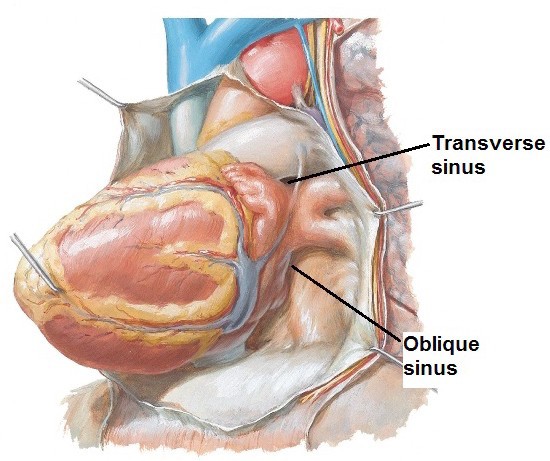
1Transverse pericardial sinus: A channel connecting the left and right sides of the pericardial cavity, posterior to the arterial end of the heart. Slide a finger behind the ascending aorta and pulmonary trunk.
Clinical correlation
A cardiothoracic surgeon could pass a clamp into the transverse sinus and around the great arteries during heart surgeries.

2Oblique pericardial sinus: This is a cul-de-sac behind the base of the heart (left atrium), surrounded by the veins entering the heart. Lift the heart up and out of the pericardium and slide your fingers behind the base of the heart. Your finger will be halted by the serous pericardium that reflects from the inside of the pericardial sac onto the pulmonary veins. Your fingers are in the oblique sinus.
Remove the heart.
Use a scalpel or large scissors to cut the great vessels. Leave about an inch of each vessel attached to the heart. Use the following sequence:
1Elevate the heart to reveal the short course of the inferior vena cava (IVC) within the pericardium. Cut the IVC above the diaphragm.
2From right to left, cut the superior vena cava, ascending aorta, and pulmonary trunk. Don’t cut the pulmonary trunk too high (where it branches into left and right pulmonary arteries), or you will damage the ligamentum arteriosum.
3Lift and rotate the heart from side to side in order to cut the left and right pulmonary veins.
4Remove the heart from the pericardium and place it on a tray.
Now, back to the pericardium: EXAMINE the parietal layer of the serous pericardium—the shiny layer on the inside of the pericardial sac that remains in the donor. Note the reflection of the serous pericardium as it forms cuffs around the pulmonary veins. Identify the location of the oblique pericardial sinus.
External Heart
EXAMINE the external anatomy of the heart.
The heart is a modified blood vessel, so it has three layers (as vessels do).
The outer layer is the epicardium (aka = visceral layer of serous pericardium). It is a serous membrane = a single layer of epithelial cells (mesothelium) supported by a layer of connective tissue (which often contains considerable adipose tissue).
Question
What are the other two layers of the heart?
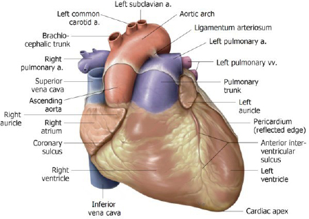
Identify these features of the external heart:
■Apex
■Base
■Sternocostal surface
■Diaphragmatic surface
■Left and right pulmonary surfaces
■Inferior border
Question
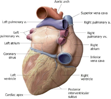
Which chambers of the heart are associated with each of the above surfaces and borders?? We’ll give you a hint and a head start: the Base of the heart is formed by the left atrium.
■Locate the left and right auricles. Clinicians sometimes call these the atrial appendages. The auricles (“ear-shaped”) are parts of the atria.
■The coronary sulcus is an external groove that separates the atria from the ventricles. It is filled with blood vessels and lots of epicardial fat.
■The interventricular sulci (anterior and posterior) are landmarks on the sternocostal and diaphragmatic surfaces, respectively. They indicate the location of the interventricular septum within the heart and can be used to demarcate the locations of the ventricles externally. They are filled with vessels.
Question
Which chamber forms most of the left border?
Question
Which chamber forms the right border?
Question
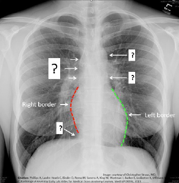
Which vessels merge with the left and right borders of the heart to complete what is called the “cardiac silhouette”?
(Example = can you see the SVC (above) and the IVC (below) merging with the right border of the heart? What structures are seen on the left side?)
Posterior Mediastinum
Posterior mediastinum
Azygos System
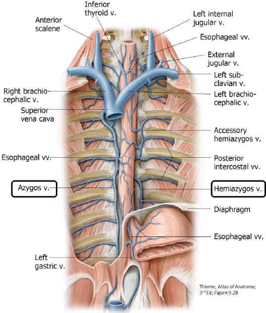
The azygos venous system is composed of two vertical venous channels = the azygos vein proper on the right and the hemi-azygos veins on the left.
On the right side, clean the azygos vein (azygos means “unpaired”). Note that right posterior intercostal veins are tributaries of the azygos vein.
Two venous channels on the left receive the left posterior intercostal veins: the hemiazygos and accessory hemiazygos veins. Clean the mediastinal pleura away from the left surface of the descending aorta. Elevate the aorta and clean along the vertebral column to locate the hemiazygos and accessory hemiazygos veins. As you might expect, they have a variable arrangement and can be small. Venous blood in these veins crosses the vertebral column and drains to the azygos vein. See Figure 9.17.
Clean and examine the sympathetic trunks.
On the left and right sides of the vertebral column, use forceps and blunt tools to clean off the loose connective tissue around the sympathetic trunks. This is tedious work so don’t worry about creating a masterpiece.
One old dissection trick is to rub the area with the blunt end of your scalpel handle to liquefy the fatty tissue a bit—then wipe the area down with paper towels to remove the liquid fatty tissue.
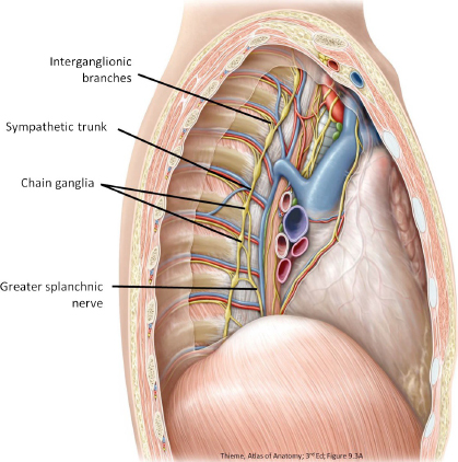
The sympathetic trunk is part of the autonomic nervous system (ANS). It is a bilateral structure composed of interconnected sympathetic (chain) ganglia.
Trace the sympathetic trunk from the superior thoracic aperture down to the diaphragm.

FIND
■Chain ganglia
■Interganglionic branches connect adjacent ganglia
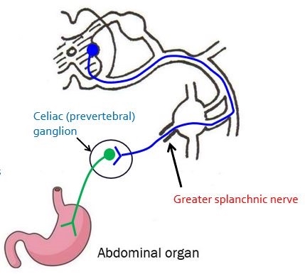
■The greater splanchnic nerve is composed of preganglionic sympathetic nerve fibers that branch medially from thoracic chain ganglia T-5 to T-9. After these medial branches merge, the greater splanchnic nerve heads medially toward the center of the body cavity and penetrates the diaphragm to enter the abdomen. The greater splanchnic nerve supplies sympathetic innervation to organs of the foregut (stomach, liver, gall bladder, pancreas).
Classroom correlation
Earlier in the term, we studied the autonomic nervous system in class. See if you can draw a 2-neuron sympathetic pathway to the stomach. (Tap to open; use your Apple Pencil to draw.)
This pathway utilizes the greater splanchnic nerve to transmit preganglionic sympathetic axons from the thoracic sympathetic trunk to the celiac ganglion (a prevertebral ganglion) in the abdominal cavity. From here, postganglionic neurons innervate the stomach. For review, see the pathway below in Figure 9.19.
CORNELIUS ROSSE, M.D., D.SC., STUDY GUIDE FOR GROSS ANATOMY AND EMBRYOLOGY, UNIVERSITY OF WASHINGTON. USED WITH PERMISSION.
Note
When you have finished this session, return the lungs and chest wall to your donor.
Checklist, Lab #9
Review and make sure you have identified each of the structures below.
Superior mediastinum
Superior thoracic aperture. Which structures form its boundaries?
Trachea
Esophagus
L & R brachiocephalic veins
Superior vena cava
Aorta: ascending, arch, and descending
Brachiocephalic artery (trunk)
L common carotid artery
L subclavian artery
Ligamentum arteriosum
L & R phrenic nerves
L & R vagus nerves
L & R recurrent laryngeal nerves
Pericardium
Fibrous pericardium
Parietal layer of serous pericardium
Visceral layer of serous pericardium
Pericardial cavity
Transverse pericardial sinus
Oblique pericardial sinus
External heart
Great vessels of heart
Superior vena cava
Inferior vena cava
Pulmonary trunk w/ pulmonary arteries (R & L)
Superior and inferior pulmonary veins (R & L)
Aorta
Heart surfaces
Anterior; diaphragmatic; left and right pulmonary
Auricles of heart (atrial appendages)
Heart borders (best seen on X-ray):
Left, right, and inferior
Coronary sulcus
Anterior & posterior interventricular sulci
Right coronary artery & branches
Right marginal artery
Posterior interventricular artery (posterior descending artery)
Left coronary artery and branches
Circumflex artery
Anterior interventricular artery (aka LAD)
Coronary sinus
Cardiac veins (great, middle, and small)
Posterior mediastinum
Azygos vein
Posterior intercostal arteries and veins
Hemi-azygos vein (and accessory hemi-azygos vein, if present)
Descending thoracic aorta
Esophagus and Esophageal plexus
Thoracic duct
Sympathetic trunk w/ chain ganglia