Lab 17: Gluteal Region, Posterior Thigh, and Popliteal Fossa

  1. Clean and identify the muscles, nerves, and vessels of the gluteal region.
  2. Identify the greater sciatic foramen and the structures that traverse it.
  3. Clean and identify the muscles, nerve, and vessels of the posterior compartment of the thigh.
  4. Identify the boundaries and contents of the popliteal fossa.

Osteology Relevant to the Regions

Hip bones

Complete anatomy

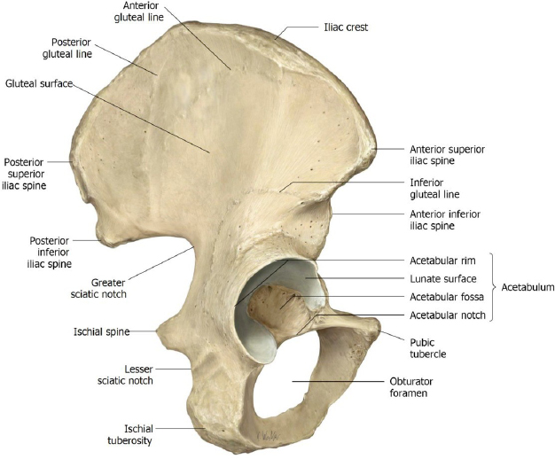
Osteology of hip bones

Iliac crest

Anterior superior and posterior superior iliac spines

Acetabulum

Gluteal lines: posterior, anterior, and inferior

Ischial tuberosity and ischial spine

Greater and lesser sciatic notches

Obturator foramen

Figure 1.

Femur

Complete anatomy

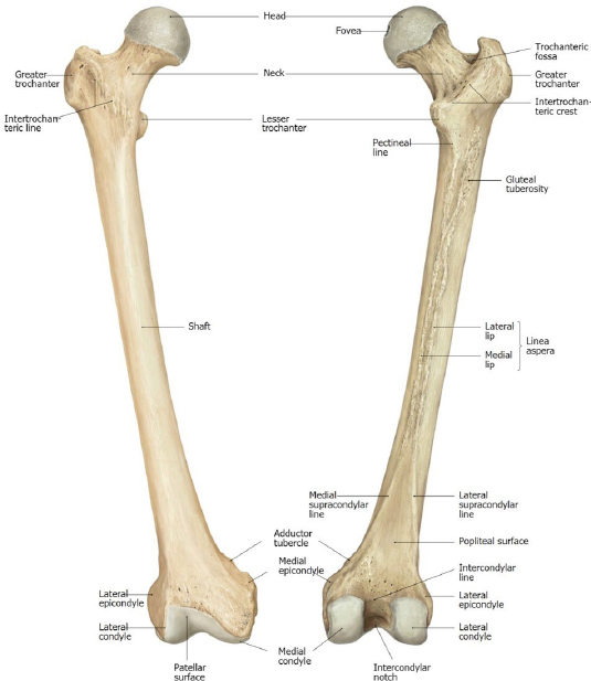
Osteology of femur

Head and Fovea capitis (for attachment of ligament of head)

Neck

Shaft

Greater and lesser trochanters

Intertrochanteric line & crest

Trochanteric fossa

Gluteal tuberosity—rough surface at the proximal end of the linea aspera, just below the lesser trochanter

Linea aspera

Popliteal surface

Medial and lateral femoral condyles

Figure 2.

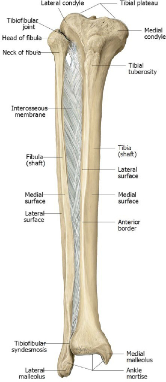
Tibia

Complete anatomy

Osteology of tibia

Anterolateral (Gerdy’s) tubercle—the distal attachment of the iliotibial tract

Medial and lateral tibial condyles

Fibula

Head

Figure 3.

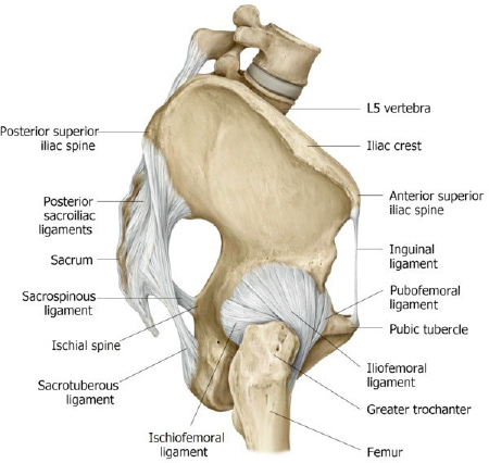
Inspect a bony pelvis or the pelvic ligament model and review the attachments and locations of the:

Sacrospinous ligament

Sacrotuberous ligament

Question

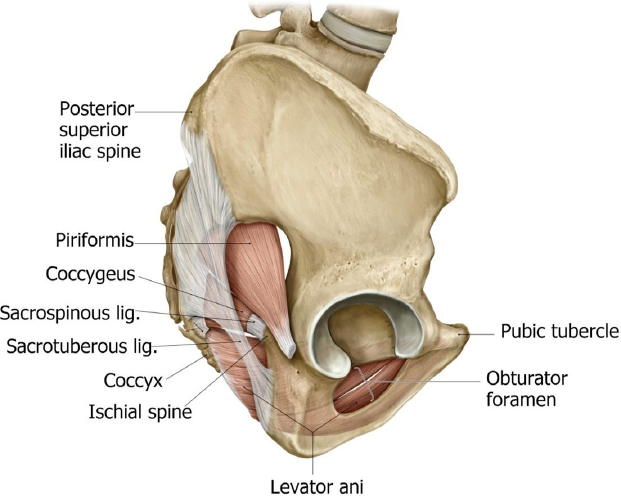
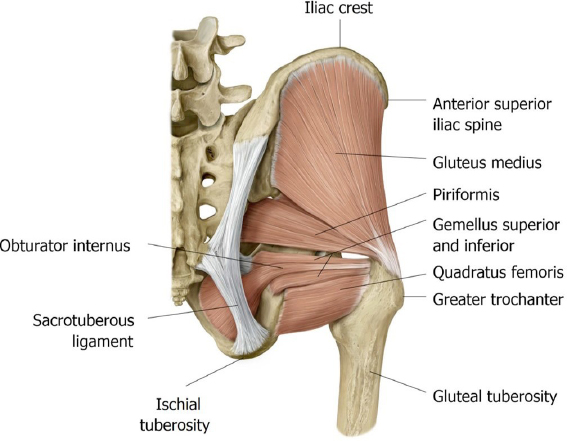
Where is the greater sciatic foramen? Where is the lesser sciatic foramen?

Review the course and attachments of the piriformis and obturator internus muscles (remember them from the pelvis?).

Question

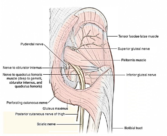
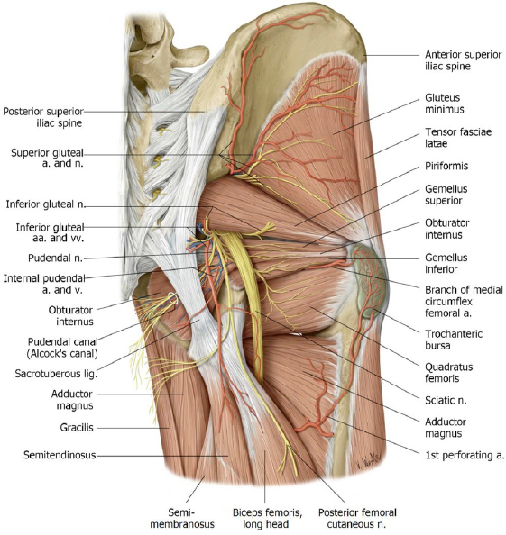
What course do the superior gluteal nerve and vessels take in order to leave the pelvic cavity? How are they related to the piriformis?

Question

What course do the inferior gluteal nerve and vessels & the sciatic nerve take in order to leave the pelvic cavity? How are they related to the piriformis?

Figure 4.
Figure 5.

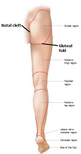
Gluteal Region: Surface Anatomy

You will need to turn the dettached leg into the prone position so we can access the gluteal region and posterior thigh.

Before cutting, see if you can locate, observe, and/or palpate the following:

Natal cleft (Intergluteal cleft)

Gluteal fold

Iliac crest

Anterior superior and posterior superior iliac spines

Greater trochanter

Sacrum and coccyx

Ischial tuberosity

Popliteal fossa

Head of fibula

Figure 6. Surface anatomy of gluteal region/posterior thigh.
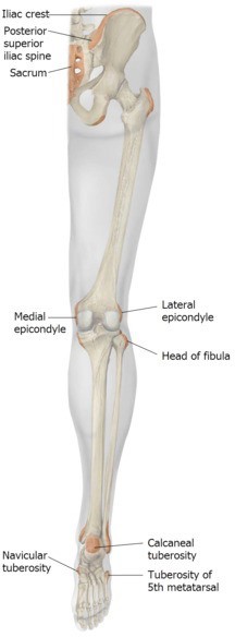
Figure 7. Palpable bony prominences of the lower limb. Right limb.
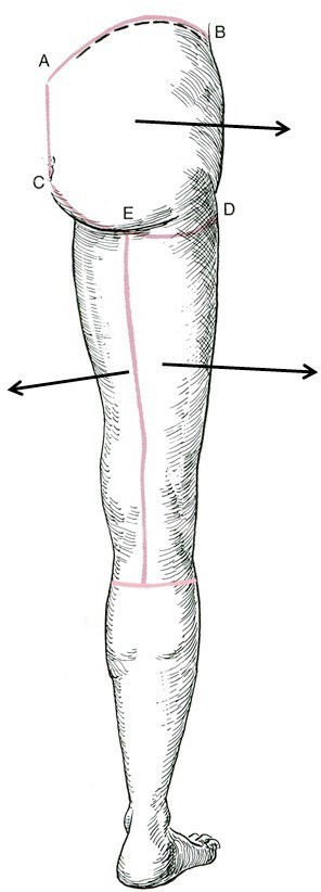
Make skin incisions.

1Start at the posterior superior iliac spine (A) and make a curved incision forward along the iliac crest to the ASIS (B).

2Now make a curved incision from medial to lateral just below the gluteal fold (C to D).

3Reflect the skin flap over the gluteus maximus laterally.

1Now make a vertical incision from superior to inferior in the posterior thigh, starting at the gluteal fold above and ending below in the proximal calf

2Now make a horizontal incision just below the knee – stay SUPERFICIAL so you don’t cut small saphenous vein entering the popliteal fossa!

3Reflect the two flaps of skin in the posterior thigh medially and laterally.

Figure 8.
Clean off gluteal muscles.

Complete anatomy

Superficial muscles of the gluteal region

1Remove the superficial fascia from the buttock in chunks.


There will be lots to remove! Clean all the superficial fascia away to reveal the deep fascia over the gluteus maximus muscle. It’s important that you clean enough away so that you can clearly visualize the superior and inferior borders of the gluteus maximus.

Note

This will be difficult along the inferior border of the gluteus maximus, where the gluteal region meets the posterior thigh.

Perseverance is the key!

2Above the superior border of the gluteus maximus will be some robust deep fascia that covers the gluteus medius. Open this fascia with a scalpel and scissors to better reveal the superior border of the gluteus maximus.

Figure 9.
Clean superficial structures of posterior thigh.

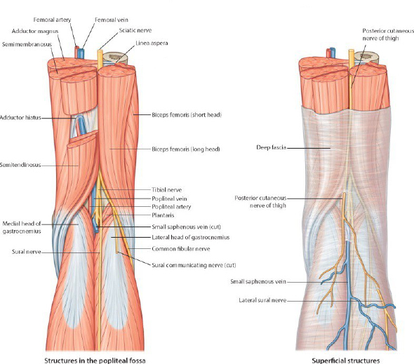
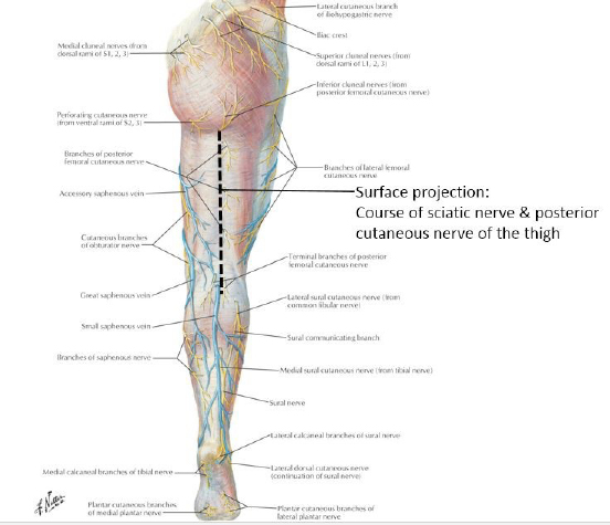
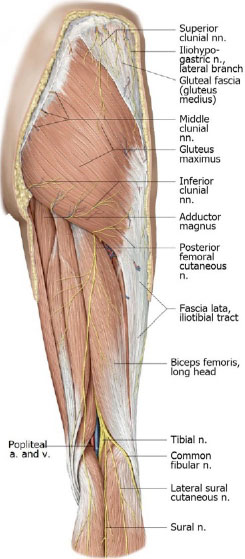
Palpate the ischial tuberosity and greater trochanter. Before doing any more cutting, look at Figure 10 and visualize the approximate course of the sciatic nerve and posterior cutaneous nerve of the thigh as they leave the pelvic cavity and pass down the center of the posterior thigh.

Figure 10.

Clean the copious superficial fascia from the posterior thigh to reveal the super tough fascia lata (deep fascia of the thigh) in the posterior thigh. The hamstring muscles are deep to this fascia.

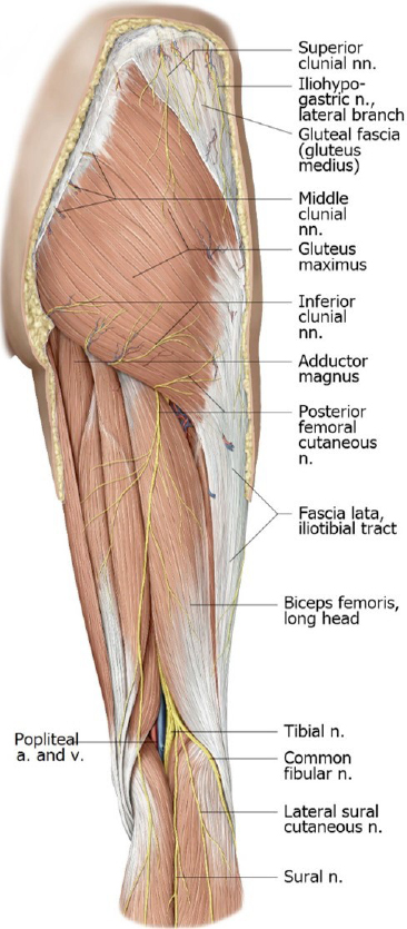
Now, with scissors carefully open up and reflect the fascia lata in the posterior thigh, looking for the posterior cutaneous nerve of the thigh running down the midline of the posterior thigh, beginning above at the midpoint of the inferior border of the gluteus maximus and ending below in the posterior calf.

The posterior cutaneous nerve of the thigh is an oddball—it courses DEEP to the fascia lata, and sends small branches through the fascia lata into the overlying superficial fascia and skin.

Figure 11.

Gluteal Region

Free up and then transect the gluteus maximus muscle.

1With blunt dissection, loosen up the superior border of the gluteus maximus muscle just below the iliac crest.

Question

What are the distal attachments (insertions) of the gluteus maximus? What is the main function of the gluteus maximus?

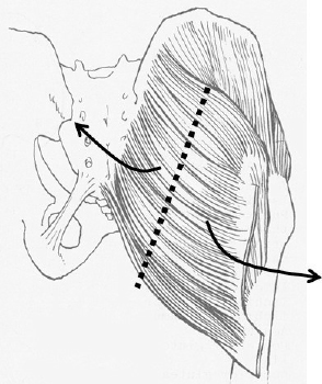
Review the anatomy of the iliotibial tract (thickening of the fascia lata) that acts as an aponeurosis for insertion of two muscles: the gluteus maximus and the tensor fasciae latae.

Question

What are the attachments of the tensor fascia latae? What are its actions?

Figure 12.

2With blunt dissection, create a plane between the gluteus maximus muscle and the underlying muscles beneath it.

Create this plane by sliding your hand beneath the gluteus maximus above its superior border, between the gluteus maximus and medius muscles.

3Transect the entire gluteus maximus from superior to inferior—from the midpoint of the superior border of the muscle to its inferior border where it crosses the ischial tuberosity.

Figure 13.

Reflect the halves of the gluteus maximus and locate the inferior gluteal nerve and vessels entering the deep side of the muscle.

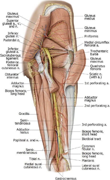
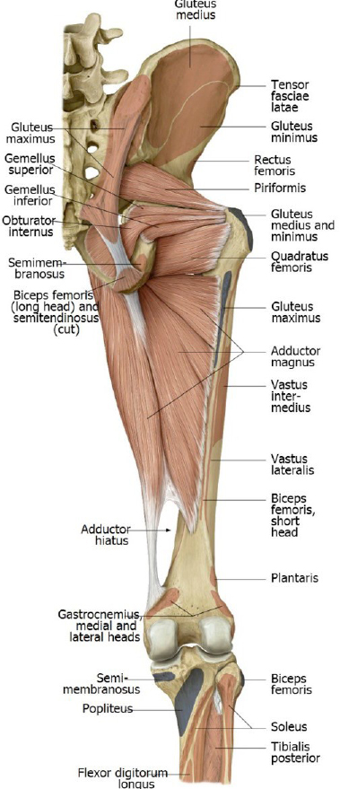
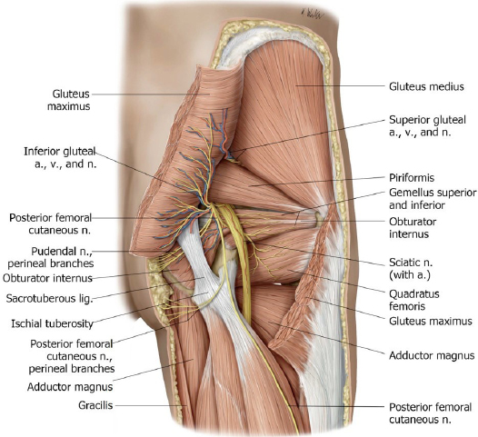
The key to the gluteal region is the piriformis muscle. Use blunt dissection to define the borders of the piriformis muscle as it emerges from the greater sciatic foramen to attach to the greater trochanter.

Below the piriformis, identify the inferior gluteal neurovascular bundle, the sciatic nerve, the posterior cutaneous nerve of the thigh, and most medially, the internal pudendal vessels and pudendal nerve.

Figure 14.

Clinical puzzler

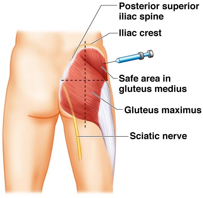
Divide the gluteal region into four quadrants: upper medial, upper lateral, lower medial and lower lateral. If you wanted to give a patient an intramuscular injection—which quadrant would be the best choice? (Injections should be done in safe places where there are no major nerves or vessels to injure.)

Figure 15.
Figure 16.

Review the course of the internal pudendal vessels and pudendal nerve as they pass from the pelvic cavity to the perineum

Clean and transect the gluteus medius.

1Use tools and blunt dissection to clean the surface of the gluteus medius muscle and separate it from the piriformis.

2Use blunt dissection to loosen up the borders of the gluteus medius.

Figure 17.

3Then cut across the gluteus medius transversely several inches above the greater trochanter. Reflect the cut edges of the muscle to expose the gluteus minimus muscle.

Note

Note that the superior gluteal nerve and vessels pass between the gluteus medius and minimus.

Figure 18.

Question

What are the attachments of the gluteus medius and minimus? Their actions?

Clinical Pearl

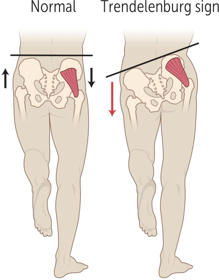
The gluteus medius and gluteus minimus abduct the thigh at the hip. Additionally, these muscles are crucial to stabilizing the pelvis when you walk or raise one limb off the ground. When one limb is raised from the ground and the other limb bears the weight of the entire body above it, the gluteus medius and minimus contract to keep the contralateral pelvis from sagging (since the lower limb on the contralateral side is not in contact with the ground, and the pelvis would therefore have the tendency to sag).

Figure 19.

Weakness or paralysis of the ipsilateral gluteus medius and minimus—the side with the limb planted on the ground—would present as sagging/drooping of the contralateral pelvis—the side with the limb raised off the ground. When tested, this sagging/drooping of the contralateral pelvis is called a Trendelenburg sign.
Clean and identify the external hip rotator muscles.

Complete anatomy

Deep muscles of the gluteal region

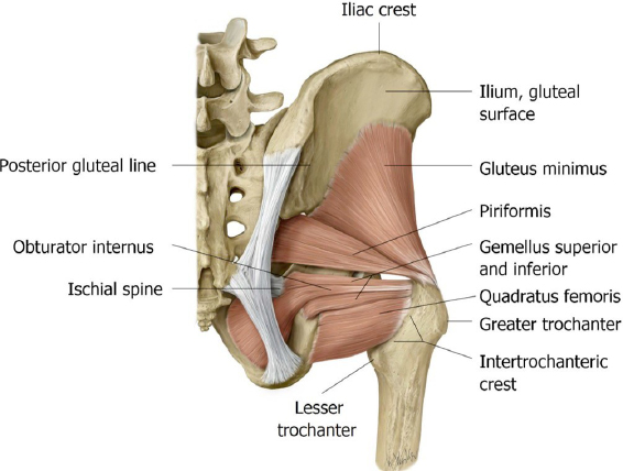
The tendon of the obturator internus muscle passes through the lesser sciatic foramen to attach to the greater trochanter. Where is the origin of the obturator internus? 

The shiny, white tendon of the obturator internus muscle is sandwiched between two small, elongated muscles that lie in the horizontal plane: The gemelli muscles (the “twins”).

Locate the gemellus superior muscle and gemellus inferior muscle above and below the tendon of the obturator internus. The gemelli are small external rotators of the hip.

Identify the quadratus femoris muscle attaching to the ischial tuberosity and the greater trochanter. Look at the relationship between the quadratus femoris and the sciatic nerve.

Figure 20.

With blunt dissection, separate the quadratus femoris from the inferior gemellus.

Summary—From superior to inferior, identify these external hip rotator muscles:

Piriformis

Superior gemellus

Tendon of obturator internus

Inferior gemellus

Quadratus femoris

Posterior Thigh

Remove the remaining fascia lata from the thigh to reveal the posterior compartment.

Complete anatomy

Posterior thigh

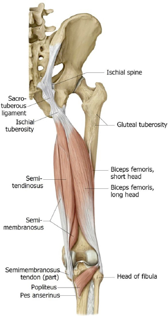
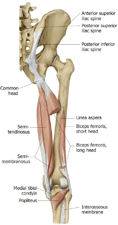
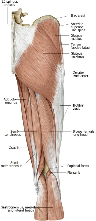
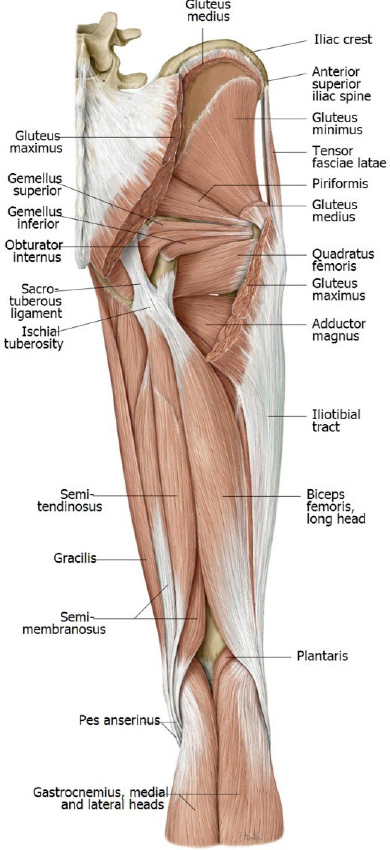
With blunt dissection, separate the three muscles that make up the hamstrings = from lateral to medial these are the:

Biceps femoris

Semitendinosus

Semimembranosus

Question

Why are they named this way?

Trace them to their insertions.

Question

What are the attachments of the hamstrings?

Note that they have a common proximal attachment, but their tendons diverge distally.

Question

Why is the lateral member of the hamstrings called the “biceps” femoris?

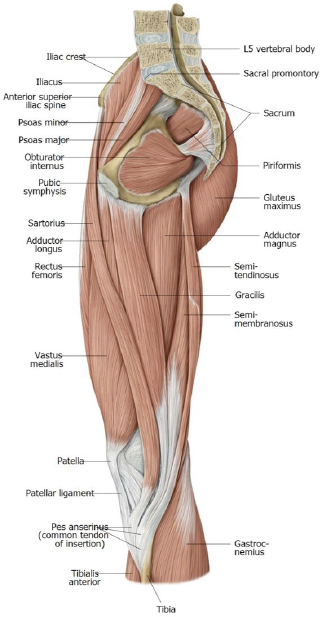
Review the anatomy of the pes anserinus. (See figures.) Although we haven’t dissected the anterior leg yet, you have heard of the pes, since it is the common distal attachment site for three muscles—two of which we examined in our last lab (sartorius and gracilis).

Question

Which of the hamstrings attaches to the pes?

Figure 24.

Trace the sciatic nerve from the gluteal region distally as it passes through the posterior thigh into the popliteal fossa. Note that it passes deep to the long head of the biceps femoris.

The tibial part of the sciatic nerve innervates these muscles:

Semitendinosus

Semimembranosus

Long head of the biceps femoris

Hamstrings portion of the adductor magnus muscle

The common fibular part of the sciatic nerve supplies the short head of the biceps femoris.

Both nerves supply all the muscles in the lower extremity below the knee.

Usually in the mid-thigh is where the sciatic nerve splits into the tibial nerve and common fibular nerve.

This is variable; it may occur as high as the gluteal region or as low as the popliteal fossa.

Figure 25.

Chalk Talk

Draw the sacral plexus and its branches - discuss the spinal levels that form the nerves seen in lab.

Complete anatomy

Lumbosacral plexus

Revisit the medial compartment to clean and identify structures related to the posterior thigh.

1Rotate the detached limb so that the medial compartment (work you did last lab) is visible. Clean the adductor magnus muscle and locate its two portions: adductor part and hamstrings part.

Review their innervations.

2Clean and identify the adductor hiatus, the distal opening of the adductor canal.

The femoral vessels change names as they exit the hiatus, becoming the popliteal artery and vein.

Figure 26.

Popliteal Fossa

Carefully clean the superficial fascia from the popliteal fossa.

Complete anatomy

Structures in popliteal fossa

1Locate the small saphenous vein (ascending from the calf region) as it enters the popliteal fossa to terminate in the popliteal vein.

2Identify the borders of the diamond-shaped popliteal fossa:

roof, the crural fascia (deep fascia of the leg)

medial inferior border, the medial head of gastrocnemius

lateral inferior border, the lateral head of gastrocnemius

lateral superior border, the long head of the biceps femoris

medial superior border, the semimembranosus

floor, the popliteus muscle, popliteal surface of the femur, and the capsule of the knee joint

You won’t see the popliteus muscle today—but know that it is located in the floor of the popliteal fossa, hidden by the two heads of the gastrocnemius: It functions to “unlock” the fully extended knee joint, so that it can be flexed.

Figure 27.

Question

Where are the tibial nerve and common fibular nerve positioned within the popliteal fossa?

3Using blunt dissection, identify from superficial to deep:

Tibial nerve

Popliteal vein

Popliteal artery (the inferior continuation of the femoral artery after it passes through the adductor hiatus)

Checklist, Lab #17

Review and make sure you have identified each of the structures below.

Bones

See the list of bony landmarks in the osteology section at the top of this page.

Muscles—know where they attach and their innervations

Gluteus maximus, medius, and minimus

Tensor fasciae latae w/ Iliotibial tract

Piriformis

Obturator internus (tendon)

Superior and inferior gemellus muscles

Quadratus femoris

Biceps femoris: short and long heads

Semimembranosus

Semitendinosus

Adductor magnus (adductor and hamstring parts)

Other

Adductor hiatus—in adductor magnus muscle

Sacrotuberous and sacrospinous ligaments

Review boundaries of popliteal fossa

Nerves and Vessels

Superior gluteal artery and nerve

Inferior gluteal artery and nerve

Posterior cutaneous nerve of thigh

Sciatic nerve: Tibial and Common fibular nerves

Popliteal artery and vein

Small saphenous vein (see it entering the popliteal vein)

Table of Contents
Headshot of David Conley, PhD · Professor, Department of Translational Medicine & Physiology
David Conley
PhD · Professor, Department of Translational Medicine & Physiology
Office: PBS 41A
envelope icon
Headshot of Shannon Helbling, PhD · Clinical Assistant Professor, Department of Translational Medicine & Physiology
Shannon Helbling
PhD · Clinical Assistant Professor, Department of Translational Medicine & Physiology
Office: PBS 41C
envelope icon

Dear students,
We would love to hear from you about your learning materials.

Page/Content Feedback Form