Lab 27: Dissection: Infratemporal Fossa and Floor of Mouth; Bony Anatomy of Pterygopalatine Fossa

  1. Identify the temporal fossa and temporalis muscle.
  2. On a skull, identify the borders of the infratemporal fossa (ITF) and the foramina that connect it to other regions in the head.
  3. Clean the masseter muscle on one side of the face and remove it from the mandible.
  4. Use an autopsy saw to cut the mandible and zygomatic arch on one side. Remove the bones to access the infratemporal fossa.
  5. Clean and identify the pterygoid muscles and major branches of the mandibular nerve (V3) and maxillary artery in the ITF.
  6. Disarticulate the temporomandibular joint on one side and study its anatomy.
  7. Remove the body of the mandible to provide a lateral view of the floor of the mouth. Clean and identify muscles, nerves, vessels, and salivary glands.
  8. With the mandible removed, identify structures in the submandibular triangle of the neck.
  9. On a skull, identify the pterygopalatine fossa and its associated foramina.

Bony Anatomy: Temporal and Infratemporal Fossae

On a skull, identify the temporal fossa, infratemporal fossa (ITF), and temporomandibular joint (TMJ).

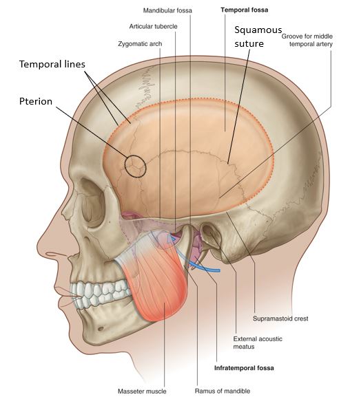
The following bony parts relate to the temporal fossa (Figure 1):

Figure 1. Temporal fossa.

■Temporal lines: These are sites of attachment for the temporalis muscle and the temporal fascia.

■Greater wing of sphenoid bone

■Squamous part of temporal bone

■Squamous suture

■Zygomatic arch: Temporal process of zygomatic bone + zygomatic process of temporal bone.

■Pterion: The thin-boned “temple” region behind the orbits, where sutures unite.
Question

What is the clinical significance of the pterion?

Question

Go to the donor and answer this: Which structure fills the temporal fossa?

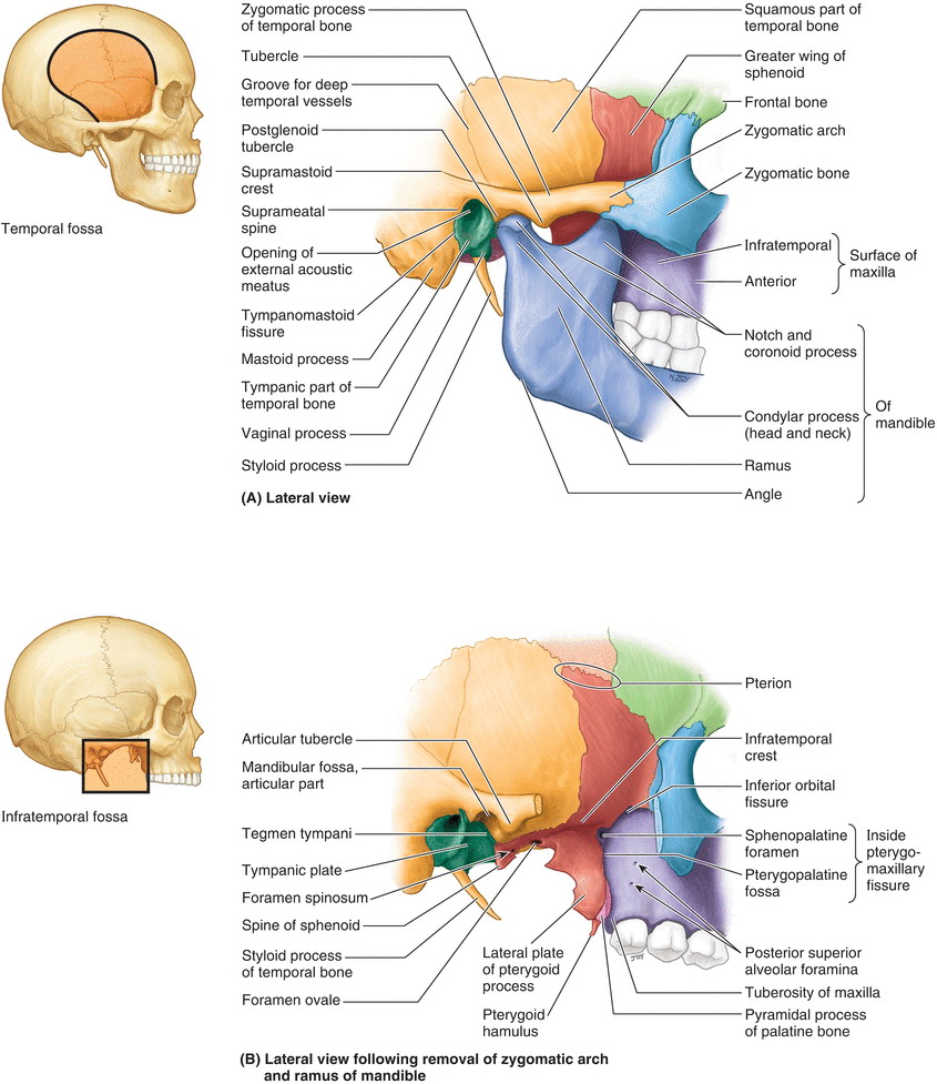
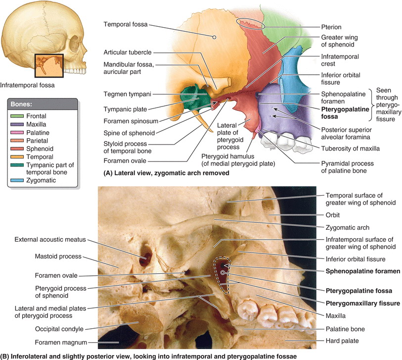
Now locate the infratemporal fossa (ITF), deep to the ramus of the mandible (removing the mandible will help). The bony borders of the ITF are (Figure 2):

■Lateral wall: Ramus of mandible

■Medial wall: Lateral pterygoid plate of sphenoid bone

■Anterior wall: Body of maxilla (contains the maxillary sinus)

■Posterior: Tympanic part of temporal bone and styloid process

■Roof: Greater wing of sphenoid bone

Using a pipe cleaner, probe these bony openings related to the ITF:

■Foramen ovale (in the roof)

■Foramen spinosum (in the roof)

■Pterygomaxillary fissure (in the medial wall)

■Inferior orbital fissure (in the anterior wall)

Figure 2. Bones of the infratemporal fossa.

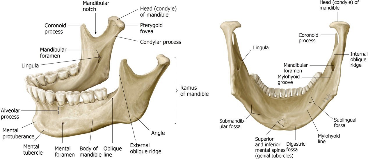
On an isolated mandible, identify the following (Figure 3):

■Ramus, body, and angle

■Coronoid process (temporalis muscle inserts here)

■Condylar process and condyle (head)

■Mandibular foramen (for the inferior alveolar nerve and vessels)

■Mylohyoid groove (for the mylohyoid nerve)


Identify the bony parts of the temporomandibular joint (TMJ) [see Figure 2 and Figure 3]:

■Condyle of mandible

■Mandibular fossa of temporal bone

Figure 3. Parts of the mandible.
Dissection of infratemporal fossa

We will dissect the infratemporal fossa on one side of the head only. Do this on the same side where the face was dissected.

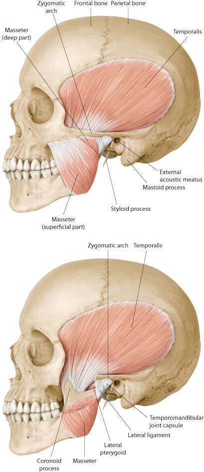
■Clean the surface of the masseter muscle.

■The masseter is a muscle of mastication. It arises above from the zygomatic bone and zygomatic arch. It inserts below into the external surface of the mandibular ramus and angle of the mandible. What is the function of the masseter?
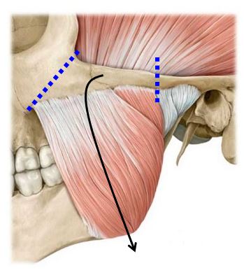
Figure 4. Reflection of the masseter muscle.

■Use an autopsy saw to make cuts through the zygomatic bone and zygomatic arch, as shown in Figure 4. Loosen up the detached segment of zygomatic bone and reflect it downward, along with the attached masseter muscle (Figure 4). As you pull down, use a chisel or scissors to scrape/cut away the masseter from its attachment to the external surface of the mandibular ramus. Leave the reflected masseter muscle and zygomatic bone fragment hanging from the mandible or remove it entirely by cutting it at its distal attachment to the angle of the mandible. The external surface of the mandible is now visible.
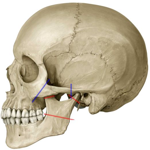
  • Use the autopsy saw to make three cuts in the mandible (RED lines shown in Figure 5).
  • Cut through the coronoid process and through the neck of the condylar process.
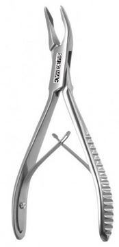
  • Deeply score the bone of the mandibular ramus horizontally as shown—but don’t saw all the way through the bone. Use a chisel and hammer to break the ramus into bone fragments. Wear eye protection!! Be careful because this may sever underlying nerves. Use bone rongeurs to pull away the bone fragments. (Click on the   at right for a picture of bone rongeurs.)
Figure 5. Bone cuts in mandible.

  • The temporalis muscle arises from the floor of the temporal fossa. It inserts on the coronoid process of the mandible and inner surface of the ramus. See Figure 6. What are the actions of the temporalis muscle?
  • Grasp the detached upper portion of the cut  coronoid process with forceps and reflect it superiorly with the temporalis. You will notice that the temporalis inserts quite far inferiorly on the inside of the mandibular ramus. The muscle is very thick behind the zygomatic bone. You will need to remove the distal part of the temporalis with scissors to get a clear view of the ITF. Take care to probe through the distal part of the temporalis muscle as you remove it, as the buccal nerve (V3) passes just deep to the temporalis on its way to the cheek.
Figure 6. Temporalis muscle attachments.

■Keep reflecting the temporalis upward until you can see the bony floor of the temporal fossa. Retain the deep temporal arteries and nerves if you can. They enter the deep surface of the temporalis, right against the bone.

After the temporalis muscle has been reflected, its inferior muscle fibers cleared, and the fragments of mandibular ramus removed, the ITF will be visible.

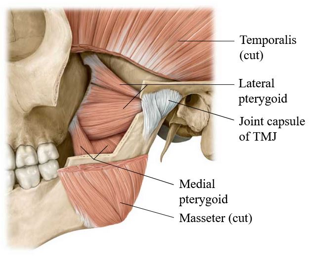
Identify and clean the lateral and medial pterygoid muscles (Figure 7).

The ptergygoid venous plexus will appear as a network of dark veins in the connective tissue surrounding the pterygoid muscles.

■The lateral pterygoid muscle arises from the lateral surface of the lateral pterygoid plate (sphenoid bone). It has upper and lower heads. Where does the lateral pterygoid insert? What are its actions?

■The medial pterygoid muscle arises from the medial surface of the lateral pterygoid plate. Where does it insert? What are its actions?

Figure 7. Pterygoid muscles.

Fun fact

The medial pterygoid is essentially the mirror image of the masseter, only it attaches to the internal surface of the mandibular ramus, not the external, as the masseter does.

Lingual and Inferior Alveolar Nerves

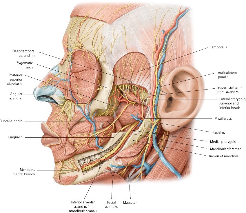
■Identify the lingual nerve and the inferior alveolar nerve on the external surface of the medial pterygoid muscle (Figure 8). They are sandwiched between the muscle and the internal surface of the mandible. The inferior alveolar nerve may have been transected when the mandible was cut.

■Separate the inferior alveolar artery from the nerve. It is a branch of the maxillary artery.

■The inferior alveolar nerve and vessels pass into the mandible via the mandibular foramen. They course through the mandible within the mandibular canal. See Figure 8. What is the function of the inferior alveolar nerve?

Figure 8. Nerves of the infratemporal fossa.

Buccal Nerve (V3)

Identify the buccal nerve (of V3) passing between the two heads of the lateral pterygoid on its way to the cheek. See Figure 8. It might be hidden by the inferior fibers of the temporalis. It is a sensory nerve supplying the skin on the external cheek and the mucous membranes inside the cheeks in the oral cavity.

Don’t get confused!

There are two buccal nerves! The buccal branch of VII is motor; the buccal nerve of V3 is sensory.

Maxillary Artery

Question

The maxillary artery is one of the two terminal branches of the external carotid. What is the other one?

Figure 9. Maxillary artery in the ITF.

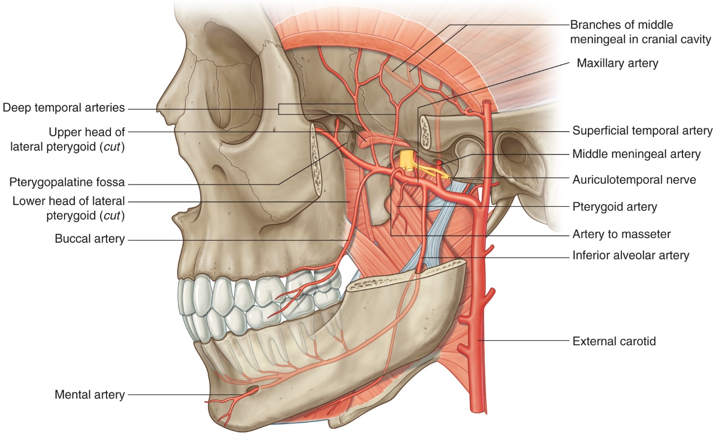
The maxillary artery (Figure 9) enters the ITF deep to the condylar process of the mandible. Once in the ITF, it passes either superficial or deep to the lateral pterygoid muscle. You will need to do some cleaning to determine which is the case in your donor. If it passes deep, you won’t see much of the artery until you remove the lateral pterygoid (instructions on this are given later).

The distal (medial) part of the maxillary artery leaves the infratemporal fossa via the pterygomaxillary fissure and enters the pterygopalatine fossa (Figure 9).

Dissection of the branches of the maxillary artery is difficult! Our goal is to trace it through the ITF to the pterygomaxillary fissure and try to identify several of its major branches. Figure 10 is a sketch that lists ALL the branches of the maxillary artery. We will seek out only a few of these today. Don’t sweat the small branches.
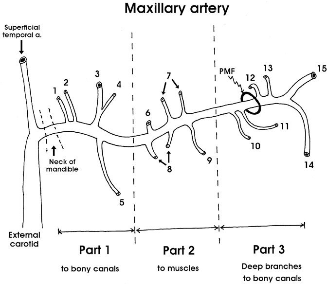
Figure 10. Branches of the maxillary artery.
(Tap the to see the labels of each part of the maxillary artery “wonder” drawing.)

Removal of lateral pterygoid muscle

To better see the nerves and vessels in the infratemporal fossa, the lateral pterygoid muscle will be removed. Carefully remove the lateral pterygoid muscle in pieces with scissors and forceps.
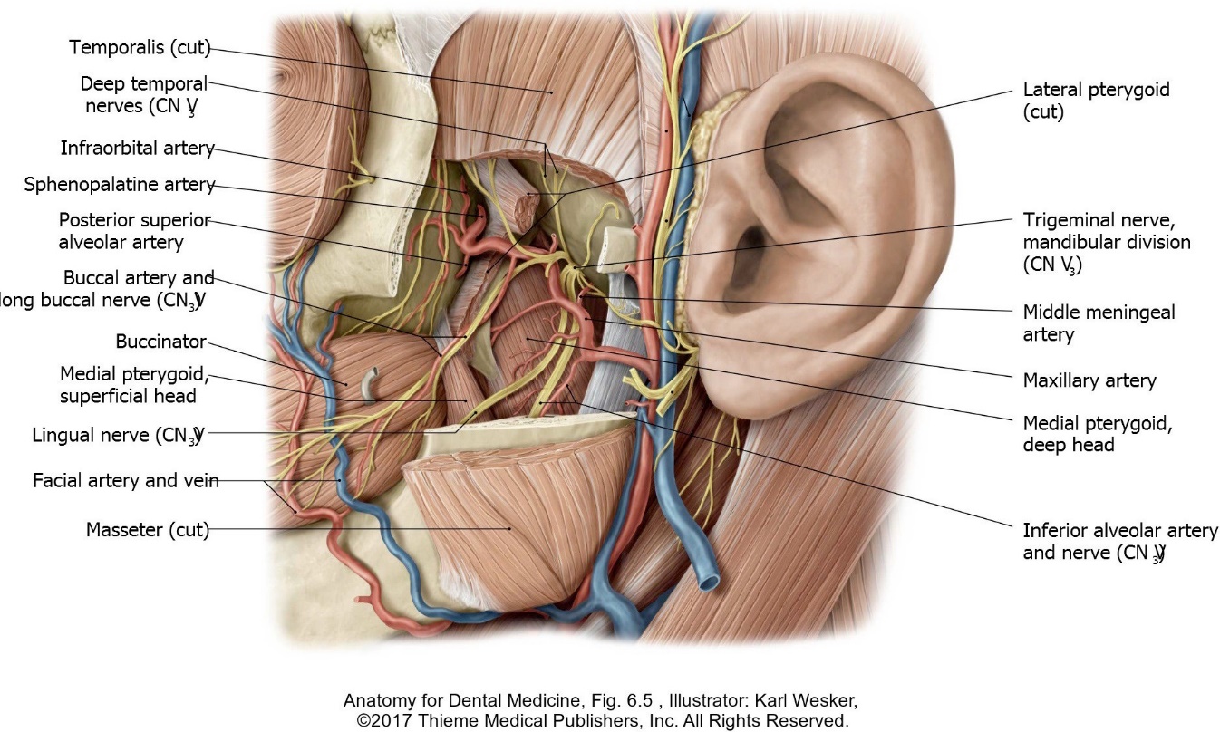
Figure 11. Nerves and arteries in the ITF.

■Identify the buccal nerve passing between the two heads of the lateral pterygoid. Preserve this nerve as the muscle is removed.

■Locate and clean the maxillary artery as you remove the lateral pterygoid. Look for these branches of the artery as you dissect (Figures 9 and 11):

■Middle meningeal artery. It exits through the roof of the ITF through which foramen? What is the function of the middle meningeal artery? What is its clinical importance in the case of skull fracture?

■Inferior alveolar artery: Accompanies the nerve of the same name.

■Deep temporal arteries: Ascend to the deep surface of the temporalis
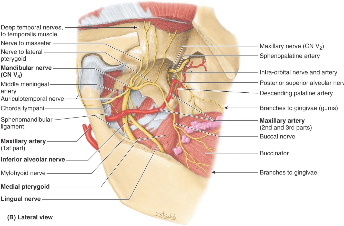
After the lateral pterygoid muscle has been removed, identify the branches of V3 in the ITF (see Figures 11 and 12):

Figure 12. Branches of the mandibular nerve in the ITF.

■Buccal nerve

■Inferior alveolar nerve

■Lingual nerve

■Deep temporal nerves

■Auriculotemporal nerve

Use the middle meningeal artery as a guide to find the auriculotemporal nerve. Oddly enough, the nerve splits around the middle meningeal, then courses laterally, deep to the parotid gland. After leaving the ITF, the auriculotemporal nerve ascends to the scalp with the superficial temporal vessels. This is a toughie to find!

Chorda Tympani Nerve (From CN VII)

■Trace the lingual nerve superiorly as high as you can.

■The chorda tympani nerve (from CN VII) leaves the tympanic cavity and enters the ITF through a small crack = the petrotympanic fissure. Once in the ITF, the tiny chorda tympani nerve joins the lingual nerve from behind. Chorda tympani carries two functional types of nerve fibers. What are they?

■The nerve fibers in the chorda tympani travel to and from their targets (salivary glands & tongue) by “hitching-a-ride” on the lingual nerve.

Finding the chorda tympani joining the lingual nerve makes you a rockstar!

Review: Innervation of the Major Salivary Glands

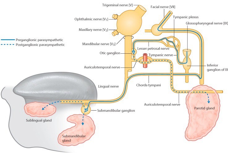
The innervation routes to the submandibular, sublingual, and parotid glands pass through the infratemporal fossa.

Chalk Talk

The pathway of parasympathetic innervation of the submandibular and sublingual salivary glands goes through the ITF. Sketch it if you can. Also sketch how taste fibers from the anterior 2/3 of the tongue are transmitted to the CNS. [Consult the interactive Conley-gram on cranial nerve VII after lab.] See Figure 13.

Chalk Talk

Discuss the pathway of parasympathetic innervation of the parotid gland. It also goes through the ITF. See Figure 13.

Figure 13. Autonomic pathways through the ITF to salivary glands.

Temporomandibular Joint (TMJ)

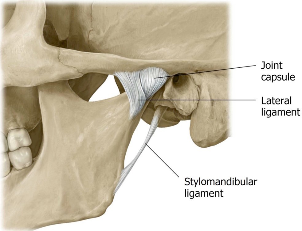
Review the anatomy of the TMJ. See Figure 14. Its bony parts are the condyle of the mandible + the mandibular fossa of the temporal bone. It’s a modified hinge joint that also has translational movements (forward and backward).

■Wiggle the stump of the condylar process of the mandible that is left in your donor, so you have an idea of the location of the TMJ.

■The TMJ is a synovial joint. It is surrounded and reinforced by an articular capsule and ligaments. Remove any remaining parts of the lateral pterygoid muscle and cut the capsule with a scalpel to open the TMJ.

■Identify the articular disc (meniscus) made of fibrocartilage. The articular disc divides the internal TMJ cavity into upper and lower joint compartments.

■The lateral pterygoid muscle inserts into the mandible, articular capsule, and articular disc. Thus, when the mandible is protruded, the articular disc moves forward with the condylar head.

Figure 14. Temporomandibular joint (TMJ).

■With forceps or rongeurs, grasp and tug hard to remove the condylar process of the mandible, thus disarticulating the TMJ. Note the articular cartilage (different from the articular disc) on the head of the condylar process. It may be eroded due to lifetime wear on the TMJ.

Question

Where is the articular disc? It may be attached to the bone fragment, or it may remain attached to the TMJ capsule in your donor. Ask your instructor for help.

Lateral Approach to the Floor of the Mouth

Now let’s trace the nerves forward from the ITF to the floor of the mouth. We will do this work on the same side as the ITF dissection.

Remove the lower lip.

■Make a vertical midline incision in the lower lip from the oral fissure to the tip of the chin. Cut away the lower lip from the mandible externally.
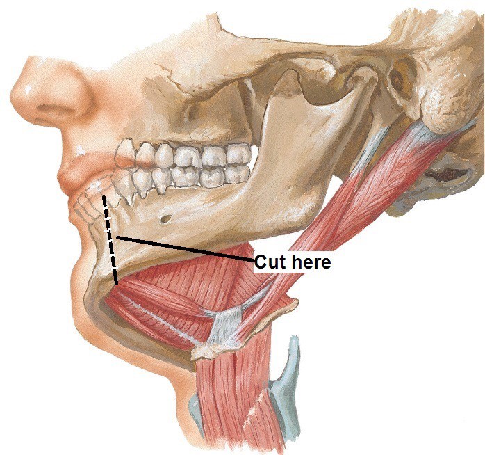
Figure 15. Removal of mandibular body.

■Transect the mandible with an autopsy saw or hacksaw, off the midline a bit, toward the side of the head with the ITF dissection (Figure 15). Cut downward vertically between the incisor teeth. Cutting off the midline will keep the geniohyoid and genioglossus muscles intact.

■With scissors, detach the anterior belly of the digastric and mylohyoid muscles from the inferior surface of the cut mandible and reflect them down.

■Pull the detached part of the mandible away from the head to apply traction, then reach into the oral cavity and use a scalpel or scissors to cut away the oral mucosa where it is attached to the lingual aspect of the lower gums.
With the segment of mandible now removed, we need some landmarks:

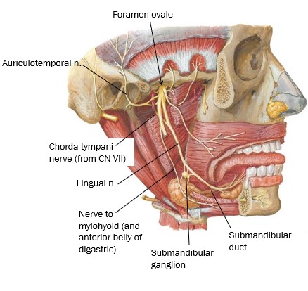
Figure 16. Lingual nerve below the tongue.

■Trace the lingual nerve forward from the ITF. Look for a wide spot as it curves below the tongue—this is the location of the submandibular ganglion (a parasympathetic ganglion). Cell bodies of post-ganglionic parasympathetic fibers that innervate the submandibular and sublingual glands are located here. Consult Figure 16.

■Trace the hypoglossal nerve forward from the neck to below the tongue.
Remember it can be found just inferior to the posterior belly of the digastric and styloyhyoid muscles.

■Identify the inferior alveolar nerve and trace it distally (it has likely been cut, so only a stump may remain). Just before it enters the mandibular foramen it gives off a thin motor branch to the neck called the nerve to mylohyoid and anterior belly of digastric, which runs inferiorly on the medial side of the mandible. See Figure 16.
Question

Its function? What else—it innervates the two muscles in its name!

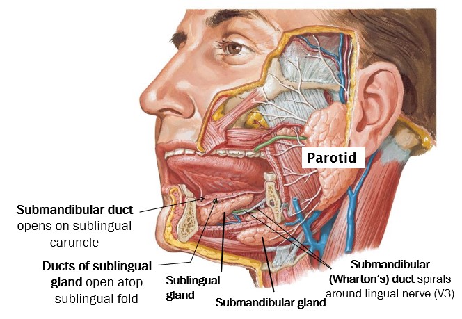
Figure 17. Salivary glands in the floor of the mouth—lateral view.

■Clean the submandibular gland and the facial artery twisting around it. Note that the submandibular gland has a large superficial part (external to mylohyoid in the submandibular triangle) and a small deep part (above the mylohyoid muscle in the floor of the mouth). The 2 parts of the gland connect by curling around the posterior “free edge” of the mylohyoid, like the letter U. See Figure 17.

■Clean the sublingual gland, located just behind the chin (Figure 17). It is a small thin gland. At first, you may not recognize it since it may not appear very glandular and can be pale in appearance.

■Starting at the deep part of the submandibular gland and using the lingual nerve as a landmark, search for the flat, thin, white duct of the submandibular gland (Wharton’s duct). It passes deep to the lingual nerve and can be tricky to find. See Figures 17 and 18.
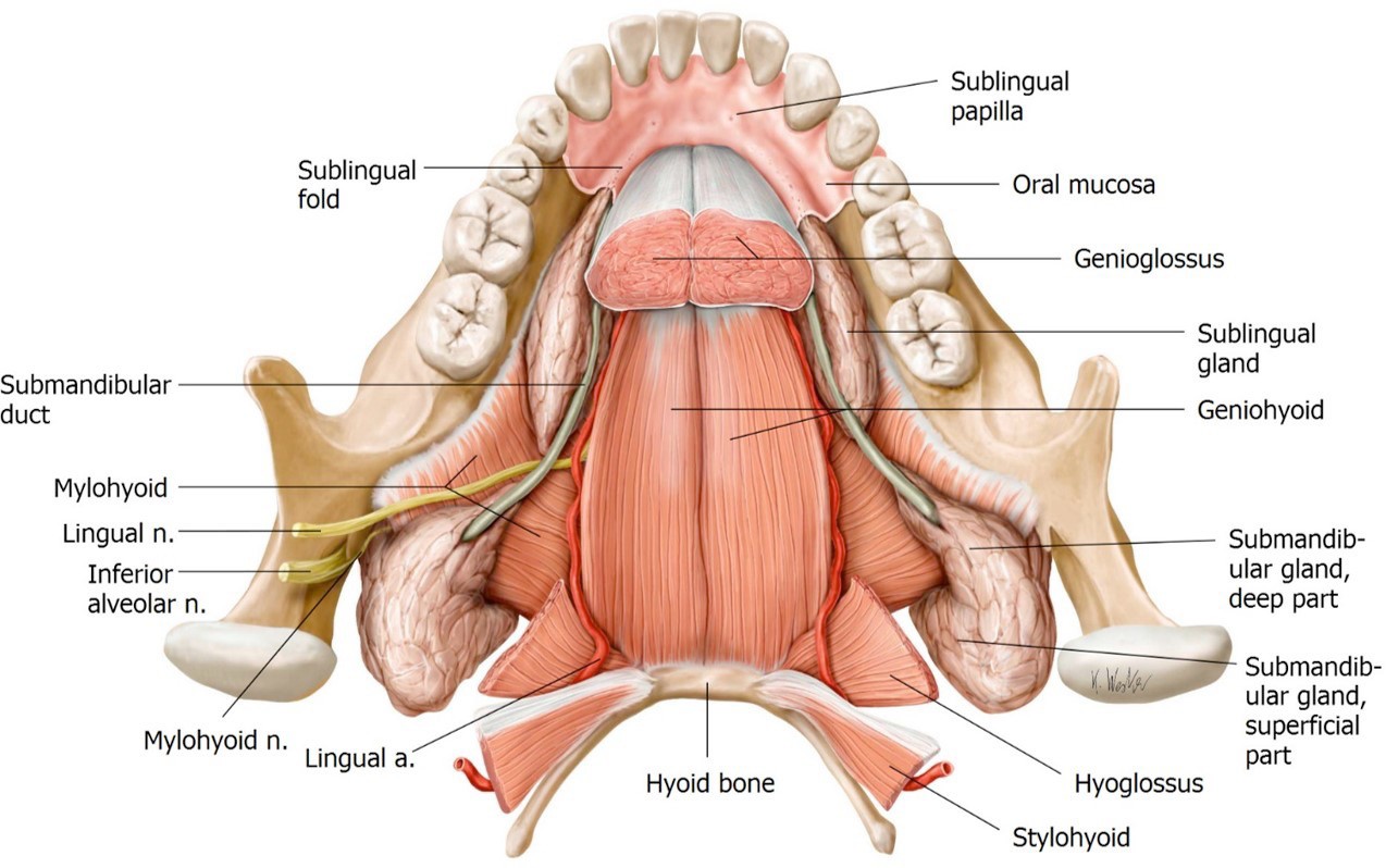
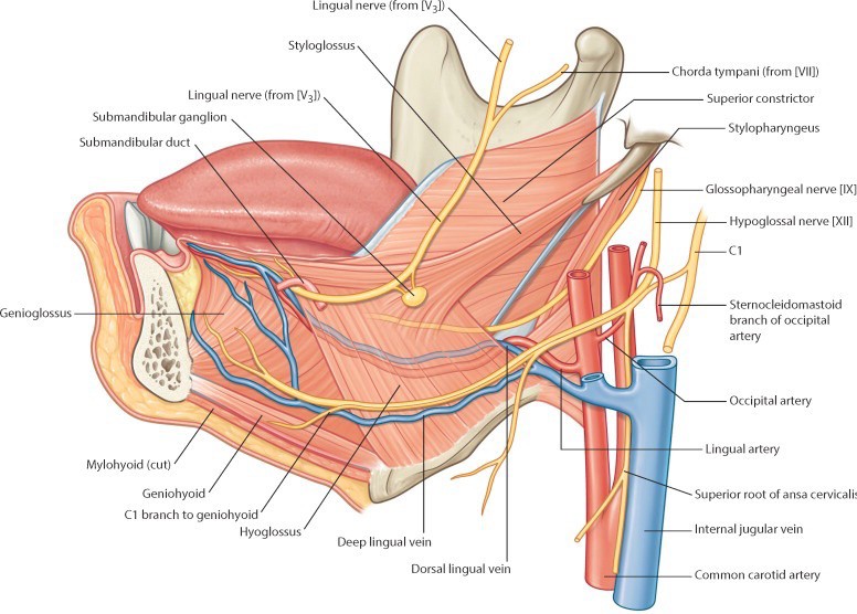
Relationships to note (Figures 16, 17, and 18):

■The lingual nerve and submandibular duct twist around one another.

■The lingual nerve and submandibular duct pass medial to the sublingual gland, between the gland and the genioglossus muscle.

■Review: Where does the submandibular duct open in the oral cavity?

Figure 18. Salivary glands in the floor of the mouth—superior view.
Cut the mylohyoid muscle down the midline and reflect it inferiorly and out of the submandibular triangle.

Now you can locate the hyoglossus muscle. It ascends from the hyoid bone to the root of the tongue. This muscle is a landmark.

■The lingual nerve, submandibular duct, and hypoglossal nerve are on the EXTERNAL surface of the hyoglossus.

■The lingual artery and glossopharyngeal nerve pass INTERNAL to the hyoglossus.

■Figure 19 summarizes these points.

Figure 19. Neurovascular structures below the tongue; related to the hyoglossus muscle.

Stylo-muscles

With a finger, palpate the styloid process by probing deeply, posterior to the infratemporal fossa and anterior to the ear.
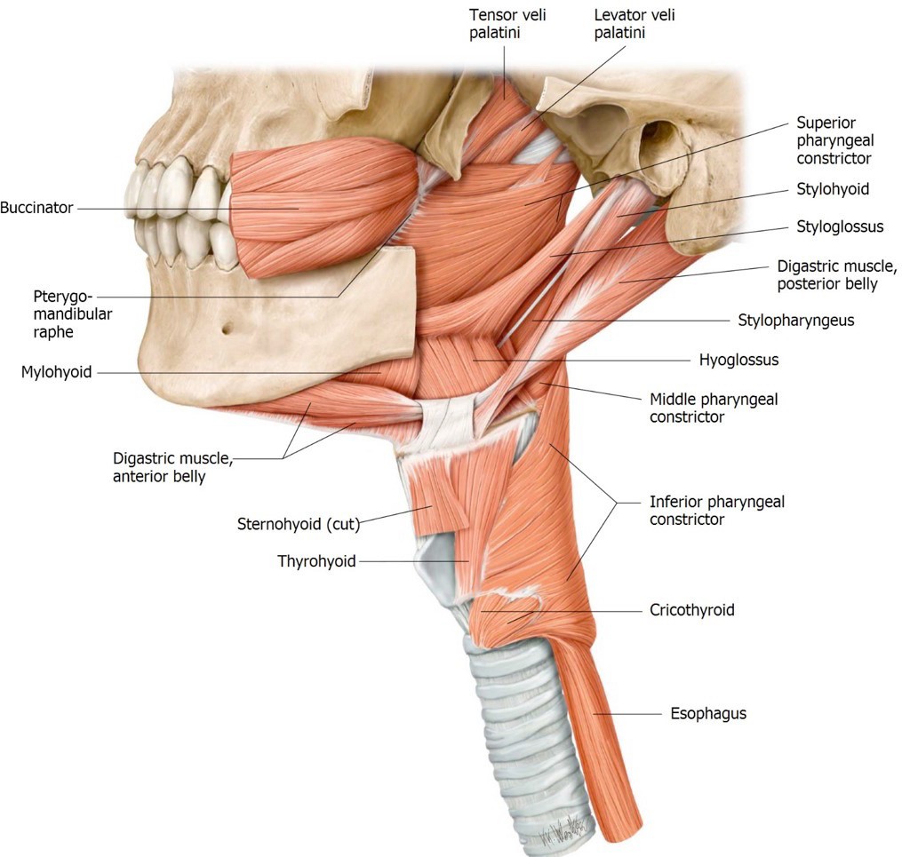
Figure 20. Muscles attaching to the styloid process.

Starting at the styloid process, clean and trace the stylohyoid, styloglossus, and stylopharyngeus muscles. They can be identified by their orientations and destinations. See Figure 20.

■The stylohyoid is intimately associated with the posterior belly of the digastric.

■The styloglossus blends with the hyoglossus in the root of the tongue.

■The stylopharyngeus is the deepest since it descends to the pharynx.

■The glossopharyngeal nerve is located on the posterior border of the stylopharyngeus (the only muscle that CN IX supplies!). It is not very large and a bit difficult to identify. See Figure 19.
You have ascended to anatomy master status if you can find the glossopharyngeal nerve!

Question

Each of the “Stylo” muscles has a different innervation. Can you name them? This also means they have different embryonic origins.

Pterygopalatine Fossa (PPF)

Grab a skull and pipe cleaners.

The pterygopalatine fossa is a small cavity, no bigger than your thumbnail, deep in the skull. It is a major crossroad for nerves and vessels supplying the oral cavity, nasal cavity, and orbit—but difficult to access clinically.

In lab, you are responsible for identifying the pterygopalatine fossa (PPF) on a skull and identifying the major bony passageways/foramina that connect the pterygopalatine fossa to other regions in the head. In the Large Group session we will discuss the contents of the PPF. However, we won’t be dissecting the fossa or identifying its contents in lab.

Identify the pterygomaxillary fissure in the skull. Recall that this fissure is in the medial wall of the infratemporal fossa. Place a pipe cleaner through the pterygomaxillary fissure—the tip of the pipe cleaner is in the pterygopalatine fossa. See Figure 21.

Figure 21. Location of the pterygopalatine fossa.

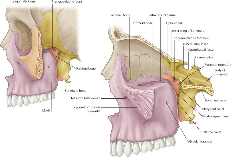
Borders of pterygopalatine fossa (Figure 22):

■The maxilla is the anterior wall (purple in Figure 22).

■The pterygoid process of the sphenoid bone is the posterior wall (yellow in Figure 22). The greater wing of the sphenoid is in the roof.

■The palatine bone is the medial wall (orange in Figure 22). The nasal cavity is on the other side of the palatine bone.

■The lateral wall of the PP fossa is open to the infratemporal fossa—via the pterygomaxillary fissure.

Figure 22. Borders of the pterygopalatine fossa.

We won’t identity these in lab, but you should know the contents of the pterygopalatine fossa:

■Pterygopalatine ganglion (a parasympathetic ganglion): The postganglionic neurons whose cells bodies are in the ganglion innervate the lacrimal gland and mucous glands in the nasal cavities.

■Maxillary nerve (V2)

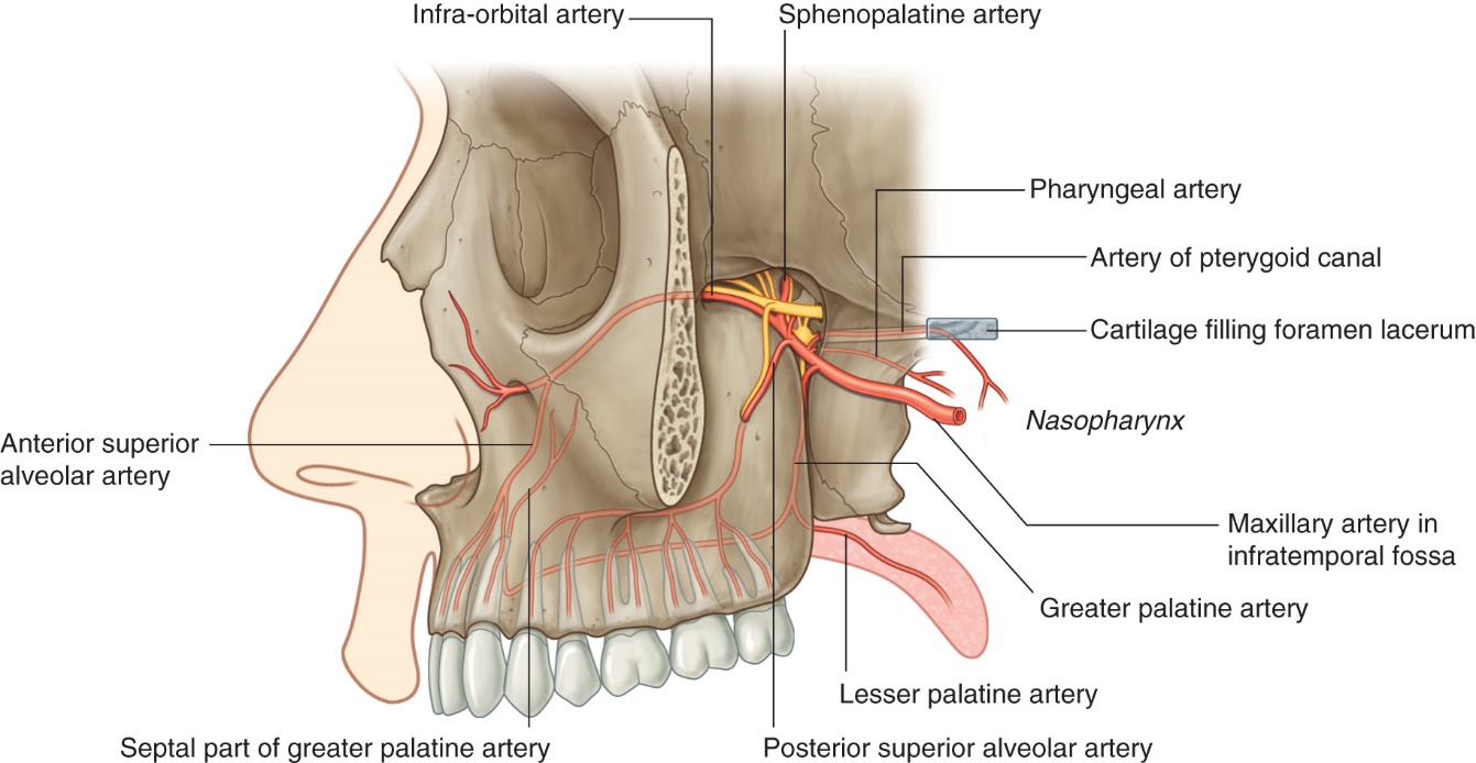
■Terminal part of the maxillary artery: The two most important branches of the maxillary artery in the pterygopalatine fossa are the descending palatine artery (to the palate) and the sphenopalatine artery (to the nasal cavity).

Figure 23. Contents of the pterygopalatine fossa.

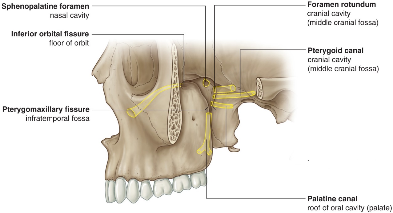
Now let’s identify some foramina that transmit nerves and vessels in and out of the PPF:

■Place a pipe cleaner from lateral to medial through the pterygomaxillary fissure—through the PPF—and into the nasal cavity. Verify this by looking in the nasal cavity through the piriform aperture. The pipe cleaner has passed from PPF to nasal cavity through the sphenopalatine foramen. The sphenopalatine artery follows this route as do branches of V2 that innervate the nasal mucosa.

■In the middle cranial fossa, place a pipe cleaner through the foramen rotundum. The tip is now in the PPF. Verify this by looking in the PPF through the pterygomaxillary fissure. The maxillary nerve (V2) enters the PPF via this route.

■In the orbit, pass a pipe cleaner backwards through the inferior orbital fissure. The tip is in the PPF. Verify this by looking in the PPF through the pterygomaxillary fissure. The infra-orbital and zygomatic nerves (branches of V2) and the infra-orbital artery (from the maxillary artery) use this route to get from PPF to orbit.

■Examine the inferior surface of the hard palate. Find the greater palatine foramen. You might be able to pass a small pipe cleaner up through the greater palatine foramen and then the palatine canal into the PPF. The palatine nerves (greater and lesser) and the descending palatine artery (from the maxillary) take this route to get from the PPF to the palate.

■Locate the foramen lacerum in the floor of the cranial cavity. Imagine a small passageway connecting the anterior wall of the foramen lacerum to the PPF. This is the pterygoid canal (Vidian canal). The greater petrosal nerve (a branch of CN VII) uses this route to reach the PPF, carrying preganglionic parasympathetic nerve fibers to the pterygopalatine ganglion, where they will synapse. Postganglionic axons emanating from the ganglion innervate the lacrimal gland as well as mucous glands in the nasal cavity.

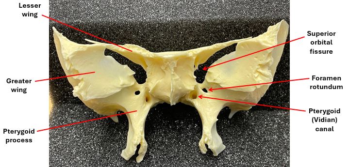
■Foramen rotundum and the pterygoid canal pass through the sphenoid bone and enter the pterygopalatine fossa through its posterior wall. Their positions are shown in the isolated sphenoid bone in Figure 24.

Figure 24.

A summary of these passageways is shown in Figure 25.

Figure 25. Foramina of the pterygopalatine fossa.

Checklist, Lab #27

Review your dissection and make sure you have identified each of the structures in the checklists that follow.

Bony Anatomy of Temporal and Infratemporal Fossae and Mandible—see the first section of this dissection guide.

Muscles

Masseter

Temporalis

Medial and lateral pterygoid

Mylohyoid

Digastric—anterior and posterior bellies

Geniohyoid

Stylohyoid

Styloglossus

Stylopharyngeus

Hyoglossus

Nerves

Buccal nerve of V3

Deep temporal nerves

Lingual nerve

Submandibular ganglion

Inferior alveolar nerve

Nerve to mylohyoid and anterior belly of digastric

Auriculotemporal nerve

Hypoglossal nerve

Glossopharyngeal (Wish list You are an anatomy god if you can find it)

Chorda tympani (Wish list A good find and definitely pin test worthy!)

Vessels

Superficial temporal artery

Maxillary artery

Middle meningeal artery

Inferior alveolar artery

Deep temporal arteries

TMJ

Articular disc (meniscus) of TMJ

Other

Submandibular gland

Duct of Submandibular gland (Wharton’s duct)

Sublingual gland

Pterygopalatine fossa
Bony canals that connect to the pterygopalatine fossa:

Pterygomaxillary fissure

Foramen rotundum

Inferior orbital fissure

Greater palatine foramen

The Pterygoid canal (Vidian canal) connects the Foramen lacerum to the Pterygopalatine fossa (see an isolated sphenoid bone)

Table of Contents
Headshot of David Conley, PhD · Professor, Department of Translational Medicine & Physiology
David Conley
PhD · Professor, Department of Translational Medicine & Physiology
Office: PBS 41A
envelope icon
Headshot of Shannon Helbling, PhD · Clinical Assistant Professor, Department of Translational Medicine & Physiology
Shannon Helbling
PhD · Clinical Assistant Professor, Department of Translational Medicine & Physiology
Office: PBS 41C
envelope icon

Dear students,
We would love to hear from you about your learning materials.

Page/Content Feedback Form