
- Goals
-
Understand the general arrangement of pelvic organs in the male and female and identify those organs that can be approached through the pelvic inlet
- Identify pelvic organs and their subparts
-
Identify muscles in the walls and floor of the pelvic cavity
-
Clean and identify the internal iliac vessels and their branches
-
Identify ventral rami of sacral spinal nerves in the pelvic cavity
-
Locate the greater sciatic foramen
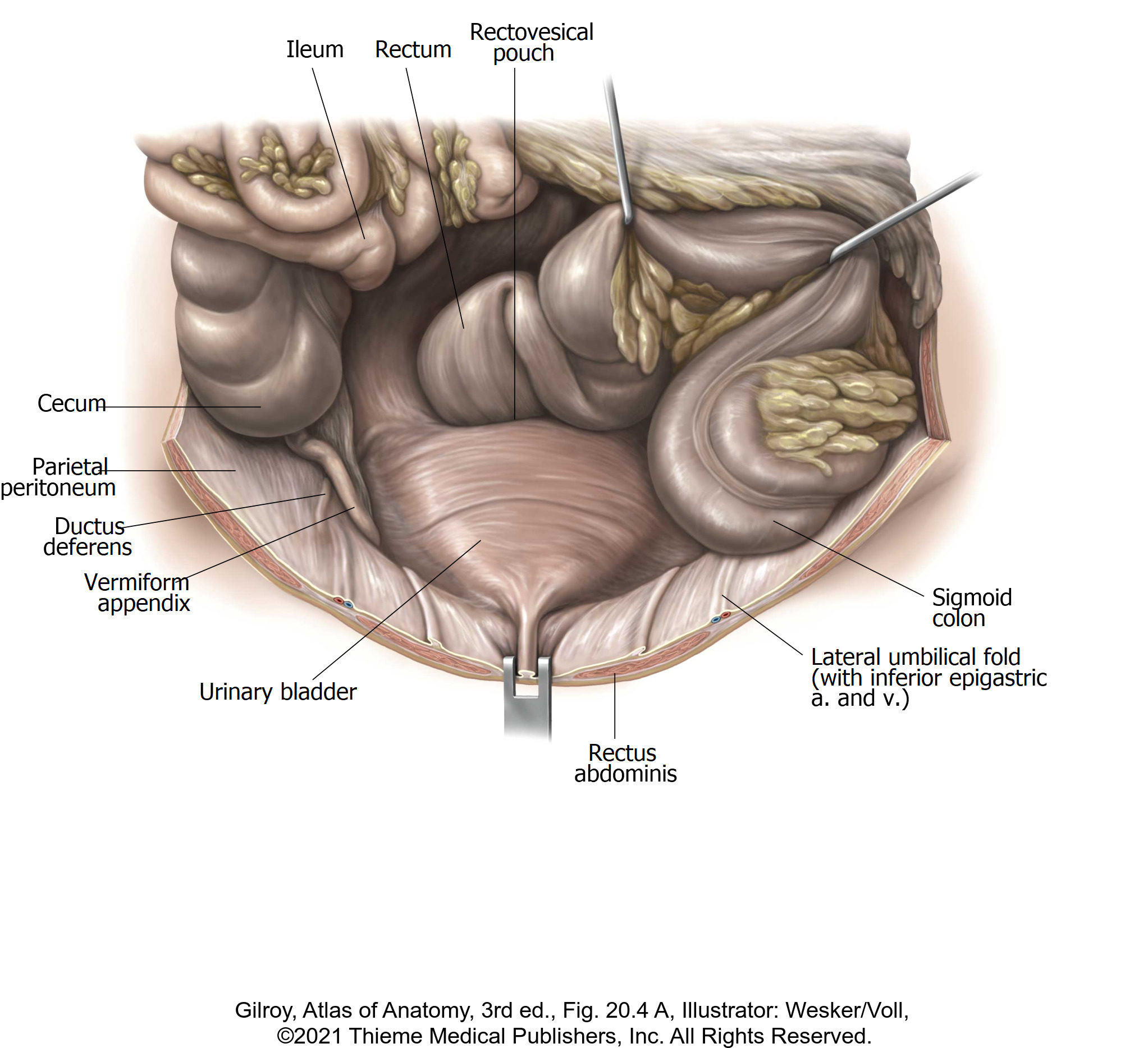
Superior View of Male Pelvic Organs
Before dissecting, locate the rectum and bladder, then identify the recto-vesical pouch. Recall that this peritoneal out pocketing is the most inferior extent of the peritoneal cavity.
Identify the following:
■Pubic symphysis
■Urinary bladder—connected to the anterior body wall by the median umbilical ligament (urachus)
■Ureters
■Pelvic part of ductus deferens (trace it from the deep inguinal ring into the pelvic cavity. Where does it terminate?)
■Testicular vessels
■Rectum
Superior View of Female Pelvic Organs
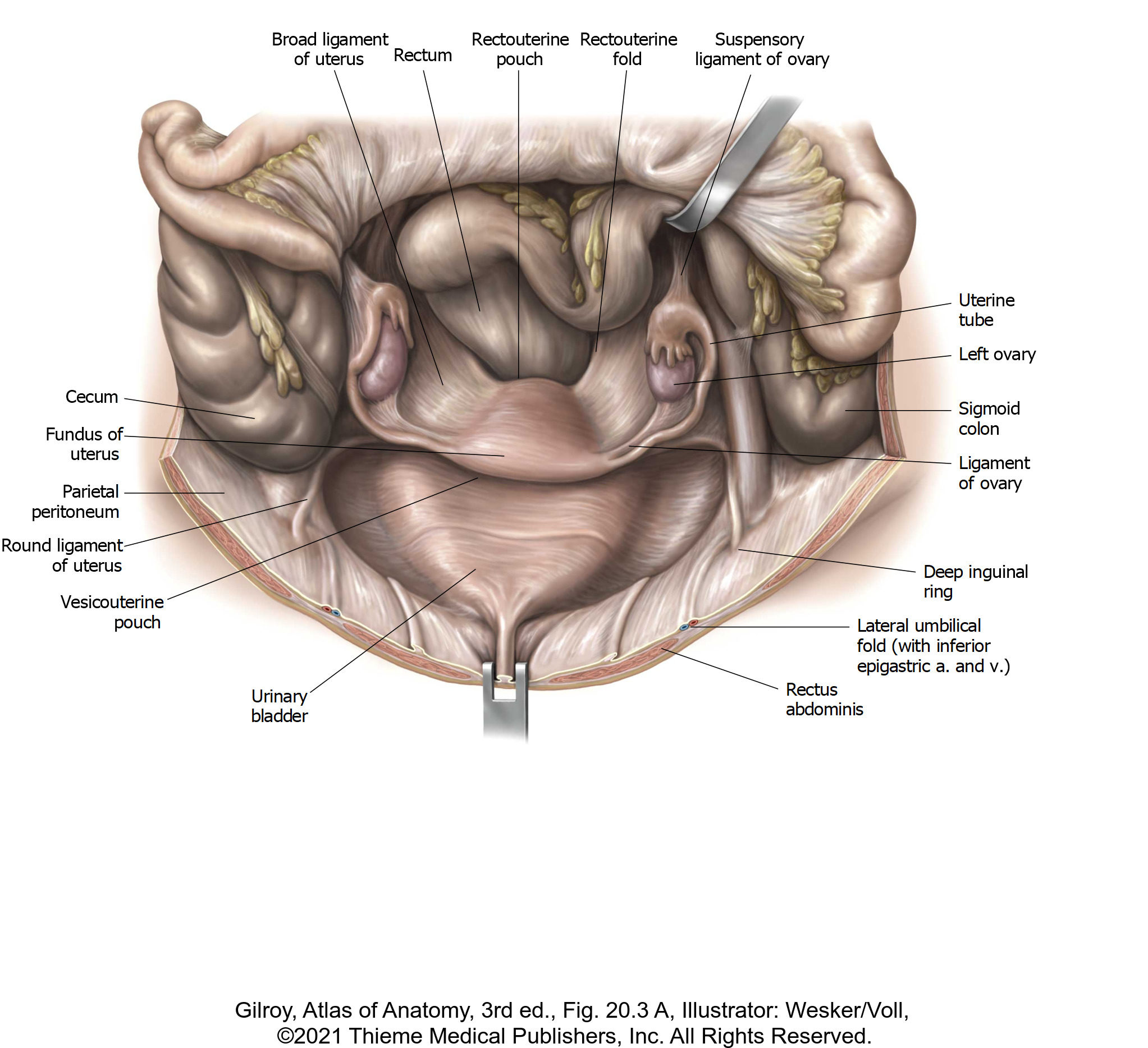
Before dissecting, locate the rectum and bladder. If your donor has a uterus, identify the recto-uterine and vesico-uterine pouches. The recto-uterine pouch is also known as the pouch of Douglas. Why is it clinically important?
Identify the following:
■Pubic symphysis
■Urinary bladder
■Ovarian vessels
If your donor has a uterus, IDENTIFY:
■Uterus (bent anteriorly over the urinary bladder).
■Uterine (Fallopian) tubes
■Ovaries
■Broad ligament = a peritoneal “tent” that is pitched over the uterus and Fallopian tubes; it also attaches to the ovaries. The three parts of the broad ligament are:
■Mesometrium
■Mesosalpinx
■Mesovarium
Through the transparent broad ligament, you may see the fibrous cord of the round ligament of the uterus stretching from the uterus to the deep inguinal ring.
Question
Externally, where do the round ligaments of the uterus attach?
The peritoneum that drapes off the pelvic wall and covers the ovarian vessels is the suspensory ligament of the ovary (infundibulopelvic ligament).
Question
Where do the ovarian arteries originate? Where do the ovarian veins terminate?
Dissection of the Pelvic Cavity and Organs
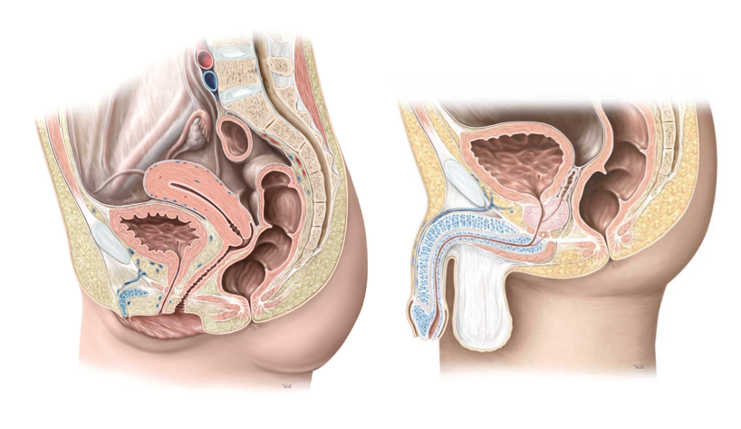
Figure 3.
The hemi-sectioned pelvis will allow your team to work on both sides of the pelvic cavity and have clear access to the pelvic floor and lateral pelvic walls.
Superficial side: Your team will identify organs and their parts on this side. Leave the peritoneum in place if possible—reflect it toward the midline in order to clean the subperitoneal space below it. The superficial side should be the side of the pelvis that is still attached to the body.
Deep side: Remove the pelvic peritoneum and reflect the organs toward the midline. Clean the pelvic walls, pelvic floor, and the branches/tributaries of the internal iliac vessels. The deep side should be the side of the pelvis that is detached with the lower limb.
Rectum and Anal Canal
Clean out feces from the rectum and anal canal with paper towels.
Dispose of these in the biohazardous waste barrel as soon as possible.
Notice that despite its name, the rectum is NOT straight. It has several lateral bends called flexures, which form internal rectal folds. Below the lowest fold is the dilated ampulla which receives and accumulates feces for defecation.
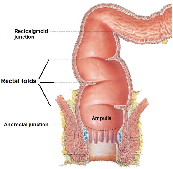
Figure 4.
Question
What two vessels supply blood to the rectum? (Hint: we saw one in the abdomen, and you will dissect one today). What does this mean for its lymphatic drainage?
Identify the anal canal. It is directed posteriorly, in the opposite direction as the rectum. The sudden bend occurs at the ano-rectal junction. Find and review the anal columns and sinuses and discuss how these relate to the pectinate line.
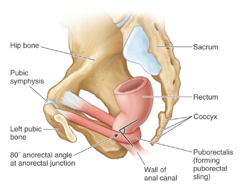
Figure 5.
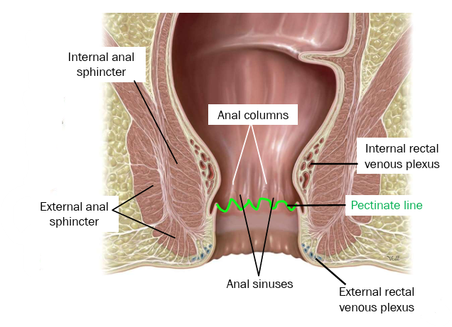
Figure 6.
Urinary Bladder
■Bladder: locate the apex, body, fundus (base), and neck.
■If you sectioned the bladder exactly in the midline, identify the internal urethral orifice (where the urethra leaves) and one of the internal ureteric orifices (where the ureter enters).
■Note the relationship of the bladder to the other pelvic organs in your donor.
■Clean the bladder and identify the ureter on both sides.
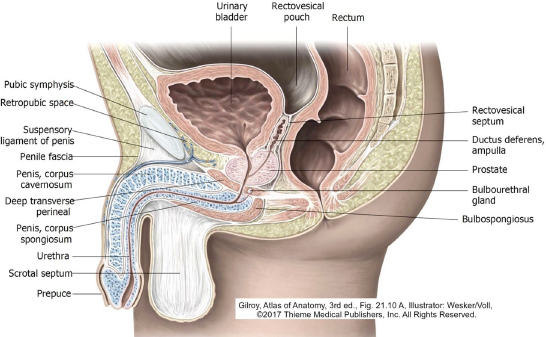
Figure 7.
Male Internal Genital Organs
Complete anatomy
Male pelvic organs
Identify the prostate immediately beneath the bladder. Study the relationships of the prostate:
■Anterior surface: faces pubic symphysis
■Posterior surface: faces the rectum
Question
Why is the relationship between prostate and rectum important?
Behind the bladder, clean away the fascia to locate a seminal vesicle. What is the function of the seminal vesicles?
■Trace the ductus deferens behind the bladder toward the seminal vesicle. On each side, the ductus deferens joins the duct of the seminal vesicle to form an ejaculatory duct.
■The ejaculatory ducts (right and left) pass through the center of the prostate. They open via tiny orifices into the prostatic portion of the urethra, atop a bump in the posterior wall of the urethra called the seminal colliculus. Semen is ejected from the ejaculatory ducts into the urethra. If you have sectioned the prostate off-center, you may see an ejaculatory duct.
■The central core of tissue in the prostate that surrounds the urethra is known as the transitional zone. What is the clinical significance of this area of the prostate?
If the penis was sectioned in the midline, locate all the parts of the male urethra: prostatic, intermediate (membranous), and spongy parts of the urethra. Locate the navicular fossa—the part of the male urethra just inside the external urethral orifice.
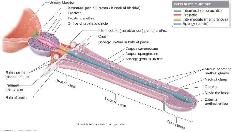
Figure 8.
Female Internal Genital Organs
Complete anatomy
Female internal organs
Let’s orient ourselves. Identify the following:
■Bladder
■Urethra = extends from bladder to vestibule
■Uterus = its major parts are fundus, body, and cervix
■Vagina = extends from cervix to vestibule. The part of the vagina that forms a blind cul-de-sac around the cervix is called the fornix.
■Uterine (Fallopian) tubes
■Ovaries
■Rectum and anal canal
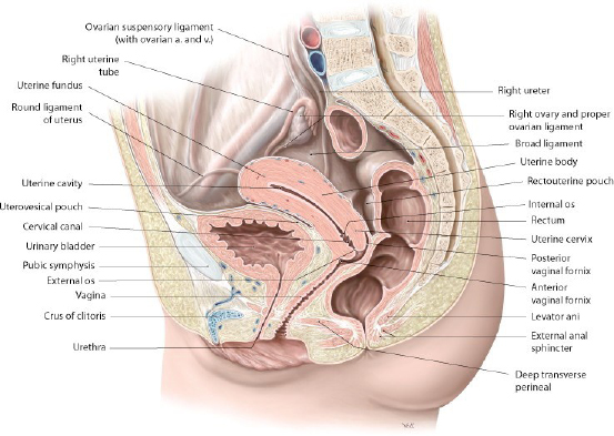
Figure 9.
Question
What is the relationship of the uterus and vagina?
Together they form the number “7” when viewed in cross-section.
question
What is the relationship of the uterus and bladder?
question
How are the urethra and vagina related? How are the cervix and vagina related to the rectum?
question
Where is the vaginal fornix located?
question
How is the posterior part of the vaginal fornix related to the recto-uterine pouch? Why would this relationship have clinical importance?
Uterus
Identify its named parts:
■Body: Superior 2/3 of uterus. The rounded, upper part of the body is the fundus.
■Uterine cavity
■Cervix: Lower 1/3 of uterus.
Uterine (Fallopian) Tubes
Identify their named parts:
■Isthmus
■Ampulla
■Infundibulum
■Fimbriae
question
How are the uterine tube and ovary related?
Ligaments of the Uterus
Peritoneal ligaments:
■Broad ligament: It’s named parts are mesometrium, mesosalpinx, and mesovarium
■Suspensory ligament of the ovary (Infundibulopelvic ligament)
Fibrous (vestigial) ligaments:
■Ligament of the ovary
■Round ligament of the uterus
question
The ligament of the ovary and the round ligament are vestiges of which embryonic structure?
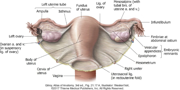
Figure 10.
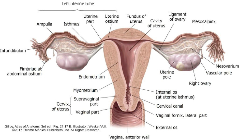
Figure 11.
question
Regarding the uterus, what do the terms anteverted and anteflexed mean?
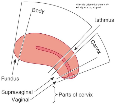
Figure 12.
Nerves and Vessels: Both Sexes
Complete anatomy
Nerves of the pelvic cavity
DEEP SIDE: Remove the peritoneum from the viscera of the deep side
On the “Deep side” (detached portion), remove all the peritoneum and dissect the structures of the lateral pelvic wall (do these steps in sequence). Conceptualize the structures of the lateral pelvic wall as being arranged in concentric layers. From internal to external, in dissection order, they are:
■Pelvic plexus of autonomic nerves
■Internal iliac vessels and their branches/tributaries
■Sacral plexus of nerves
■Muscles of pelvic wall (pelvic diaphragm, obturator internus, and piriformis)
Autonomic Nerves
Locate the bifurcation of the aorta. A network of thin nerve fibers, the superior hypogastric plexus hangs off the bifurcation.
Follow the superior hypogastric plexus down on to the sacrum as it as it splits into the right and left hypogastric nerves. These are the main source of sympathetic fibers to the pelvic organs.
Recall that the source of parasympathetic fibers to pelvic organs are the pelvic splanchnic nerves (from S-2, S-3, and S-4 segments of the spinal cord).
Nerve fibers in the hypogastric nerves and pelvic splanchnic nerves, along with visceral afferent fibers, meet and mix in the pelvic plexuses (inferior hypogastric plexuses). These are deep to the peritoneum, on the internal surfaces of the internal iliac arteries.
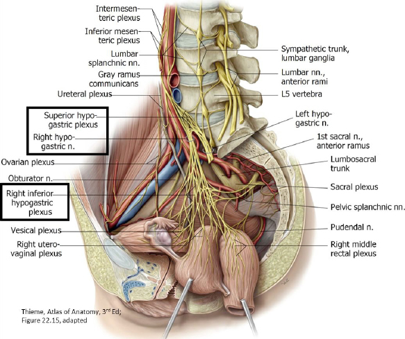
Figure 13.
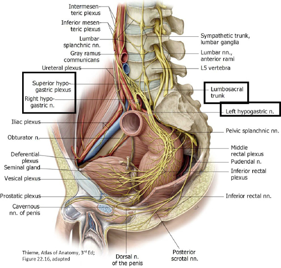
Figure 14.
Internal Iliac Artery
Complete anatomy
Internal iliac artery
DEEP SIDE: Clean off and identify branches of the internal iliac artery
Before we start,
consider that the internal iliac artery has an extremely variable branching pattern, so don’t be surprised by what you find.
This is best done on the detached “deep side”.
Clean and follow the common iliac arteries until they bifurcate into internal and external iliac arteries.
Follow the main trunk of the internal iliac artery as it descends into the pelvic cavity anterior to the sacroiliac joints. It is crossed anteriorly by the ureter.
Remove the internal iliac vein and its tributaries in order to get a better look at the artery and its branches. Veins get no respect!
Understand that the internal iliac artery is the major source of blood to the pelvic cavity, but it also supplies the perineum and the lower limb (gluteal region and medial thigh).
■Normally, the first branch from internal iliac artery is the umbilical artery. It passes lateral to the bladder, and then ascends on the internal surface of the anterior abdominal wall toward the umbilicus.
■Clean the obturator nerve in the lateral pelvic wall and see if you can find the companion obturator artery and vein. Follow the trio into the obturator canal.
■Locate the greater sciatic foramen. You will recognize it as it contains the large ventral rami of sacral spinal nerves that are joining here to form the sciatic nerve.
Note
Many of the branches of the internal iliac artery arise near the greater sciatic foramen.
Continue to clean the artery and identify as many branches as you can. Use the figures below to help. Trace the branches as far as you can and use their destinations as a guide to their identification.
The internal iliac artery splits into anterior and posterior divisions before giving off its branches. Some students find it useful to learn the branches that arise from each division.
Here is the layout:
■From the posterior division:
■Iliolumbar artery: Ascends out of the pelvic cavity toward the iliacus muscle.
■Lateral sacral arteries: Enter sacral foramina.
■Superior gluteal artery: Leaves pelvic cavity via the greater sciatic foramen ABOVE the piriformis muscle.
Complete anatomy
Posterior division of internal iliac
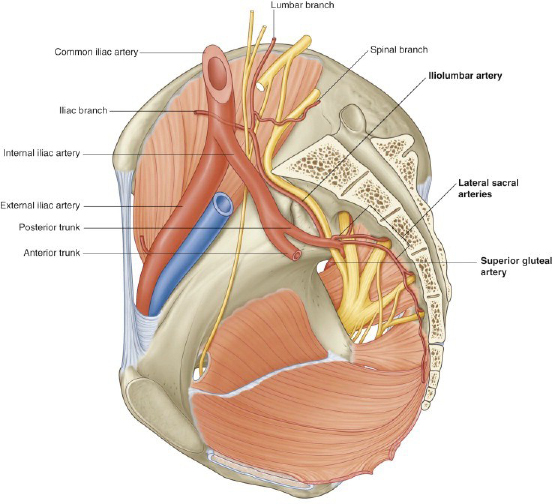
Figure 15.
■From the anterior division:
■Umbilical artery (patent and obliterated parts):
■The patent part is the proximal part—it carries blood as far as the bladder, where it gives off multiple superior vesical arteries
■Distal to the patent part is the obliterated part. It ascends on the anterior abdominal wall toward the umbilicus as a fibrous cord called the medial umbilical ligament.
■Obturator artery: Find its companion, the obturator nerve, and you are golden! Leaves pelvic cavity via the obturator canal
■Inferior gluteal artery: Leaves pelvic cavity via greater sciatic foramen BELOW the piriformis muscle
■Internal pudendal artery: Accompanies inferior gluteal artery out of pelvic cavity via greater sciatic foramen, circles around the ischial spine and enters the ischio-anal fossa via the lesser sciatic foramen. Crazy!
■Middle rectal artery: Branches off internal iliac artery near the pelvic diaphragm
■Uterine artery: Passes medially toward the cervix ABOVE the ureter in the cardinal ligament
Note
Do your best to identify these branches in your donor. There is a prosection to study from as well.
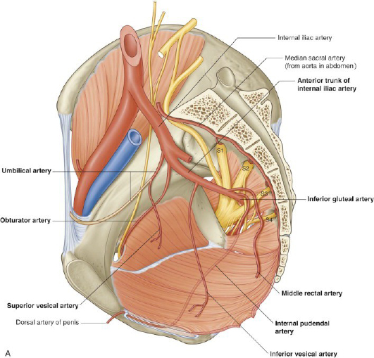
Figure 16.
Clean and examine the sacral plexus.
Identify and clean the piriformis muscle. Find the anterior rami of S1, S2, and S3 exiting the anterior sacral foramina and passing onto the piriformis.
■Trace the lumbosacral trunk (L4,5) from the posterior abdominal wall down into the pelvic cavity as it joins the S1–S3 rami to form the sciatic nerve. The sciatic leaves the pelvic cavity to enter the posterior thigh via the greater sciatic foramen.
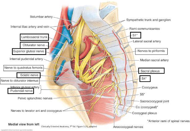
Figure 17.
We will continue to learn about the branches of the sacral plexus as we move into the lower limb.
Checklist, Lab #15
Review and make sure you have identified each of the structures below.
Rectum
Rectal folds
Ampulla
Anal canal
Anal columns
Anal sinuses
Pectinate line
Urinary bladder and named parts:
Apex, Fundus, Neck, and Body
Internal ureteric orifices
Internal urethral orifice
Trigone
Prostate
Seminal vesicles
Ductus deferens
Male urethra: Prostatic, intermediate, and spongy parts
Ovaries
Uterus
Fundus, Body, Cervix, Cervical canal, and External os
Broad ligament of uterus w/ three named parts: Mesometrium, Mesosalpinx, and Mesovarium
Round ligament of uterus
Ligament of the ovary
Suspensory ligament of ovary (Infundibulopelvic ligament)
Vagina
Fornix of vagina (posterior part is most important)
Female urethra
Cardinal ligament—you won’t see it, but know its location and importance to support of the cervix
Common iliac artery and vein
External iliac artery and vein
Internal iliac artery (see prosections, too)
Superior gluteal artery
Inferior gluteal artery
Internal pudendal artery
Obturator artery
Umbilical artery (patent and obliterated parts)
Superior vesical arteries
Uterine artery—note relationship to ureter
Middle rectal artery
Obturator nerve
Lumbosacral trunk (L-4 and L-5 spinal nerves)
Ventral rami of sacral spinal nerves (S-1 to S-4) forming sciatic and pudendal nerves → exiting the greater sciatic foramen
Hypogastric nerves (R&L)—the major source of sympathetic nerves to pelvic organs ( if you find these, they could be extra credit items!)
Pelvic splanchnic nerves = these are tiny branches from the ventral rami of S-2, S-3, and S-4 spinal nerves that carry parasympathetic nerves fibers to pelvic organs ( extra credit item!)
Greater sciatic foramen
Piriformis muscle