- Goals
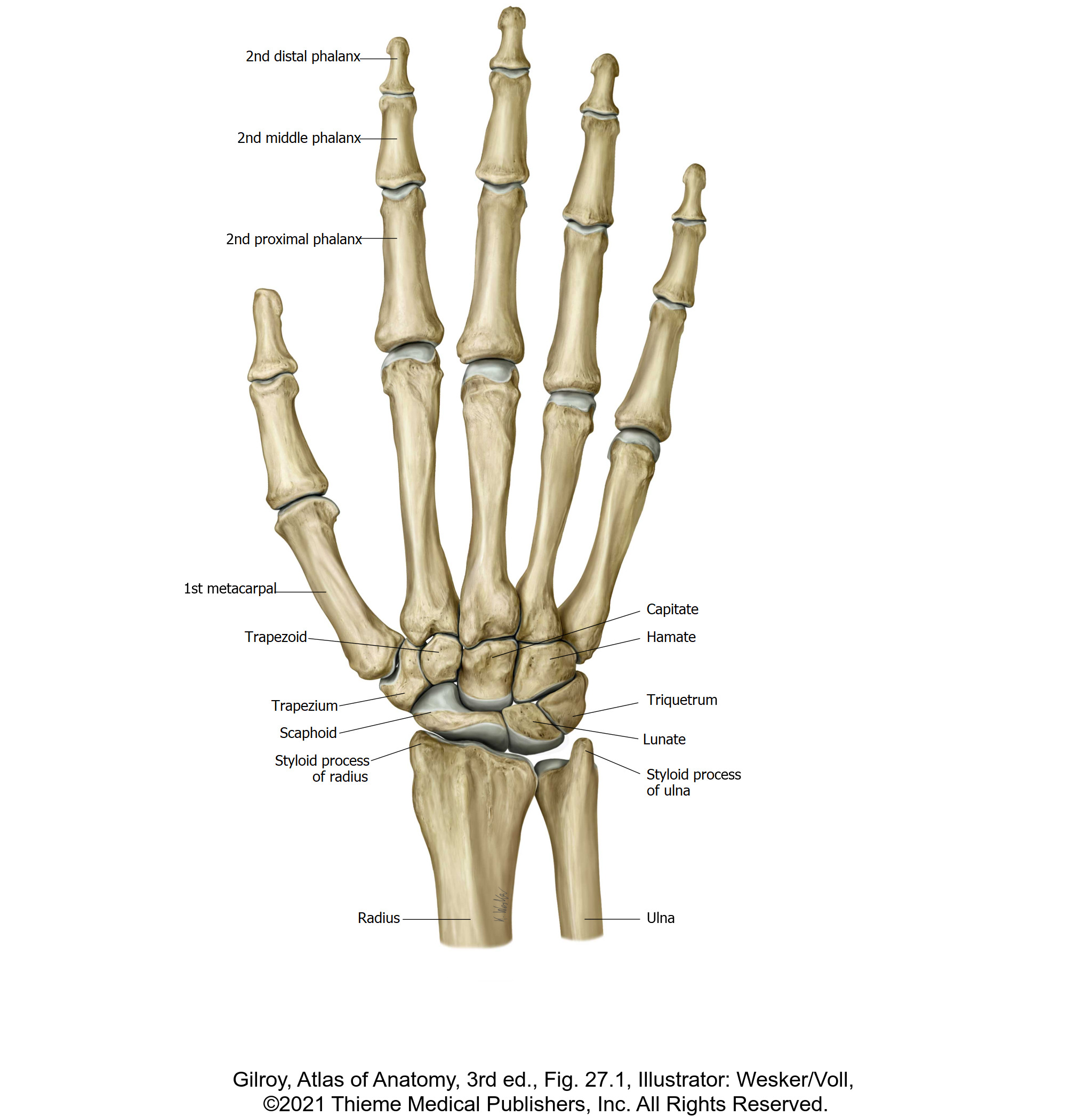
- Identify the bones associated with the wrist and hand.
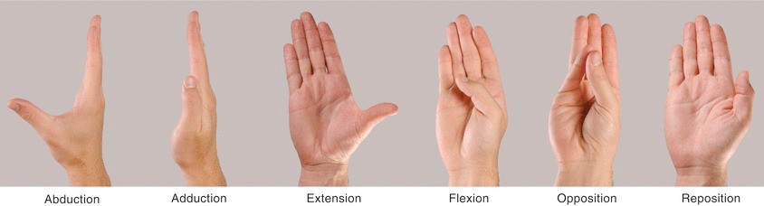
- Demonstrate and describe the movements of the thumb.
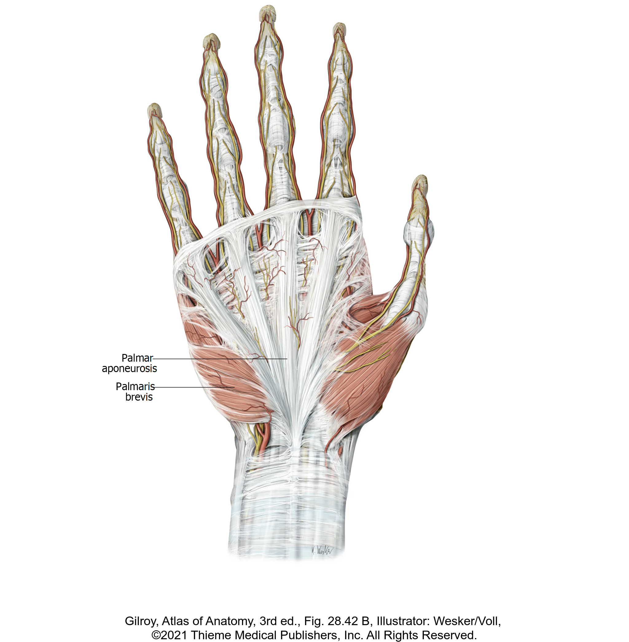
- Clean and identify the palmar aponeurosis.
- Identify the boundaries and contents of the ulnar canal and carpal tunnel.
- Trace the long flexor tendons from the anterior forearm to the palmar surface of the digits.
- Open one fibrous digital sheath and examine the relationships of tendons within it.
- Identify the intrinsic muscles and neurovascular elements of the palm.
Osteology of the Hand
Bony landmarks of the hand
Joints of the hand
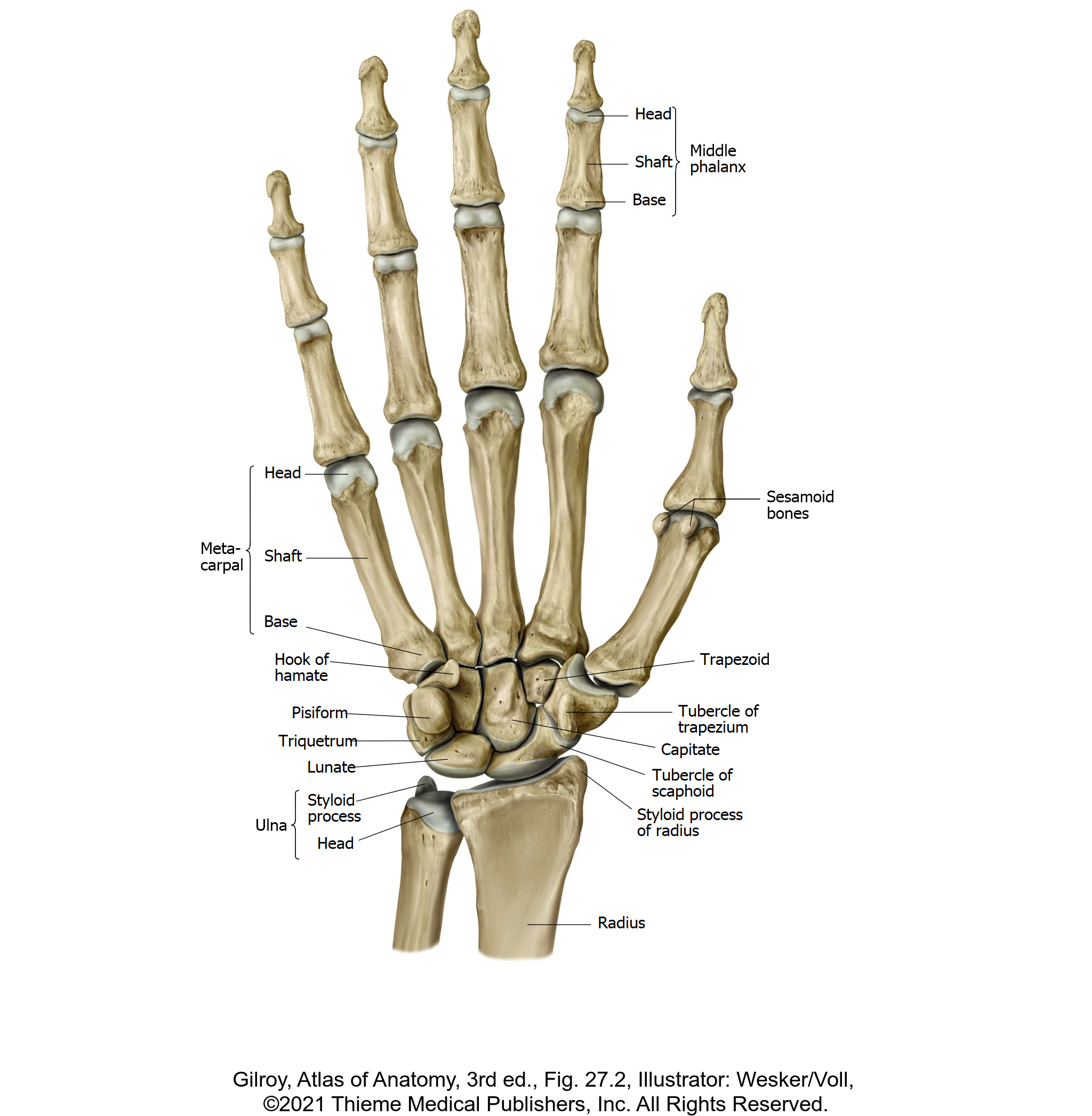
Identify the eight carpal bones:
■Scaphoid, Lunate, Triquetrum, Pisiform, Trapezium, Trapezoid, Capitate, and Hamate
■Locate the hook of the hamate bone.
Mnemonic
Here are some clever mnemonics to help you remember the carpal bones:
- An oldie-but-goodie that Cougs will appreciate: So Long To Pullman, Time To Come Home (starting on the thumb side for both proximal and distal rows)
- So Long To Pinky, Here Comes The Thumb (start with proximal row on thumb side, then move clockwise, finishing the distal row at the thumb)
- Remember: Trapezium sounds like Thumb—so it is nearer to the thumb = the lateral-most bone in the distal row!
Identify the five metacarpal bones. Each has a base, shaft, and head. They are numbered first to fifth, starting from the thumb side.
Identify the phalanges = the bones in the digits. Each digit has three phalanges (proximal, middle, and distal), except the thumb, which has two (proximal and distal).
Joints of the Hand
Identify each of the following joints and describe the joint types and movements:
■Intercarpal joints
■Carpometacarpal (CMC) joints
■Metacarpophalangeal (MCP) joints
■Interphalangeal (IC) joints
Question
The first carpometacarpal joint (the CMC joint of the thumb) is a special type of joint. Can you name the joint type and describe the special movement that it allows in the hand?
Movements of the Thumb
Work with your team to demonstrate and describe the movements of the thumb in Figure 3.
Superficial Palm
Superficial structures of the hand
Do this work on both hands.
Both sides: Remove the skin from the palmar surface of the hands and fingers.
Scrape away the fat on the palm until you see the shiny surface of the palmar aponeurosis. If present in your donor, identify the long tendon of the palmaris longus in the forearm and trace it past the wrist to the palm—it attaches distally to the palmar aponeurosis.

The palmar aponeurosis spreads out as fibrous bands onto the palmar surfaces of the digits.
The palmar aponeurosis protects the nerves and vessels of the palm. Around the edges of the palmar aponeurosis you will see some of the digital branches of the ulnar and median nerves—observe how they are vulnerable to superficial lacerations, as they are unprotected by the aponeurosis.
Both sides: Next, cut and reflect the palmar aponeurosis.
Detach it from the tendon of the palmaris longus and carefully reflect it away from the palm of the hand distally, leaving it attached only by the thin digital expansions of the aponeurosis, such that it is hanging by only 2 or 3 bands from the distal ends of the digits.
As you do this, you will remove the relatively unimportant palmaris brevis muscle that inserts into the ulnar side of the palmar aponeurosis (see figure 4 above).
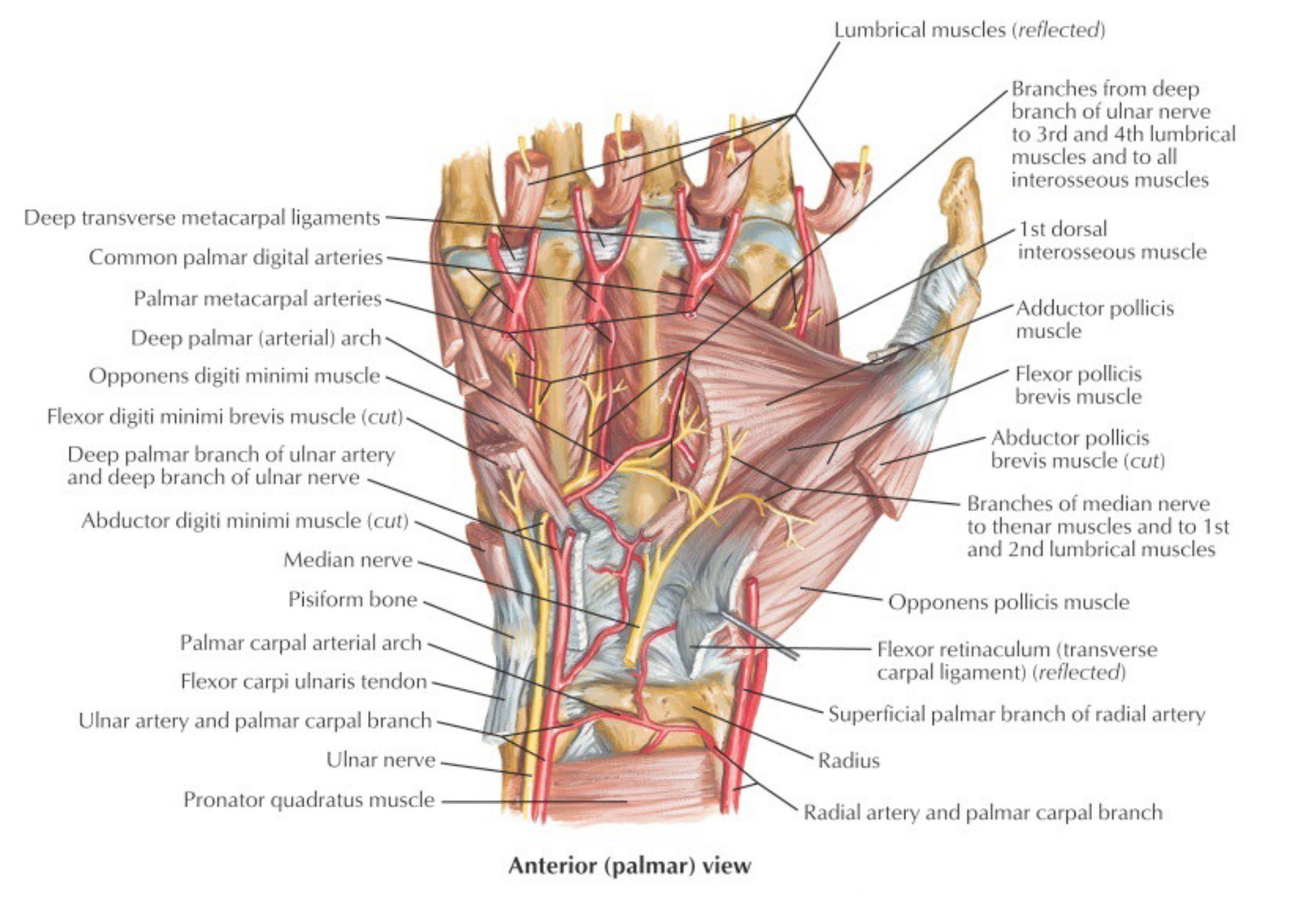
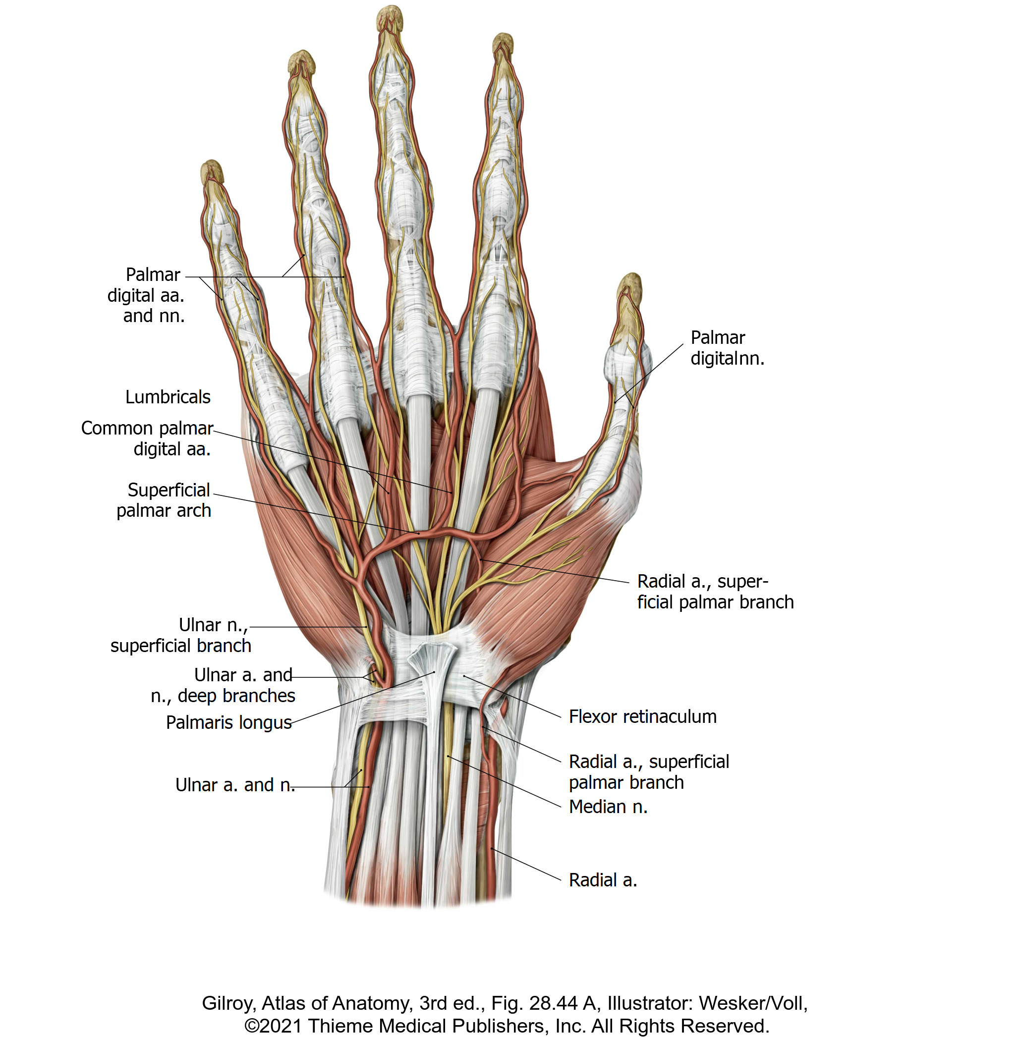
Deep to the palmar aponeurosis, in the palm of the hand, identify the arteries and nerves of the palmar hand. Be sure to find all the bolded structures from the descriptions below.
Arteries
Arteries of the hand
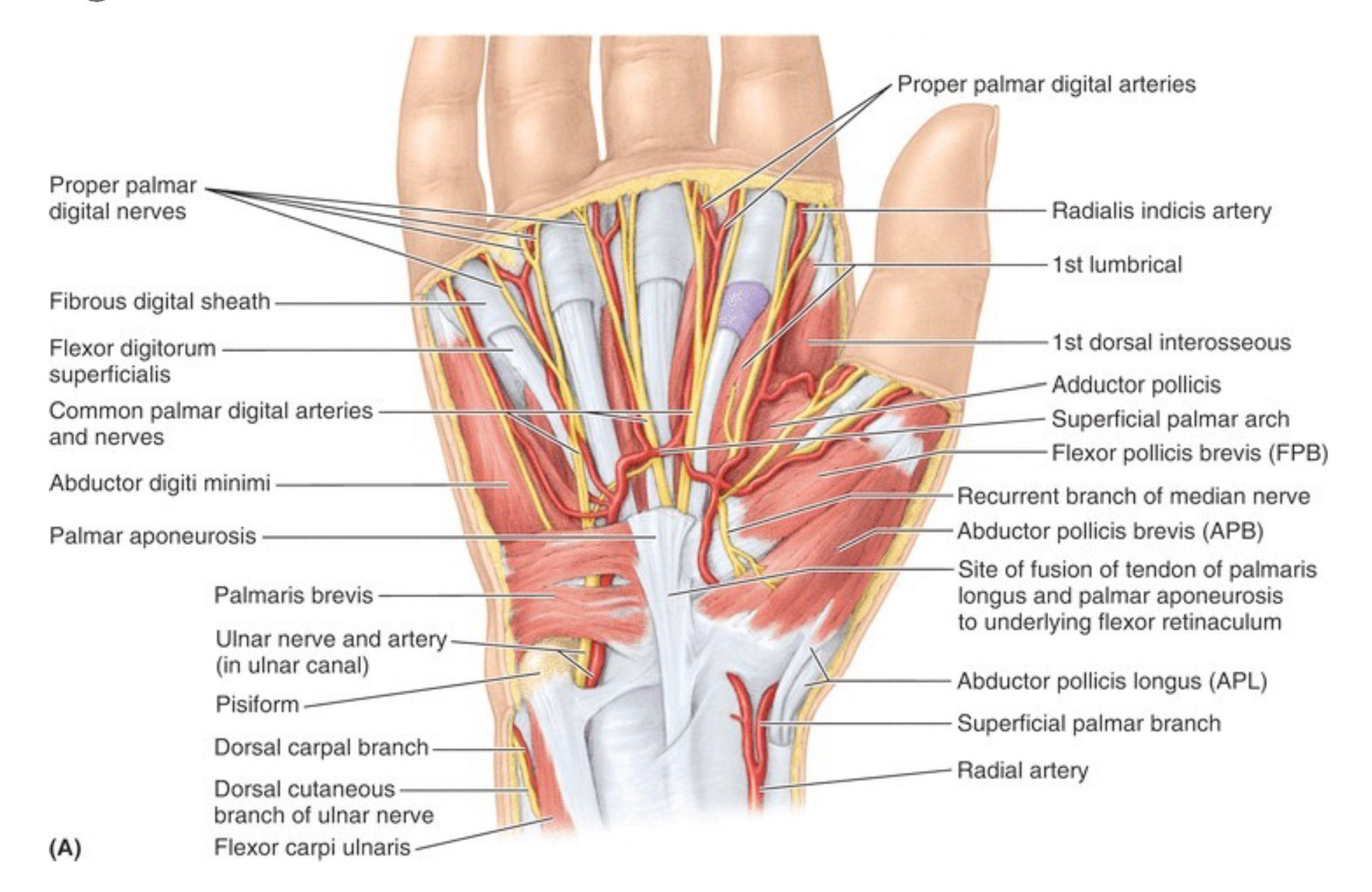
The superficial palmar arch is an arterial anastomosis crossing the palm of the hand from medial to lateral. It is the direct continuation of the ulnar artery in the hand. The superficial palmar arch is completed by the superficial palmar branch of the radial artery in the hand.

Branching from the superficial palmar arch are three common palmar digital arteries. Upon reaching the spaces between the bases of the fingers, these arteries split, giving rise to proper palmar digital arteries that continue distally along the medial and lateral borders of digits 2–5. The radial (lateral) border of digit 2 and the ulnar (medial) border of digit 5 receive proper palmar digital arteries directly from the superficial palmar arch, and not from the common palmar digital arteries. The thumb receives separate arteries from both the superficial and deep palmar arches.
Nerves
Nerves of the hand
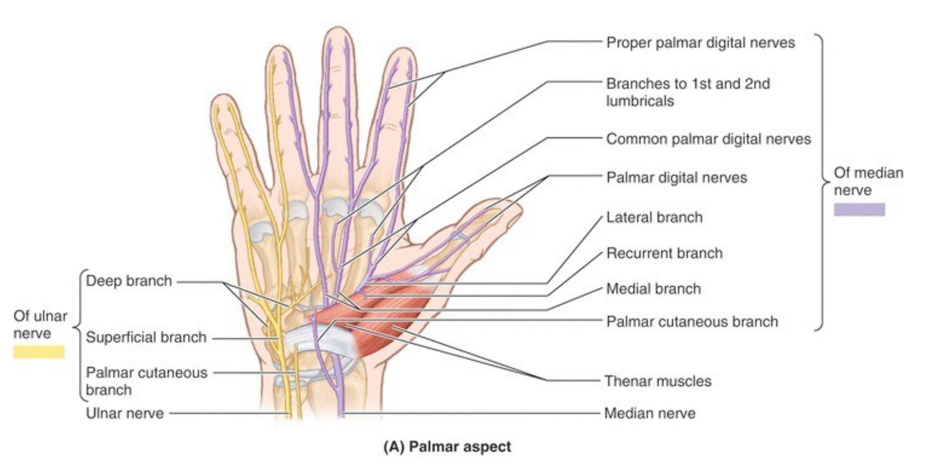
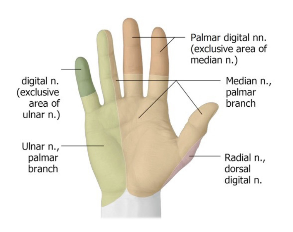
Common palmar digital nerves branch from both the median and ulnar nerves in the palm of the hand. Like the arteries, these divide at the bases of digits 2–5, giving off proper palmar digital nerves to the medial and lateral borders of these digits.
Note
The median nerve supplies digital nerves to the thumb, digit 2, digit 3, and half of digit 4. The ulnar nerve supplies nerves to half of digit 4 and all of digit 5.
The ulnar nerve splits into superficial and deep branches in the hand. The digital nerves to the 4th and 5th digits are branches of the superficial branch of the ulnar nerve.
Ulnar Nerve and Ulnar (Guyon’s) Canal
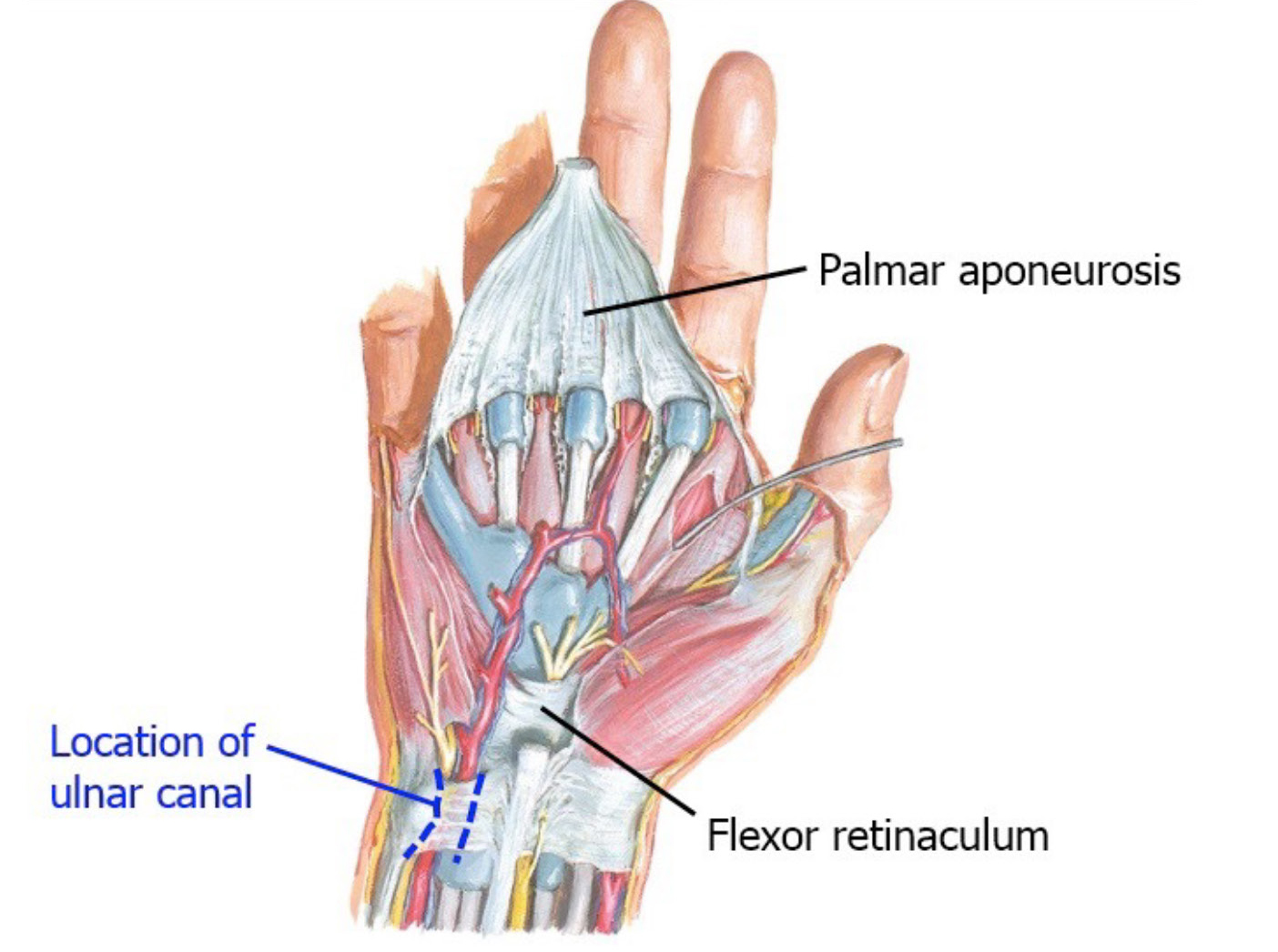
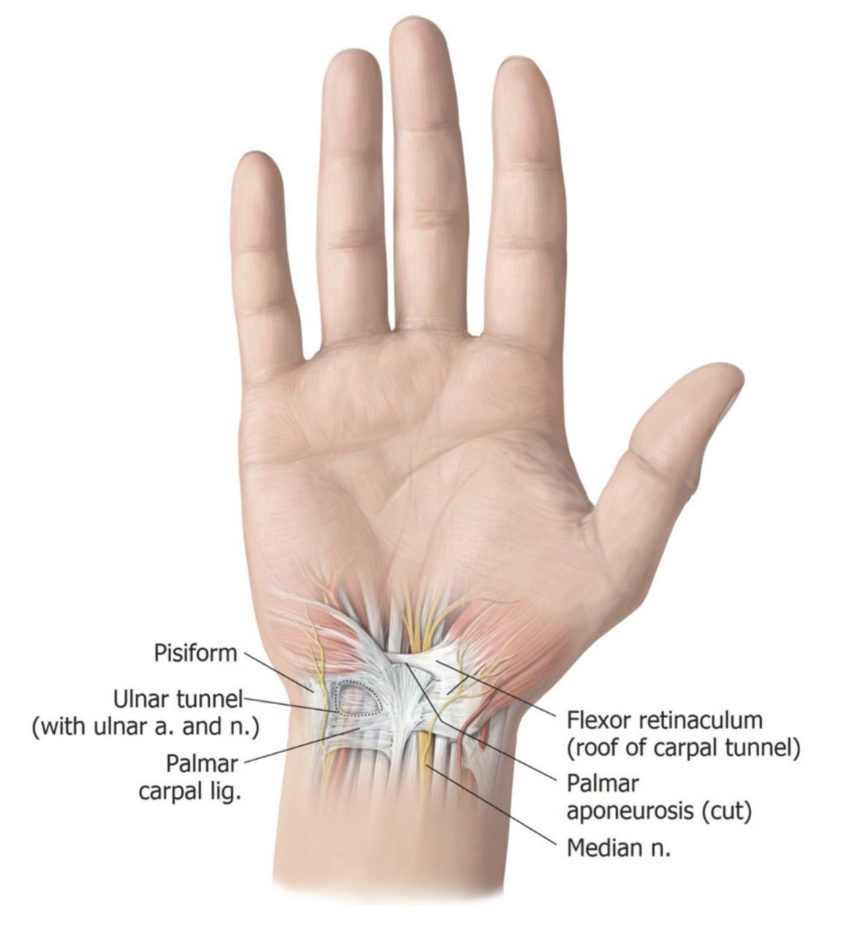
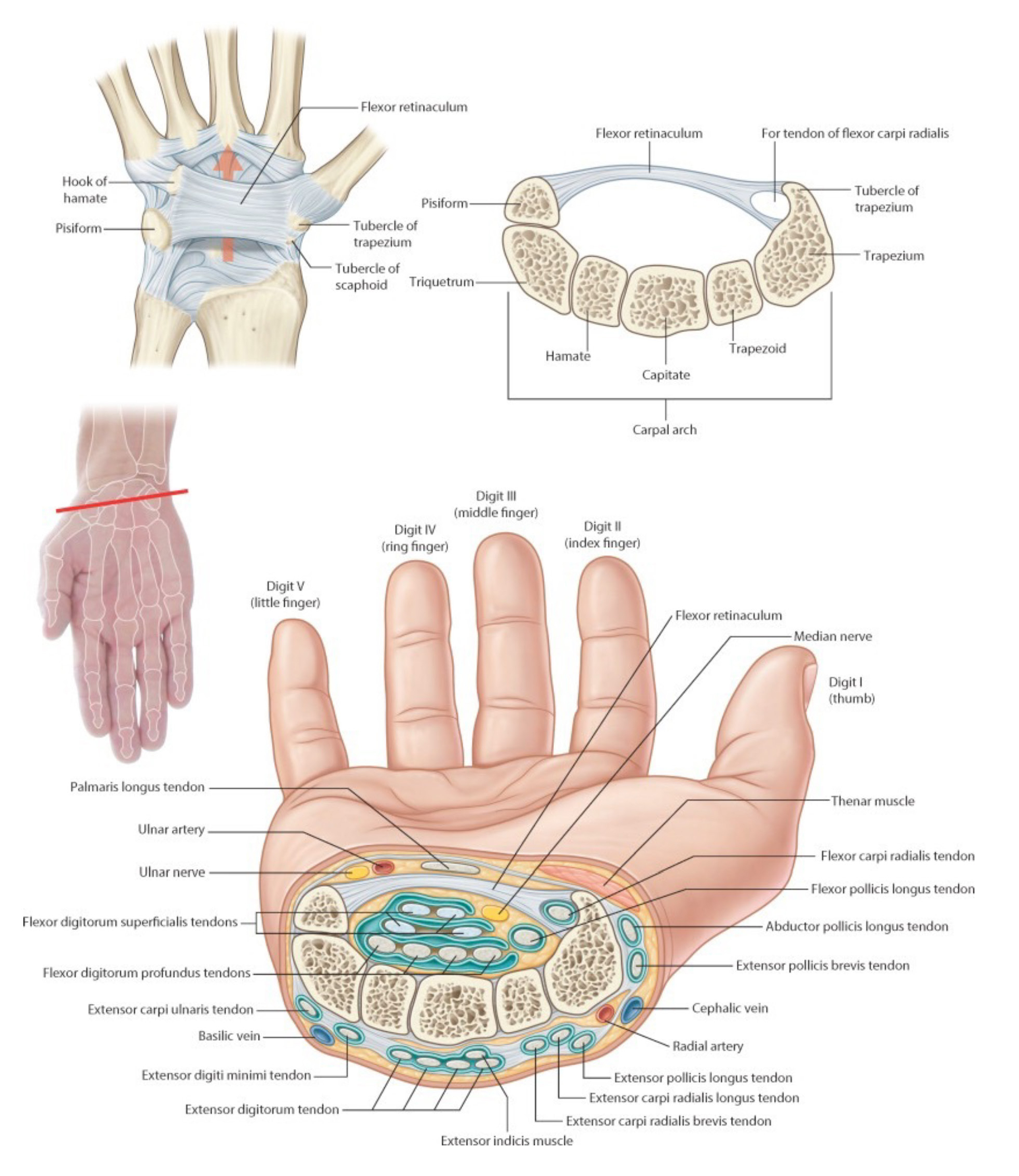
Trace the superficial palmar arch backwards to the wrist area. Note that the ulnar artery and accompanying ulnar nerve disappear under deep fascia of the palmar wrist. The ulnar artery and nerve enter the palm of the hand from the forearm through the ulnar canal (Guyon’s canal). See Figure 8. This is located between the pisiform bone and the hook of the hamate bone. The roof of the ulnar canal is a thickened band of deep forearm fascia, and not the flexor retinaculum. The ulnar nerve and ulnar artery therefore DO NOT enter the hand through the carpal tunnel.
Now, designate one hand as the superficial dissection and one hand as the deep dissection.
Pay attention to the instructions below and do deep dissections only where instructed.
Deep Dissection Side
Deep dissection: Open the ulnar canal with scissors to reveal the ulnar nerve and artery.
Make the cut parallel to the artery and nerve – TAKE CARE not to cut them. See Figure 8.
Carpal Tunnel
Carpal tunnel
Question
What is a retinaculum?
A retinaculum is a special modification of the deep fascia containing transverse collagen fibers. They function to hold tendons close to the joints they cross so they don’t “bowstring” away when the joint is moved.
Clean off the flexor retinaculum (transverse carpal ligament). This is the roof of the carpal tunnel.
The flexor retinaculum attaches to the hook of the hamate and pisiform bone medially and to the scaphoid and trapezium bones laterally.
Deep Dissection Side
Deep dissection: Open the carpal tunnel as instructed below.
Slide a metal probe through the carpal tunnel (deep to the flexor retinaculum) from proximal to distal, so that the tip of the probe appears in the palm of the hand. Using the probe as a guide, open the carpal tunnel by cutting the flexor retinaculum over the probe with a scalpel. This technique will protect the structures within.
Ten (10) structures pass through the carpal tunnel.
■Identify the median nerve.
■Identify the 9 flexor tendons that run through the carpal tunnel. Within the tunnel, the tendons are surrounded by a synovial sheath, which lubricates the tendons so they slide without friction.
Question
Which muscles do these nine tendons belong to?
Clinical correlation
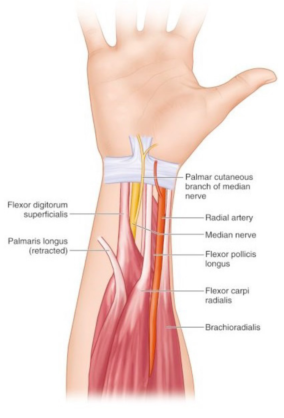
Carpal tunnel syndrome is caused by pressure on the median nerve within the carpal tunnel. Its symptoms appear gradually, usually beginning with numbness and tingling of the thumb, index, and middle fingers. Weakness of thumb muscles may occur later. Interestingly, sensory symptoms don’t occur in the center of the palm of the hand distal to the carpal tunnel as you might expect. The anatomic explanation for this is that the palmar cutaneous branch of the median nerve that supplies sensation to the palm of the hand branches from the median nerve in the distal forearm, proximal to the carpal tunnel. The palmar cutaneous branch enters the palm via the superficial fascia that is external to the flexor retinaculum. See Figure 12.
Chalk Talk
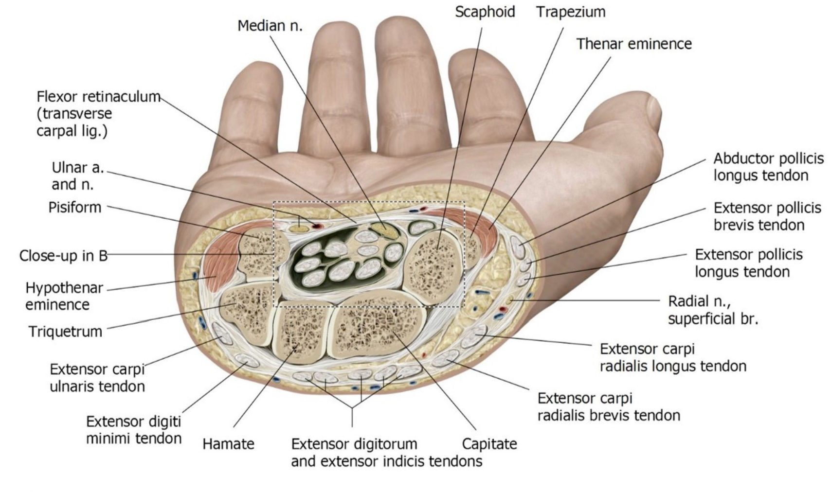
Draw a cross section of the wrist (similar to Figure 13), noting the locations of the ulnar nerve and artery in Guyon’s canal and the contents of the carpal tunnel: median nerve, tendons of flexor digitorum superficialis, flexor digitorum profundus, and flexor pollicis longus.
Question
Is the tendon of the flexor carpi radialis within the carpal tunnel? If not, where is it in relation to the carpal tunnel?
Muscles of Thenar Eminence
Superficial muscles of the hand
The thenar eminence is the fleshy mass on the palmar surface of the hand at the base of the thumb.
Both sides: Clean and separate the three thenar muscles.
Review their actions and innervation.
1Abductor pollicis brevis
2Flexor pollicis brevis
3Opponens pollicis
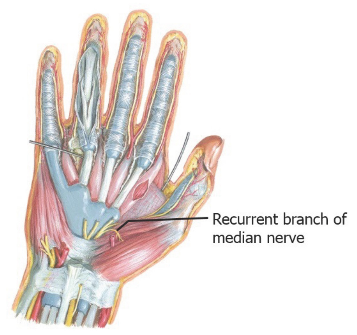
Identify the small recurrent branch of the median nerve = the motor branch that supplies the thenar muscles. It leaves the median nerve just distal to the flexor retinaculum. It is a short nerve that curls backward on itself (thus its name) to the enter the muscles of the thenar eminence.
Note
Note how superficial the recurrent branch is on the thenar eminence. A relatively shallow laceration to this area could result in a great deal of disability.
Muscles of Hypothenar Eminence
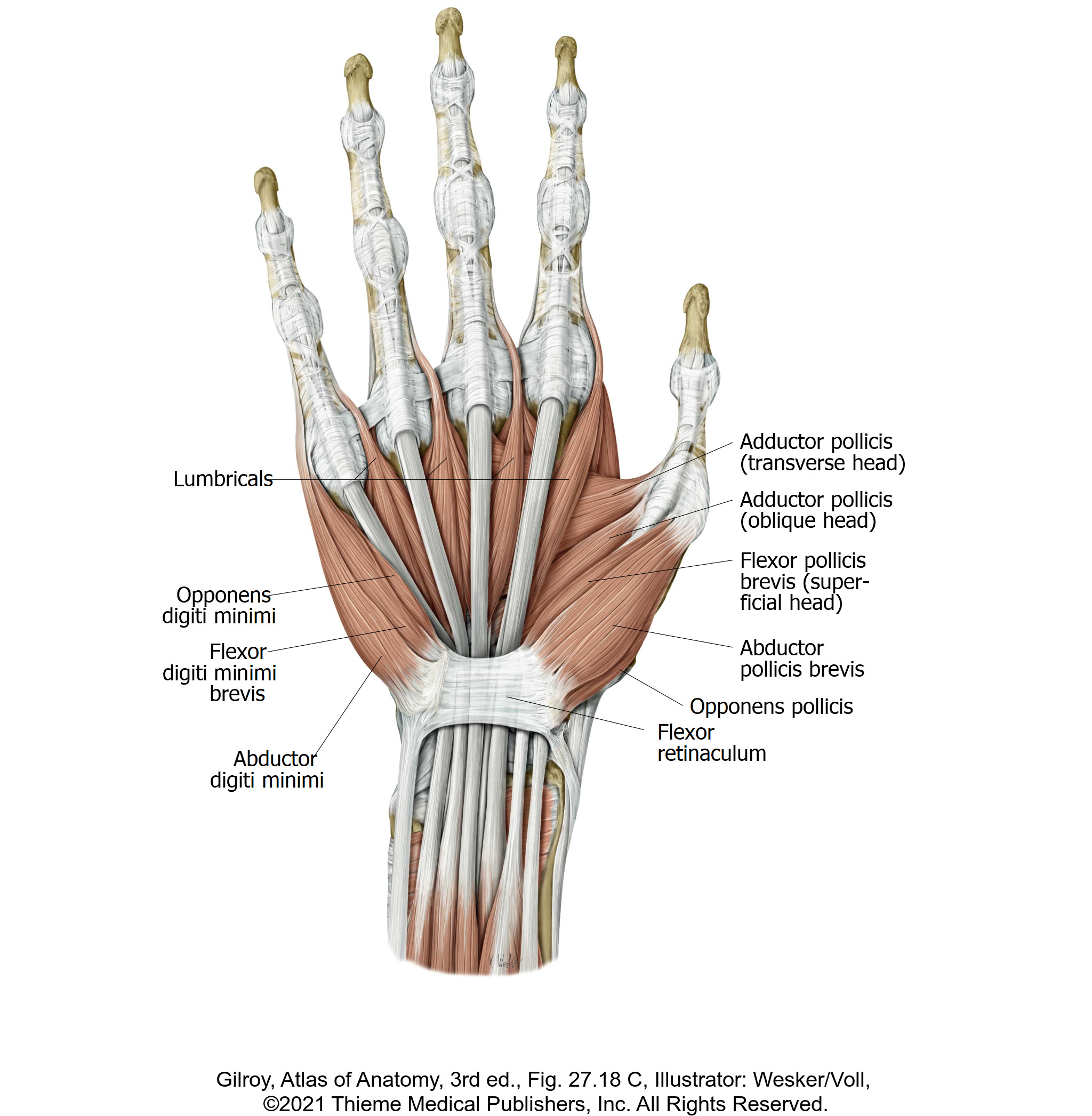
The hypothenar eminence is the fleshy outcropping on the palm of hand, along the medial border at the base of the 5th digit. (See Figure 14.)
Both sides: Clean and separate the three hypothenar muscles.
Review actions and innervation.
1Abductor digiti minimi
2Flexor digiti minimi brevis
3Opponens digiti minimi
Question
The muscles of the hypothenar eminence are innervated by the deep branch of the ulnar nerve. Is this the same nerve that innervates the muscles in the thenar eminence?
Lumbrical Muscles
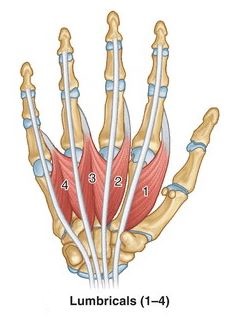
Both sides: In the palm of the hand, clean and identify the four lumbrical muscles.
Question
What does the word lumbrical mean?
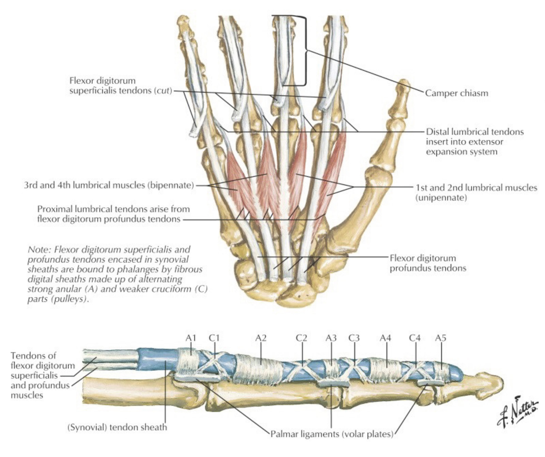
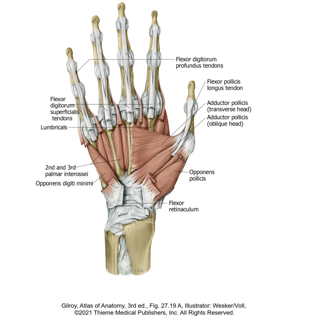
Note that they arise from the four tendons of the flexor digitorum profundus. How odd!
They insert into the extensor hoods (dorsal digital expansions) on the dorsal side of the digits.
Question
How are the lumbricals innervated?
Question
What are the actions of the lumbricals? They act on two joints: the metacarpophalangeal (MCP) joints and the interphalangeal (IP) joints.
Deep dissection: Cut away and remove any skin and remnants of the palmar aponeurosis that might still be attached to the palmar side of the digits and examine the fibrous digital sheaths.
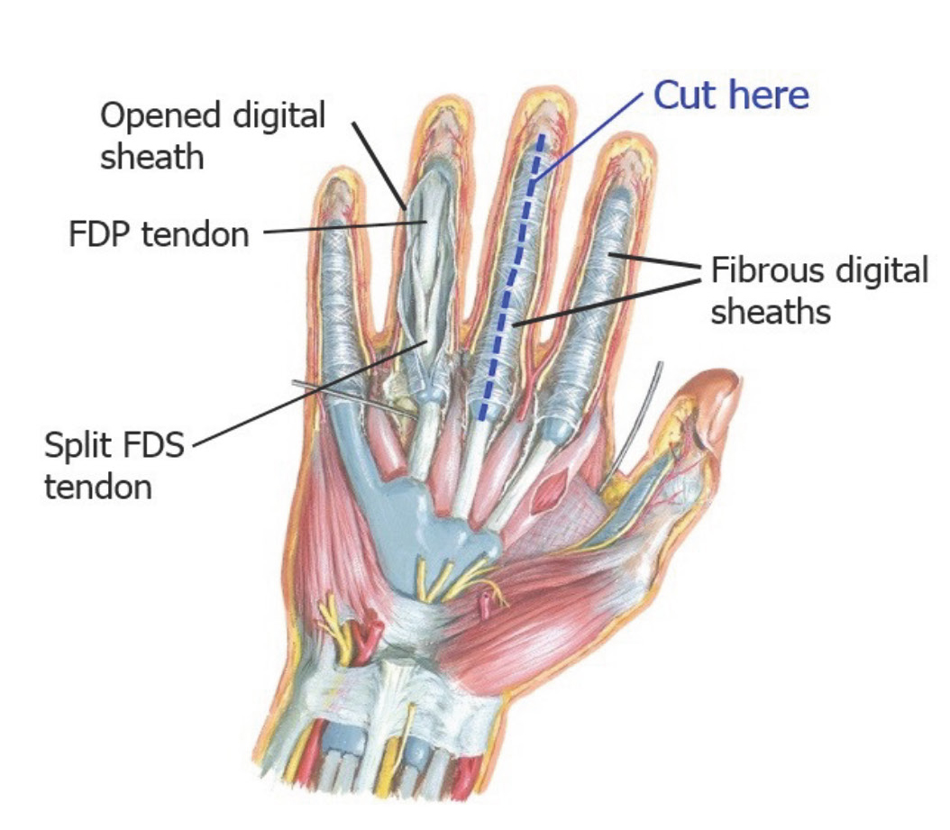
The fibrous digital sheaths are osseo-fibrous tunnels located on the palmar surfaces of the digits, formed from arched layers of dense connective tissue connecting to the metacarpal bones and phalanges. Each sheath contains a tendon of the flexor digitorum superficialis (FDS) and the flexor digitorum profundus (FDP), plus the surrounding synovial sheaths that lubricate them. The fibrous digital sheaths prevent “bowstringing” of tendons when the fingers are flexed.
Question
The tendon of the FDS attaches to the middle phalanx, while the tendon of the FDP attaches to the distal phalanx. Since the tendon of the FDP is deep to the tendon of the FDS, how can it reach the distal phalanx?
The answer is elegant: the tendon of the FDS splits to allow the tendon of FDP to pass through and reach the distal phalanx. The gap in the FDS tendon where its splits is called a chiasm.
Deep dissection: With a scalpel and scissors, open the sheath of one finger (middle finger is easiest).
Note
Note how the flexor digitorum profundus tendon attaches to the distal phalanx as it passes through the gap (chiasm) in the flexor digitorum superficialis tendon (attaches to the middle phalanx).
For you aspiring hand surgeons, the fibrous digital sheaths are constructed of a series of alternating ligamentous structures called annular (A) pulleys and cross-shaped cruciform (C) pulleys. See Figure 18.
Deep Dissection Side
Complete anatomy
Deep palmar hand
Deep dissection: Cut the flexor tendons of FDS and FDP at the knuckles and reflect them proximally into the forearm. This will expose the deep palm.
Follow the ulnar nerve from the forearm into the ulnar (Guyon’s) canal.
At the level of the pisiform bone, the ulnar nerve divides into two branches: superficial and deep.
The superficial branch of the ulnar nerve gives rise to the digital nerves—thus it is sensory to the skin of the 5th digit and half of the 4th digit.
The deep branch of the ulnar nerve is motor—it supplies the hypothenar muscles, the medial two lumbricals, the adductor pollicis, and all the interosseous muscles (palmar and dorsal).
Deep dissection: Clean and follow the deep branch of the ulnar nerve.
Observe how the deep branch of the ulnar nerve passes between the flexor digiti minimi brevis and abductor digiti minimi muscles. It then curls around the hook of the hamate bone and passes across the palm of the hand, accompanied by the deep palmar branch of the ulnar artery.
See if you can find the deep palmar arch. The deep palmar arch is an arterial anastomosis—much like the superficial palmar arch, but smaller and deeper. The deep palmar arch is formed from the distal part of the radial artery in the hand, with a small contribution from the deep palmar branch of the ulnar artery.
Muscles in the Deep Palm
Complete anatomy
Deep muscles of the hand
Identify the adductor pollicis muscle and its two heads (oblique and transverse). See Figure 20.
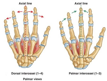
Identify the three palmar interosseous muscles. These are on the palmar surfaces of the metacarpals in the deep hand. The four dorsal interosseous muscles are located between the metacarpals and are best seen on the dorsal side of the hand (next lab). See Figures 20 and 21.
Review the actions of the palmar and dorsal interosseous muscles.
Memory aid
PAD = Palmar interosseous muscles ADduct
DAB = Dorsal interosseous muscles ABduct
Checklist, Lab #6
Review and make sure you have identified each of the structures below.
Bones
Carpal bones
Scaphoid
Lunate
Triquetrum
Pisiform
Trapezium
Trapezoid
Capitate
Hamate with hook
Metacarpal bones = base, shaft, head
Phalanges = proximal, middle, distal (except for thumb)
Joints
Intercarpal
Carpometacarpal (CMC)
Metacarpophalangeal (MCP)
Proximal and Distal Interphalangeal (PIP and DIP)
Muscles
Abductor pollicis brevis m.
Flexor pollicis brevis m.
Opponens pollicis m.
Adductor pollicis m.
Abductor digiti minimi m.
Flexor digiti minimi brevis m.
Opponens digiti minimi m.
Lumbrical muscles (4)
Palmar interosseous muscles (3)
Dorsal interosseous muscles (4)
Tendons of flexor digitorum superficialis, flexor digitorum profundus, and flexor pollicis longus muscles in carpal tunnel
Nerves
Median nerve
Recurrent branch of median nerve
Common palmar digital nerves (from median and ulnar nerves)
Proper palmar digital nerves (from median and ulnar nerves)
Palmar cutaneous branch of median nerve (branches from median nerve proximal to carpal tunnel)—WISH LIST item
Ulnar nerve
Superficial branch of ulnar nerve
Deep branch of ulnar nerve (you’re a superstar if you can find it!)
Common palmar digital nerves (from median and ulnar nerves)
Proper palmar digital nerves (from median and ulnar nerves)
Vessels
Superficial palmar arch (Ulnar artery + superficial palmar branch of radial artery)
Deep palmar arch (Radial artery + deep palmar branch of ulnar artery)—this is a toughie to dissect
Common palmar digital arteries
Proper palmar digital arteries
Other
Palmar aponeurosis
Flexor retinaculum (transverse carpal ligament)
Carpal tunnel
Ulnar (Guyon’s) canal
Fibrous digital sheaths (on palmar surfaces of digits)