
Dissector instructions
Tap the Dissector Instructions menu on the left side of your screen to open the steps of your dissection. It's sticky, so it will scroll with you as you progress through the lab.
Need to log in to view the dissector?
Tap here for instructions on how to show the keyboard on the displays in the anatomy lab
- Goals
- Identify the boundaries and contents of the axilla.
- Learn the architecture of the brachial plexus.
- Identify the muscles, nerves, and vessels of the arm.
Axilla
Before you dissect, discuss the boundaries of the axilla.
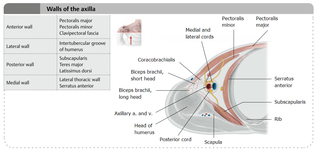
Note that it is pyramid-shaped, with its base facing the skin of the axillary fossa (“armpit”, between the anterior and posterior axillary folds), and its apex projecting toward the neck, between the first rib and the clavicle. The walls of the axilla are summarized in Figure 1.
Remove skin from the arm in preparation for dissection of the axilla.
TAKE CARE not to cut the superficial veins of the arm—
review the course of the cephalic and basilic veins BEFORE making the incisions.
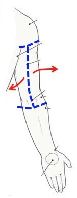
- Make a shallow incision in the midline skin of the arm, from the shoulder superiorly to just distal to the elbow inferiorly. See Figure 2.
- Make a shallow circular incision in the skin of the upper forearm, just below the elbow joint. Carry the incisions as far posterior as you can, both medially and laterally. (Watch out for the median cubital veins!)
- Reflect the skin as two shallow flaps: Medial and Lateral.
Contents of the Axilla
Dissect the axilla on BOTH sides of the donor.
Keep the veins on one side;
remove the veins on the other.
Abduct the arm to about 45° and keep it there throughout the dissection.
Use blunt dissection with fingers and sharp scissors (open-and-close technique) to pry open the axillary fascia.
Cut away fat and connective tissue to expose the axillary vessels and the brachial plexus.
Axillary Vein
Axillary vein
On one side of the donor, clean and identify the veins in the axilla.
Identify the axillary vein, formed by union of the basilic vein (a superficial vein) and the paired brachial veins = the venae commitantes (companion veins) of the brachial artery. It changes its name proximal to the first rib, where it becomes the subclavian vein.
As you clean the axillary vein, look for lymph nodes. Sometimes these are small and hard to find, but it is said there are 20 or 30 axillary lymph nodes.
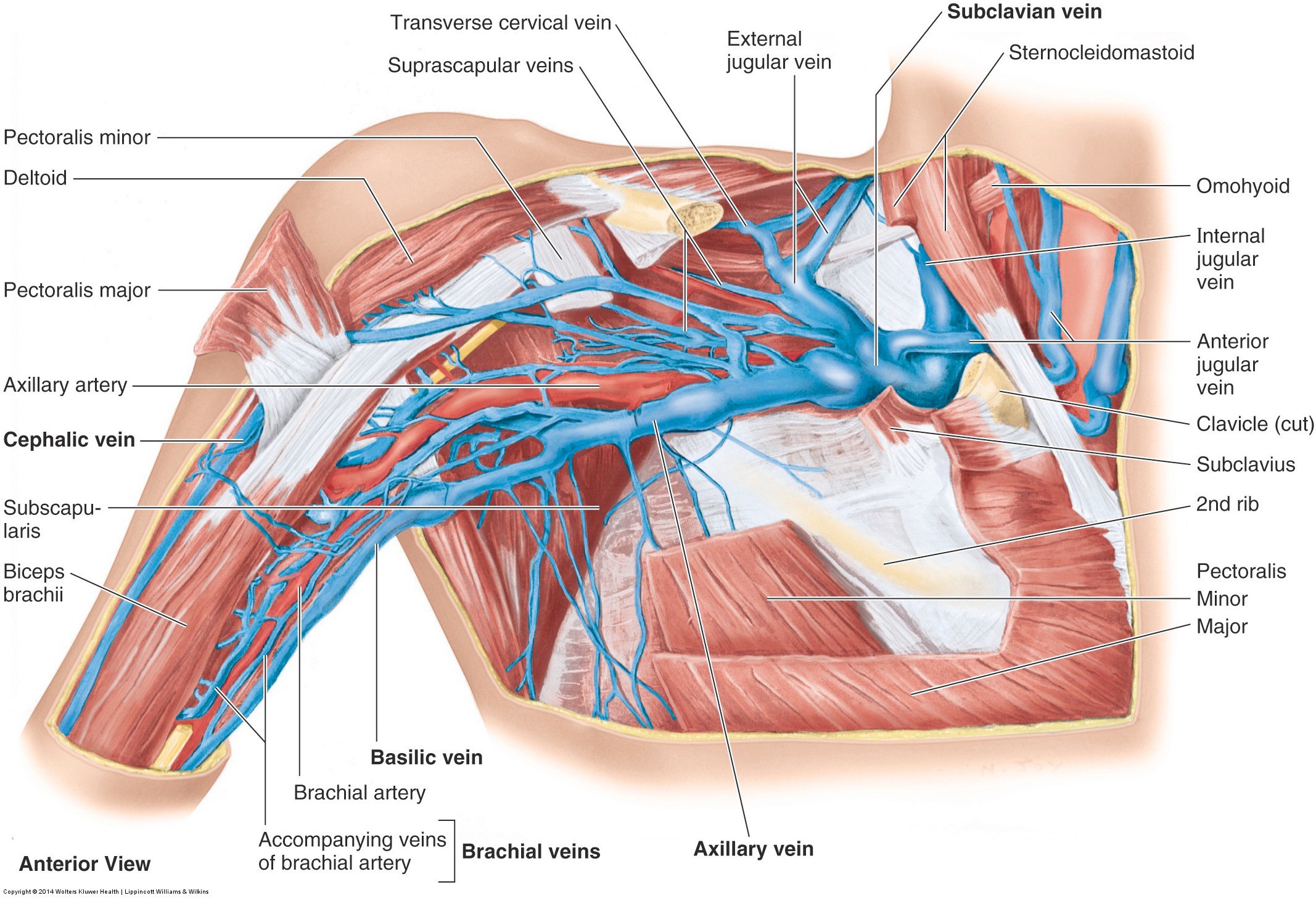
The tributaries of the axillary vein are the cephalic, basilic, and brachial veins.
Axillary Artery
Axillary artery
On one side of the donor, remove the axillary vein.
Cut the large axillary vein close to where it crosses the first rib and then distally as it enters the arm. Carefully peel the vein away, removing its branches as you go. This will allow you easier access to the underlying artery.
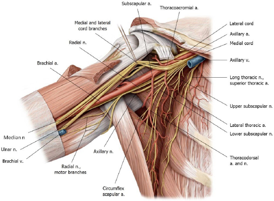
Clean the axillary artery. Proximal to the first rib it becomes the subclavian artery. Distally at the teres major muscle it becomes the brachial artery.
Return the reflected pectoralis minor muscle to its anatomic position and note that it crosses the axillary artery. The pectoralis minor divides the axillary artery into 3 parts:
- Part 1: Proximal to pectoralis minor
- Part 2: Posterior to pectoralis minor
- Part 3: Distal to pectoralis
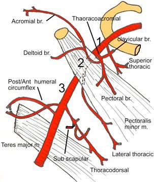
Identify the three parts of the artery and look for these branches from each.
- Part 1:
- Superior thoracic artery (not important to find)
- Part 2:
- Thoraco–acromial trunk and lateral thoracic artery (we examined these in the last lab, remember?)
- Part 3:
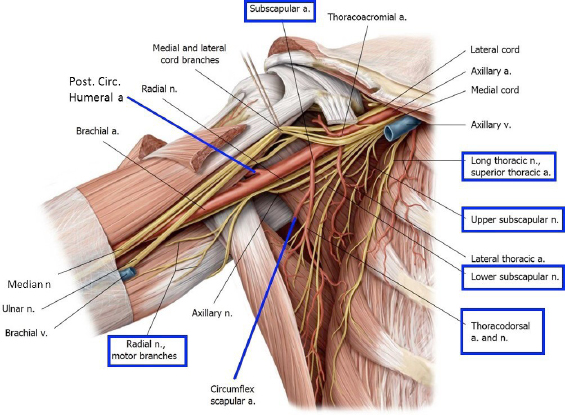
- Subscapular artery and the posterior and anterior circumflex humeral arteries (the posterior is the larger of the two).
- Part 1:
The posterior circumflex humeral artery loops around the posterior humerus with the axillary nerve and enters the quadrangular space.
A branch of the subscapular artery—the circumflex scapular artery—curls around to the posterior surface of the scapula via the triangular space.
Brachial Plexus
Structure of the brachial plexus
Draw the Brachial Plexus on the whiteboard as you perform this part of the lab.
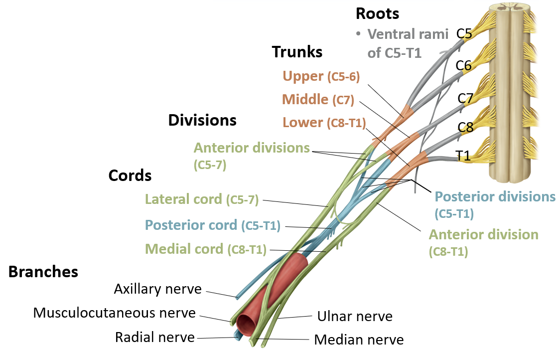
Use the "Rule of 3s," as demonstrated in the slides at Learn the Brachial Plexus in 5 Minutes.
To study the architecture of the brachial plexus you will use both dissection and prosection. Because the brachial plexus begins in the neck you will need to use the prosection to study the proximal structures of the brachial plexus. While you should focus on identifying the roots, trunks, and divisions on this specimen, it’s also a great time to review the cords and branches.
Prosection: Use the prosection to identify the roots, trunks, and divisions of the brachial plexus.
- Start proximally in the neck and identify the roots (ventral rami of spinal nerves) = C–5, C–6, C–7, C–8, and T–1. They emerge between the anterior and middle scalene muscles.
- Next identify the upper, middle, and lower trunks and determine which roots form them
- Each trunk splits into an anterior and posterior divisions
Question
Which cord is formed from the posterior divisions?
Which cord is formed from the anterior divisions of the upper and middle trunks?
Which cord is formed from the anterior division of the lower trunk?
Dissect and identify the cords and branches of the brachial plexus.
Structures of the axilla
You will study the distal parts of the brachial plexus through dissection. The best way to tackle the brachial plexus is by starting with the terminal branches and working back towards the cords.
This work can be done on both sides of the donor,
but it is best to do thoroughly on the side with the veins removed.
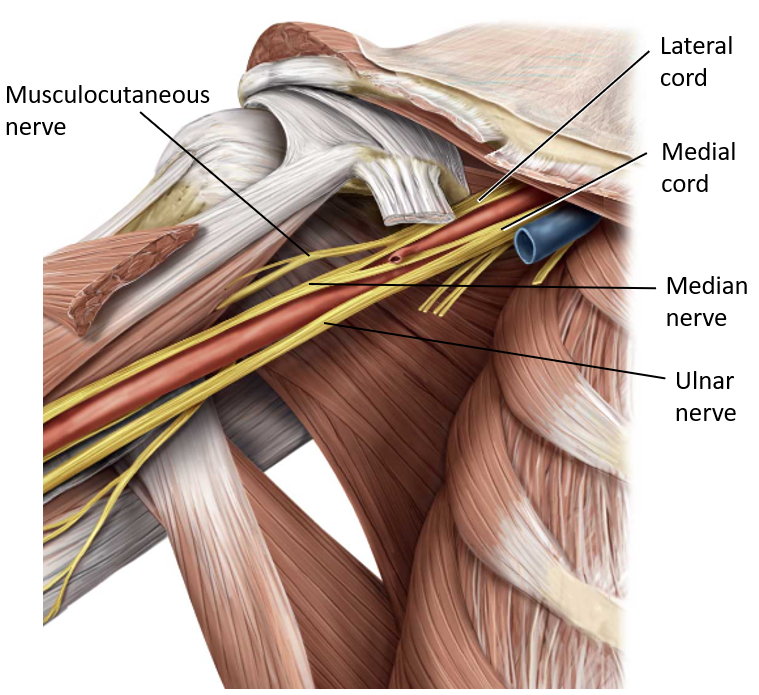
The musculocutaneous nerve, a terminal branch, pierces the coracobrachialis muscle (verify this). Follow it proximally to find the lateral cord of the brachial plexus lateral to the axillary artery.
Note
The cords of the brachial plexus are named based on their relationships to the axillary artery.
Once you have identified the lateral cord, clean and trace its other terminal branch: the lateral root of the median nerve.
Pick up the median nerve and trace its medial root proximally to find the medial cord of the brachial plexus (Note: This cord sits medial to the axillary artery!).
Question
Can you see that the median nerve is a terminal branch of the brachial plexus made by contributions from the lateral and medial cords?
Finally: trace the medial cord distally, it becomes the ulnar nerve.
Fun fact
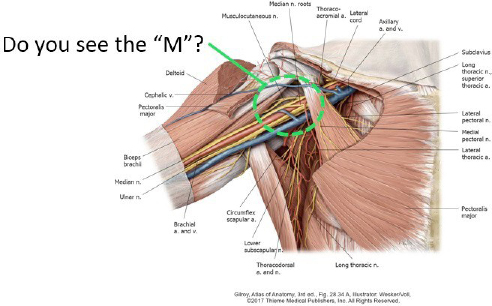
The terminal branches discussed above are configured in the shape of the letter “M”: the musculocutaneous nerve, the medial and lateral roots of the median nerve, and the ulnar nerve. Lots of “M’s”: musculocutaneous nerve, median nerve, and medial cord—all contribute to the “M”.
Let’s go deeper—find the posterior cord and its terminal branches.
Now grab the medial cord, lateral cord, and the axillary artery and retract them just enough to expose the posterior cord of the brachial plexus, posterior to the axillary artery.
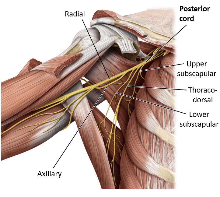
Clean the posterior cord and all of its branches. The posterior cord is large and continues distally as the radial nerve, a terminal branch. It leaves the posterior wall of the axilla with the deep brachial artery (profunda brachii)—a branch of the brachial artery—via a gap in the triceps muscle.
Trace the posterior cord higher and locate the thick axillary nerve. The axillary nerve and posterior circumflex humeral artery leave the posterior wall of the axilla via the quadrangular space (they are travel buddies!).
Clinical correlation
Radial nerve + deep brachial (profunda brachii) artery spiral around the shaft of the humerus together.
Fractures of the midshaft of the humerus can damage the radial nerve and/or deep brachial artery. What symptoms might be seen in a patient with damage to the radial nerve because of a midshaft fracture of the humerus? The triceps muscle would be spared . . . but not the muscles on the posterior side of the forearm.
Both sides: Identify side branches of the brachial plexus.
Branches of the brachial plexus
Hint
Branches from the proximal parts of the brachial plexus (branches from roots and trunks) will need to be identified on the prosection.
- From the roots of the plexus:
- Dorsal scapular nerve: from C-5 (hard to find—look for it near the middle scalene muscle, then piercing the levator scapulae muscle, which it innervates).
- Long thoracic nerve: from C-5, C-6, and C-7, to serratus anterior. You identified this nerve on the external surface of the serratus anterior in the last lab session. On the prosection, trace it back proximally to its origin from the brachial plexus.
- From the upper trunk:
- Suprascapular nerve
- From the lateral cord:
- Lateral pectoral nerve
- From the medial cord:
- Medial pectoral nerve
- Medial cutaneous nerve of arm
- Medial cutaneous nerve of forearm (often the two cutaneous nerves travel together as one trunk—one branch given off to arm and one continuing into the forearm). What does the word “cutaneous” tell you about these nerves?
- From the posterior cord:
- Upper subscapular nerve
- Thoracodorsal nerve
- Lower subscapular nerve
Note
A mnemonic for remembering all the branches of the posterior cord is ULTRA:
Upper subscapular
Lower subscapular
Thoracodorsal
Radial
Axillary
(These are not in the correct topographic order however—oh well!)
The Arm
Superficial veins of the arm
Anterior Compartment of the Arm
The skin was reflected from the anterior compartment earlier in the lab session.
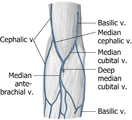
In the superficial fascia, look for the two superficial veins of the arm: basilic and cephalic veins.
The cephalic vein (“bodybuilder’s vein”) is on the preaxial border of the arm. It gets its names since the preaxial border is closer to the head in the embryo.
The superficial vein of the postaxial border is the basilic vein (it gets its name because it courses along the “base” of the upper limb = its postaxial border).
Muscles of the shoulder and arm
Bony landmarks of the arm
Identify the three muscles in the anterior compartment of the arm:
■ Biceps brachii
■ Coracobrachialis
■ Brachialis
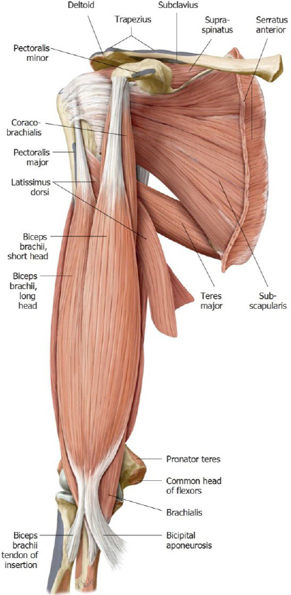
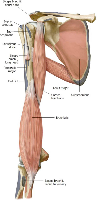
Review the two proximal attachments of the biceps brachii on the scapula:
■Long head: courses through the intertubercular (bicipital) groove of the humerus, enters and passes through the glenohumeral joint, and inserts on the supraglenoid tubercle. You won’t see this attachment in your donor unless you open the glenohumeral joint. Find the supraglenoid tubercle on a skeleton.
■Short head: attaches to the coracoid process. You will see this attachment in your donor; palpate the coracoid process.
The biceps has two distal attachments:
■Via the bicipital aponeurosis to the ulna. The bicipital aponeurosis is a thick layer of deep fascia that forms a protective layer over the roof of the cubital fossa.
■ Via the biceps tendon to the radial tuberosity. Identify this on a skeleton.
Note
The brachialis muscle is the “workhorse” of elbow flexion, producing the most power during this movement. The biceps contributes to forearm flexion, but perhaps its most important function is as a supinator of the forearm. The biceps is the power supinator—a very useful muscle when using a screwdriver or opening the lid of a jar.
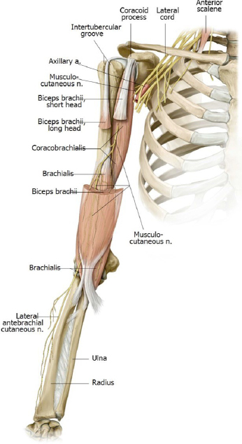
Note that the musculocutaneous nerve pierces the coracobrachialis muscle, then passes deep to the biceps between it and the brachialis. It innervates all the muscles in the anterior compartment of the arm.
Clean the fascia from the biceps and explore the plane between the biceps and brachialis and follow the musculocutaneous nerve.
Nerves and arteries of the arm
The musculocutaneous nerve will eventually leave this plane and the anterior compartment of the arm to enter the superficial fascia of the forearm as the lateral cutaneous nerve of the forearm.
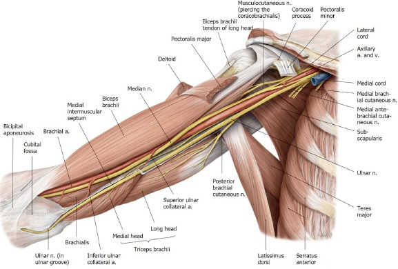
Use blunt dissection to separate the muscles of the anterior and posterior compartments, on the medial side of the arm.
There is lots of good stuff here, in the groove between the biceps and triceps = the basilic vein (a superficial vein), the brachial artery (the distal continuation of the axillary artery), the median nerve, and the ulnar nerve.
Posterior Compartment of the Arm
We won’t dissect the posterior compartment today. It contains the triceps brachii muscle. You saw this in a previous lab session. Study the triceps on the prosected upper limb specimen. Review the three heads. Know their origins and insertion and the innervation of the triceps.
Checklist, Lab #4
Review and make sure you have identified each of the structures below.
Bones
Scapula
Coracoid process (which muscles attach here?)
Supraglenoid and infraglenoid tubercles (which muscle attaches to each?)
Distal humerus
Capitulum
Trochlea
Proximal radius
Head of the radius
Radial tuberosity (what muscle attaches here?)
Proximal ulna
Olecranon process (what muscle attaches here?)
Coronoid process (what muscle attaches here?)
Trochlear notch
Muscles
Pectoralis major
Pectoralis minor
Serratus anterior
Biceps brachii (long and short heads)
Triceps brachii (long, lateral, and medial heads)
Brachialis
Coracobrachialis
Nerves
Parts of brachial plexus
Roots (C-5, C-6, C-7, C-8, and T-1)
Trunks (upper, middle, and lower)
Divisions (anterior and posterior)
Cords (medial, lateral, and posterior)
Terminal branches
Musculocutaneous nerve
Ulnar nerve
Median nerve (made from medial and lateral roots)
Axillary nerve
Radial nerve
Side branches
Long thoracic nerve
Suprascapular nerve
Lateral pectoral nerve
Medial pectoral nerve
Medial cutaneous nerves of arm and forearm
Upper subscapular nerve
Thoracodorsal nerve
Lower subscapular nerve
Vessels
Axillary vein
Basilic vein
Cephalic vein
Brachial veins
Axillary artery
Thoraco-acromial trunk—with pectoral arteries
Lateral thoracic artery
Subscapular artery
Circumflex humeral arteries (anterior and posterior—posterior is the largest)