Lab 23: Face, Parotid Gland, and Superficial Neck

  1. Identify the bones associated with the face and parotid gland.
  2. Identify surface landmarks on the face.
  3. Clean and identify muscles of facial expression.
  4. Identify branches of the facial nerve on the face.
  5. Locate the major sensory nerves of the face.
  6. Identify the facial artery and vein.
  7. Identify the parotid gland and parotid duct.
  8. Remove the parotid gland on one side; identify the nerves and vessels within the gland.
  9. Reflect the skin from the neck and identify the platysma muscle.
  10. Study a prosection of the face.

Osteology

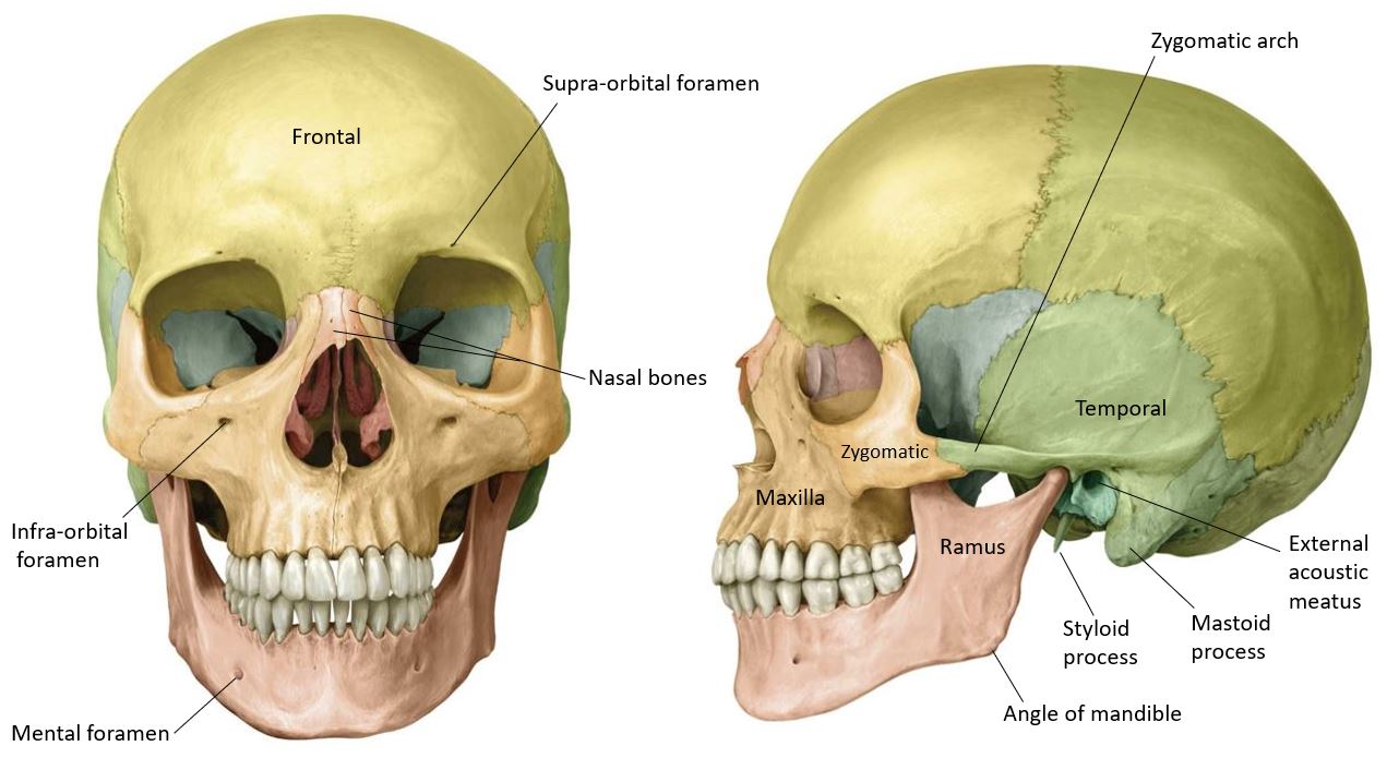
Before beginning the dissection, grab a skull and locate the following (see Figure 1):

Frontal, nasal, and zygomatic bones

Maxillae and mandible

Orbits with supraorbital and infraorbital margins

Supraorbital notches (or foramina)

Infraorbital foramina

Mental foramina

Angles and rami of mandible

Mastoid processes and styloid processes of the temporal bones

Stylomastoid foramina

Zygomatic arches

Temporomandibular joints

External acoustic meatuses

Figure 1. Bones associated with the face dissection.

Facial Landmarks

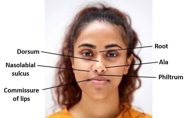
Figure 2.

On the donor, identify these landmarks on the face:

Forehead

Nose: root, apex (tip), dorsum (bridge), alae, nares (nostrils)

Upper lip with philtrum (in the center)

Lower lip

Commissures of the lips (angles of mouth): Where the upper and lower lips meet

Chin

Cheeks (buccal regions) = each contains a buccal fat pad

Nasolabial sulci: These are deep creases in the facial skin that separate the two cheeks from the upper lip.

Dissection of the face: Prep work

Before you cut, place a plastic block under your donor’s upper back; this will allow the neck to extend back toward the table, and provide a greater area for working.

Important note

Each group will work on only ONE SIDE OF THE FACE. Groups will perform slightly different tasks on their face, either a SUPERFICIAL dissection or a DEEP dissection. PAY CLOSE ATTENTION TO THE INSTRUCTIONS.

ALL Groups on ONE side: Skin incisions

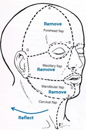
Using a sharp scalpel, make shallow incisions in the skin on the undissected side of the face and neck as shown in Figure 3.

Do NOT cut into the eyelids, the lips, or the nose. Make circular incisions around their margins, as shown in the figure. Do not remove the nose. We will want to study it later.

Figure 3. Skin incisions.

1Midline incision in forehead—down to the root of the nose (A to B). Note: The scalp has already been peeled forward, so this incision has likely been made.

2Circular incision around the orbit, leaving the eyelid intact; then, a horizontal incision from the lateral side of the orbit to the ear (B to G). Remove the forehead flap of skin.

3Incision around the nose, then down the midline of the philtrum (B to C).

4Incision around half of the oral fissure, leaving the lips intact (C to D)—then, a horizontal incision from the angle of the mouth to the mastoid process behind the ear (H).

5Midline incision from lower lip to chin (D to E).

6Incision along edge of mandible—from chin to mastoid process (E to H).

7Midline incision in neck, from chin to suprasternal notch (E to F).

8Vertical incision just in front of the ear (G to H).

ALL groups: Skin flaps

This will take some time—patience is a virtue!

Use a sharp scalpel to peel back the skin, from anterior to posterior—make shallow skin flaps and leave the superficial fascia intact.

1Remove the forehead, maxillary, and mandibular skin flaps.

2Reflect the skin of the neck as far posterior as possible. Leave the skin flap attached posteriorly.

Fascia of the Face

The superficial fascia of the face is copious and unique because it contains skeletal muscles. Clinicians refer to it as the superficial muscular aponeurotic system (SMAS). Recall that skeletal muscles in other areas of the body are invested by deep fascia. The superficial fascia of the face connects the muscles of facial expression to the dermis of the skin. These muscles couldn’t produce facial expressions if they were isolated from the skin by the deep fascia.

True deep fascia in the face invests the parotid glands and the masseter muscles.

ALL GROUPS: Clean the muscles of facial expression.

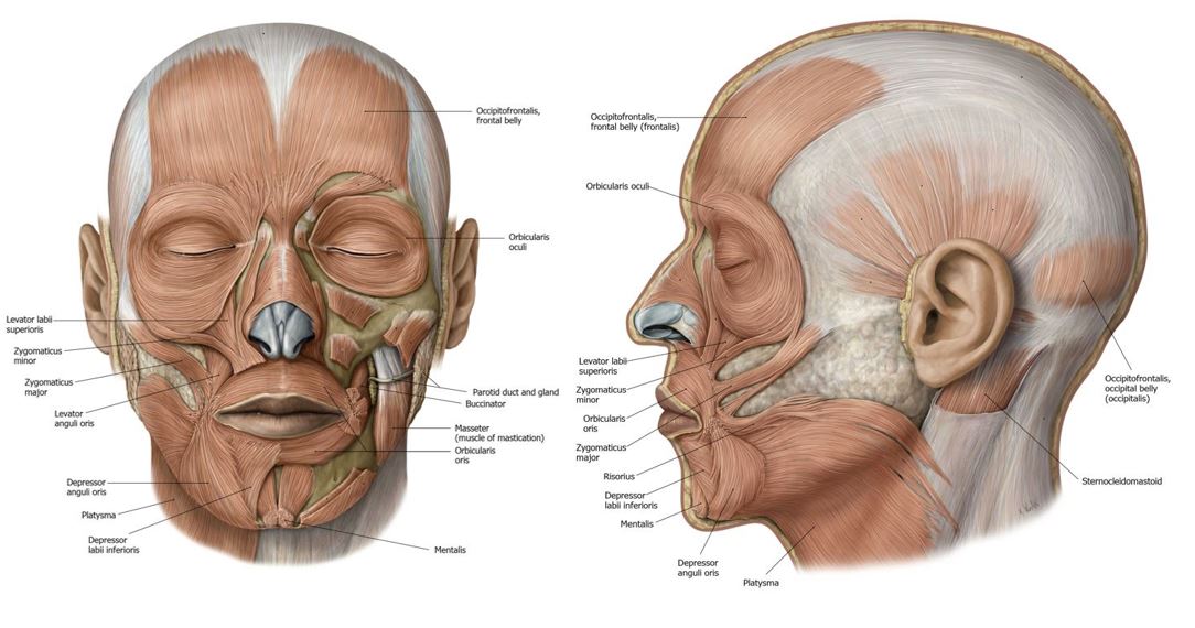
Use scissors to probe through and trim away the superficial fascia to find muscles of facial expression (a.k.a. mimetic muscles). Review the general arrangement of the muscles in an atlas or in Figure 4. We will focus on the muscles that surround the orbit and mouth in the list below. Think about the names—they are clues to their attachments and actions.

This is a difficult task—it requires determination and patience!

Figure 4. Muscles of facial expression.

The major muscles of facial expression are listed below. Examine these on a prosection as well.

Frontal belly of occipitofrontalis (the muscle of surprise!)

Orbicularis oculi: Has two parts: orbital and palpebral (this muscle is the sphincter of the eyelids—used in the “blink” reflex)

Zygomaticus major (smile wide!)

Levator anguli oris (grinning muscle)

Levator labii superioris (snarling muscle)

Orbicularis oris (pucker up! = the sphincter of the oral fissure)

Depressor anguli oris (frowning muscle)

Depressor labii inferioris (muscle of disbelief!)

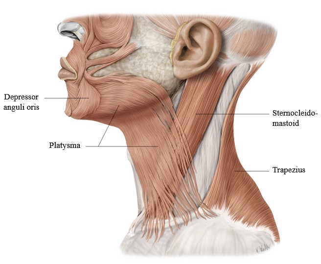
Platysma: Extends from the angle of mouth into the superficial fascia of the neck (grimacing muscle = tenses the skin of the neck)

The buccinator is a muscle of facial expression (“trumpeter’s muscle” = compresses the cheeks): We won’t see it yet since it is hidden deep to the buccal fat pad. We will expose it a little bit later.

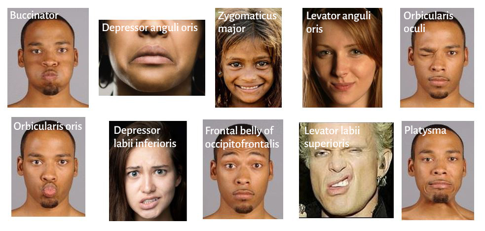
Test your knowledge!

Which muscles produce the facial expressions shown here? (Tap for answers.)
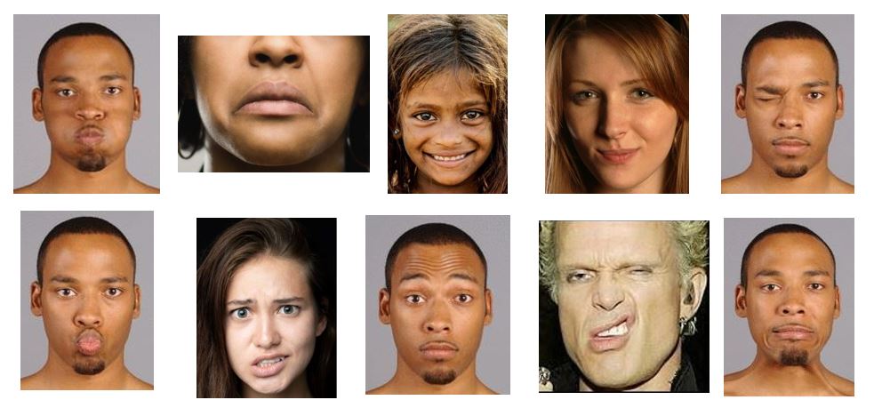
 

Figure 5. Facial expressions.

Answers


ALL groups: Expose the parotid gland.

Question

What kind of gland is the parotid? What does the word parotid mean?

It is anterior to the ear; sandwiched between the mandible and sternocleidomastoid muscle.

It is invested by a capsule of dense connective tissue (deep fascia).

The parotid covers the outer surface of the masseter muscle. This muscle is a chewing muscle, not a muscle of facial expression.

Remove the subcutaneous fat that overlies the parotid gland and expose the deep fascia covering the gland. Do your best to remove the deep fascia from the surface of the gland, but DO NOT cut around the borders of the gland with a scalpel. Get a sense of the size of the gland and approximate where its superior, anterior, and inferior borders are. 

We are now going to locate a few structures that emerge from the anterior border of the parotid. Using a probe or small scissors, begin at the top of the gland and carefully BLUNT dissect along its anterior border for nerves and other structures that emerge anteriorly.

Because these structures course horizontally across the face, probing through the fascia with scissor should be done in the transverse direction, NEVER VERTICALLY.

Now it's decision time!
TALK with the groups around you:

At this point, it would be ideal if the groups could split the work and alternate tasks, such that half the groups in each lab continue with a SUPERFICIAL dissection while half complete a DEEP dissection. Check with your instructors and the groups at the tables next to yours and make a plan.

If your dissection displays nice facial muscles and you can identify numerous branches of the facial nerve exiting the parotid gland, your group will continue with a superficial dissection.

This will require careful, meticulous work.

If fewer facial muscles can be identified and you aren’t able to find branches of the facial nerve emerging from the parotid gland, your group will perform a deep dissection.

This will require a little more cutting of tissue and a coarser dissection to remove the parotid gland, but will be important for preparing the area for work in future labs on the neck and infratemporal fossa!

SUPERFICIAL Groups: dissection of the face

Continue the detailed work of cleaning around the borders of the parotid gland and following the nerves and other structures that emerge:

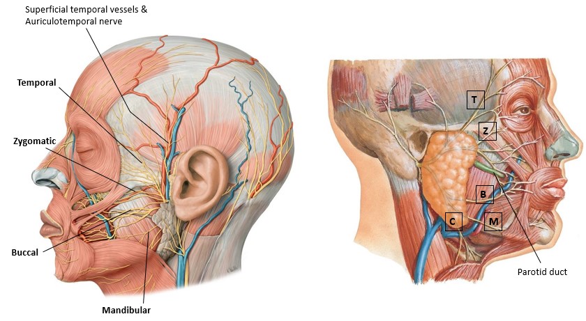
Identify the large parotid duct (Stensen’s duct) leaving the anterior border of the gland. It runs across the masseter, parallel to the earlobe. See Figure 6. Trace it anteriorly until it vanishes into the buccal fat pad, then through the buccinator muscle. Sometimes the parotid duct is obscured by accessory parotid glandular tissue. Where does the parotid duct open in the oral cavity?

Parallel and just below the parotid duct you should be able to identify a large nerve (travelling towards the corner of the mouth) = the buccal branch of the facial nerve. There may be multiple branches.

Parallel and superior to the parotid duct is the zygomatic branch of the facial nerve (travelling towards the corner of the eye). Again, there is often more than one.

Figure 6. Parotid gland and duct. The buccal fat pad has been removed.

Keep working along the superior, anterior, and inferior borders of the gland, searching for the following nerves and vessels:

Branches of the facial nerve—probe above and below the parotid duct to find the following facial nerve branches on the face (Figure 6):

Temporal (to forehead)

Zygomatic (to upper cheek, below the orbit)

Buccal (to the fleshy central cheek)

Mandibular (often quite thin—runs along the margin of the mandible and dives under the platysma).

The Cervical branch descends from the lower part of the parotid into the neck, deep to the platysma. It may be difficult to identify at this point in the dissection.

Note

To remember the branches of the facial nerve on the face, try this old-school, yet catchy mnemonic: To Zanzibar By Motor Car

Superficial temporal artery and vein: These emerge from the upper part of the parotid, anterior to the ear, and course superiorly across the “temple” region toward the scalp. Deep to the vessels is the auriculotemporal nerve, a sensory branch of V3 that supplies the scalp and ear. See Figure 6.

Sensory Nerves of the Face

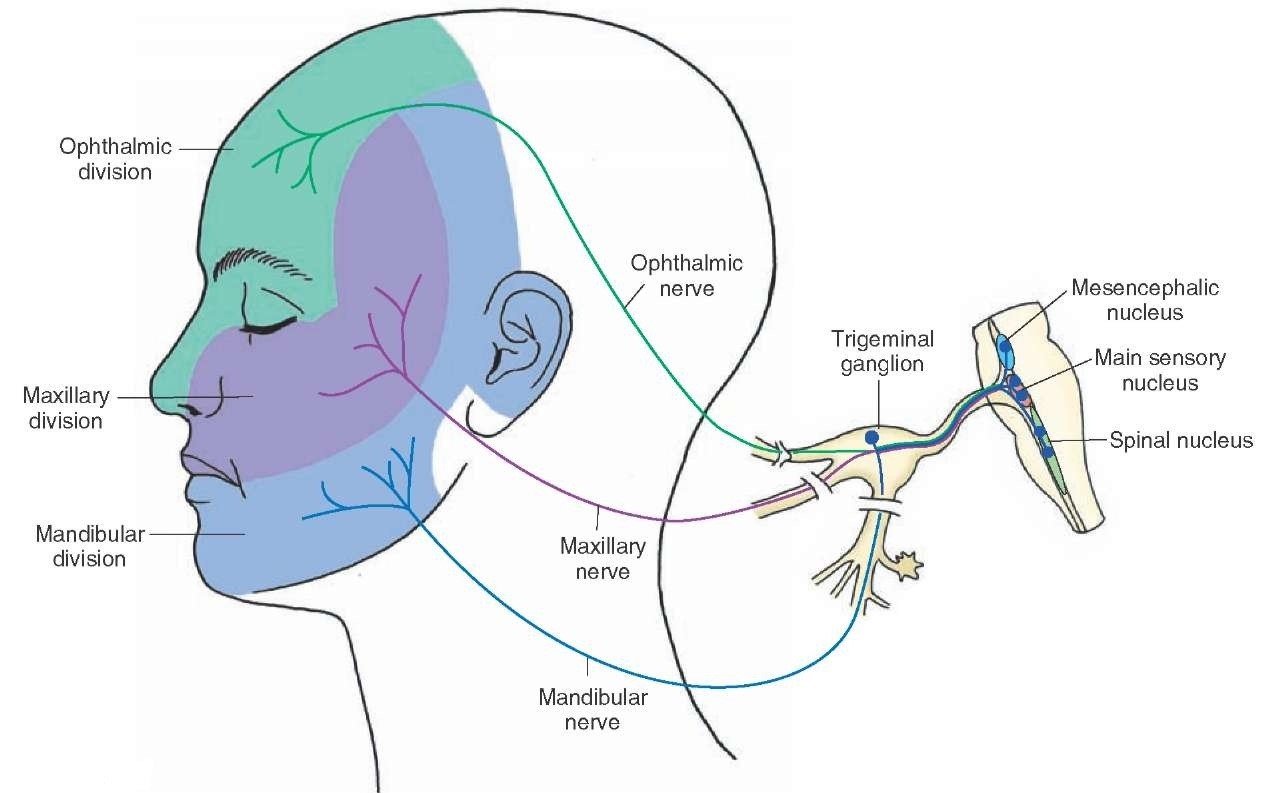
The skin of the face is supplied by branches of the three divisions of the trigeminal nerve. Each division supplies a characteristic swath of skin (dermatome) that reflects the embryonic development of the face (Figure 7). A small area of skin over the angle of the mandible is supplied by the great auricular nerve (from the cervical plexus).

Figure 7. Dermatomes of the divisions of the trigeminal nerve.
DEEP Groups: Reveal the sensory branches

Branches of CN V leave bony openings on the face and pass through the superficial fascia and facial muscles to reach the skin. Some of the facial muscles will have to be sacrificed on the deep dissection to find these nerves.

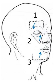
Using a scalpel, make deep incisions in the superficial fascia and muscles of the face and reflect the flaps of tissue in the directions indicated in Figure 8.

Figure 8. Tissue flaps for locating major sensory nerves of the face.

Locate and clean these nerves as they emerge from the skull bones (see Figure 8):

1From Ophthalmic (V1): Supra-orbital nerve.

Reflect the scalp flap (made when the scalp was cut in the previous lab) on the side of the face where you are working downward toward the orbit, exposing the frontal bone. As you tug the flap down to the level of the supra-orbital margin, you should see the supra-orbital nerve emerging from its foramen (or notch)above the orbit.

2From Maxillary (V2): Infra-orbital nerve.

Make a V-shaped incision (to the bone) in the upper cheek from the medial and lateral corners of the eyelids down to an apex at the level of the nostrils.

Elevate this flap of tissue upwards toward the orbit to locate the infra-orbital nerve leaving the infra-orbital foramen. It is a large nerve.

3From Mandibular (V3): Mental nerve.

Starting at the midpoint of the lower lip, make a deep incision (to the bone) down and then along the margin of the mandible to a location lateral to the angle of the mouth. Elevate this square flap of tissue to locate the mental nerve leaving the mental foramen.

DEEP Groups: dissection of the parotid gland

The parotid has a capsule of deep fascia and the gland itself is dense and tough—so dissecting through it will be difficult. Use scissors to remove the gland in pieces.

Several structures traverse the gland in layers. From superficial to deep these are:

1Facial nerve branches

2Retromandibular vein

3Terminal part of the external carotid artery

We will tackle the first two layers in this dissection.

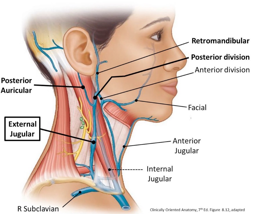
The retromandibular vein is a wide, short vein. At the inferior border of the parotid, it splits into two divisions:

Posterior division: Joins the posterior auricular vein to form the external jugular vein.

Anterior division: Joins the facial vein to form the common facial vein = a tributary of the internal jugular vein.

Layer 1: Facial nerve and its branches

The facial nerve emerges from the skull through the stylomastoid foramen, then enters the parotid gland. It typically divides into two trunks within the gland. See Figure 9.

From the upper trunk arise temporal and zygomatic branches

From the lower trunk arise buccal, mandibular, and cervical branches.

Let’s find the facial nerve within the parotid gland using this method:

See if you can locate a zygomatic and buccal branch of the facial nerve on the face above and below the parotid duct, anterior to the gland. Dissect them back to the gland, then remove pieces of the gland with forceps and scissors and follow the branches into the gland until you reach the trunks of the nerve. Follow the upper and lower trunks posteriorly to where they merge: this is the facial nerve, entering the parotid gland from the stylomastoid foramen.
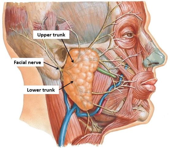
Figure 9.

Layer 2: Retromandibular vein (Figure 10)

This vein is formed by the union of the superficial temporal vein (from the scalp) and maxillary vein (from the infratemporal fossa—deep to the mandible). The veins merge within the parotid gland.

Best bet: Locate the superficial temporal vein in the lower scalp and trace it down into the parotid. Remove parotid tissue as you clean the vein from superior to inferior.
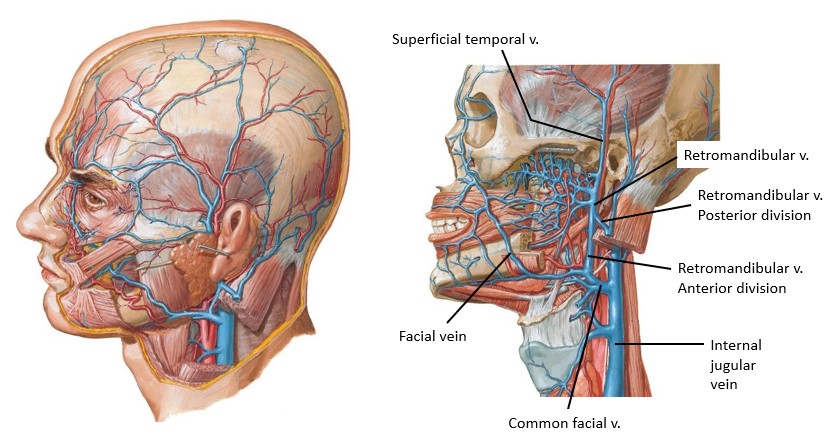
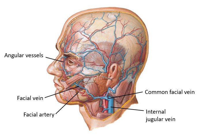
Figure 10. Retromandibular vein.

Facial Vessels

The major source of blood to the face is the facial artery—a branch of the external carotid artery in the neck. The facial artery loops around the submandibular salivary gland then curls over the mandible on to the face.

Try this

Clench your jaw and palpate the taut masseter muscle. Place a finger on the margin of the mandible just anterior to the masseter. Feel a pulse? That’s the facial artery.

Figure 11. Facial vessels—the buccal fat pad has been removed to reveal the buccinator.
ALL GROUPS: Find and clean facial vessels

BOTH groups can look for the facial vessels—but the SUPERFICIAL GROUPS especially should take the time to carefully clean and trace them

The facial artery has an oblique course across the face (Figure 11). It is tortuous (twisty), allowing it to have some slack when the facial muscles contract and contort the face. It winds around the facial muscles and through the buccal fat pad. When it passes lateral to the nose, it changes its name to the angular artery. The angular artery terminates at the medial corner of the eye where the upper and lower eyelids meet.

The angular vein arises at the medial corner of the eye and descends along the nose and through the buccal fat pad. Below the nose it becomes the facial vein. It is not as tortuous as the facial artery. In the cadaver, it may be collapsed and inconspicuous. The facial vein usually joins the anterior division of the retromandibular vein to form the common facial vein.

Identify the facial artery as it crosses the mandible anterior to the masseter. Trace it through the buccal fat pad to the angle of the mouth. Look for the inferior and superior labial arteries branching from the facial artery and entering the lips. The facial vein is usually posterior to the facial artery in the buccal fat pad.

Remove the buccal fat. You will now be able to identify the buccinator muscle in the cheek, deep to the facial vessels (Figure 11). It is a muscle of facial expression, but also is useful when chewing, as it contracts to compress the cheeks and keep the food in your mouth between your teeth, rather than collecting in the oral vestibule.

Clinical correlation

Patients with Bell’s Palsy often lose the functions of the orbicularis oris and buccinator, in which case food may dribble from the corner of the mouth.

ALL Groups: Superficial neck

Like the face

We will do this dissection on ONE SIDE.

Skin incisions: If you haven’t done this yet, make a shallow midline incision from the chin to the suprasternal notch. Reflect a thin skin flap on the same side as your face dissection as far posterior as possible. Leave the superficial fascia.

Carefully clean the superficial fascia in the anterior neck. Identify the thin platysma muscles [platysma = flat, like the bill of a platypus]. The origins of the muscles are in the superficial fascia of the upper chest, over the clavicles. They extend upward through the neck to insert on the mandible and the angles of the mouth. Since the thoracic wall has been removed in your cadaver, you won’t be able to visualize the attachment of the platysma below.

Figure 12. Platysma muscle.

The platysma is a muscle of facial expression, derived from the same mesoderm that forms the muscles of facial expression on the face. Like the muscles in the face, the platysma is unique in that it is located within the subcutaneous (superficial) fascia, inserts into the skin, and it is innervated by the facial nerve.

Optional dissection—if there is time.

Reflect the platysma muscle superiorly toward the face. Grasp the thin muscle with forceps and carefully peel it away from the subcutaneous tissue, using a scalpel or scissors. Leave the remaining superficial fascia in the neck intact.

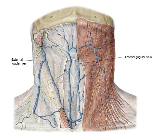
Identify the superficial veins of the neck. These have a variable arrangement.

Anterior jugular vein: Begins just inferior to the chin, runs vertically just off the midline, ducks under the sternocleidomastoid.

External jugular vein: Arises from the posterior division of the retromandibular vein near the angle of the mandible. Descends across the external surface of the sternocleidomastoid muscle toward the midpoint of the clavicle. It is a tributary of the subclavian vein.

Figure 13. Superficial veins in the neck.
Figure 14.

Clinical correlation

Patients with heart problems often have distended neck veins—some donors may have large veins with clotted blood.

Checklist, Lab #23

Review and make sure you have identified each of the structures below.

Osteology

See the list of relevant bony structures in the Osteology section above.

Facial Landmarks

See the list of landmarks in the Facial Landmarks section above.

Muscles

Identify as many of these as possible in your donor or other student dissections. Look at the prosection for help.

Frontal belly of occipitofrontalis

Orbicularis oculi (orbital and palpebral portions)

Zygomaticus major

Orbicularis oris

Levator labii superioris

Levator anguli oris

Depressor labii inferioris

Depressor anguli oris

Buccinator & Buccal fat pad

Platysma

Masseter (this is a chewing muscle on the face—not a muscle of facial expression)

Parotid

Parotid gland and duct

Vessels

Facial artery and facial vein

Superior and inferior labial arteries (wish list items!)

Superficial temporal artery and vein

Retromandibular vein

Nerves

Temporal, zygomatic, buccal, mandibular, and cervical branches of VII (see prosection if you can’t find them—the cervical branch is tough to find)

Supra-orbital nerve (from V1)

Infra-orbital nerve (from V2)

Mental nerve (From V3)

Auriculotemporal nerve (from V3): Accompanies the superficial temporal artery on the scalp (wish list item!)

Superficial Neck

Platysma muscle

External jugular vein

Table of Contents
Headshot of David Conley, PhD · Professor, Department of Translational Medicine & Physiology
David Conley
PhD · Professor, Department of Translational Medicine & Physiology
Office: PBS 41A
envelope icon
Headshot of Shannon Helbling, PhD · Clinical Assistant Professor, Department of Translational Medicine & Physiology
Shannon Helbling
PhD · Clinical Assistant Professor, Department of Translational Medicine & Physiology
Office: PBS 41C
envelope icon

Dear students,
We would love to hear from you about your learning materials.

Page/Content Feedback Form