- Goals
- Identify the layers of the SCALP.
- Examine the dura mater and the dural septae.
- Remove the brain and study: meningeal coverings, gross features, and arterial supply (circle of Willis).
- Examine the internal aspect of the base of the skull and the cranial fossae.
- Identify cranial nerves and blood vessels that exit/enter the skull through foramina in the cranial base.
- Identify the dural venous sinuses.
- Open the cavernous sinuses and examine the structures that pass through the sinuses, or are located in their walls.
Scalp
The scalp has been cut into 4 flaps and reflected downward.
Layers of the Scalp
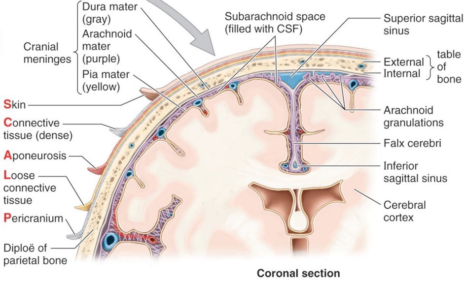
The 3 external-most layers of the scalp are the “scalp proper.” Study the flaps in cross-section to identify the layers:
- S
Skin
- C
Connective tissue: Contains fatty tissue and a lot of dense connective tissue with cutaneous vessels and nerves.
- A
Aponeurosis (epicranial aponeurosis): A flat tendon that connects anteriorly and posteriorly to skeletal muscles (the occipitofrontalis—frontal belly and occipital belly attach on either end of the aponeurosis).
- L
Loose areolar connective tissue: Allows the scalp proper to glide over the skull when it is moved by the occipitofrontalis. The looseness of this layer makes it a “potential space” where fluids can accumulate should the scalp be injured or infected.
- P
Pericranium: Scrape the external surface of the skull cap. The pericranium is the external periosteum of the skull. It is firmly attached to the connective tissue in the skull sutures.
Nerves and Arteries of the Scalp
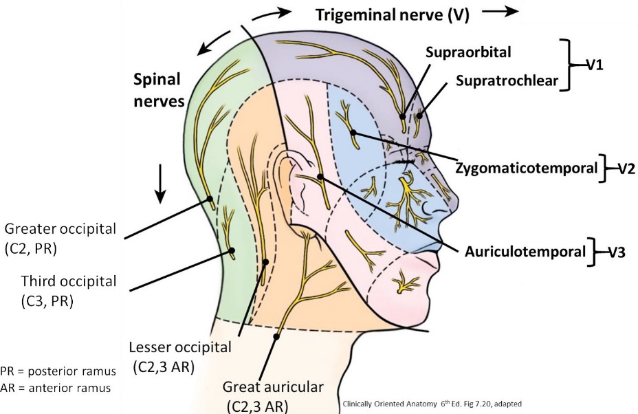
Draw an imaginary line between the two ears passing through the vertex/summit of the skull.
The anterior scalp is innervated by branches of the trigeminal nerve (CN V), while the posterior scalp is supplied by cervical spinal nerves.
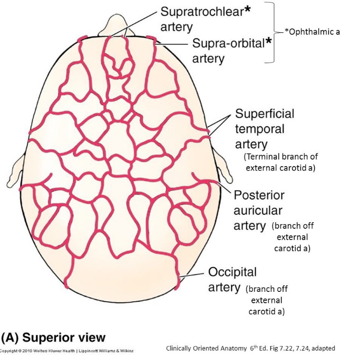
The anterior-most scalp receives blood from branches of the ophthalmic artery (which is itself a branch of the internal carotid artery), while the rest of the scalp is supplied by branches of the external carotid artery.
Cranial Cavity and Brain
The dura mater has an outer endosteal layer (the internal layer of periosteum of the skull cap) and an inner meningeal layer (the dura mater proper).
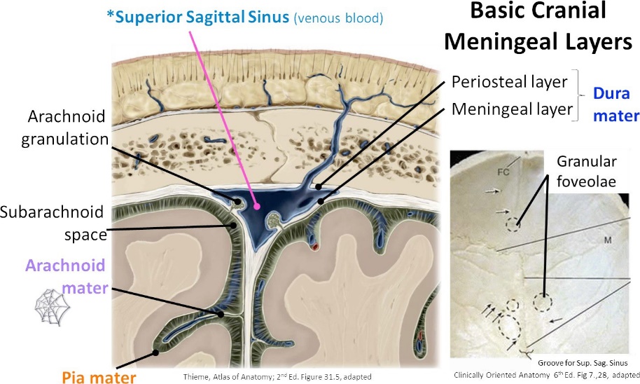
Figure 4.
The endosteal and meningeal layers of dura are fused except in certain places where they are separated to form venous sinuses (e.g., superior sagittal sinus) and where the meningeal layer departs to form dural septa (e.g., falx cerebri).
The dura was likely peeled off when the calvaria was removed — if not go ahead and peel it off— then examine the internal surface of the calvaria.
■Observe the midline impression in the bone made by the superior sagittal sinus.
■Find scattered pits in the bone = these are impressions in the bone made by large arachnoid granulations.
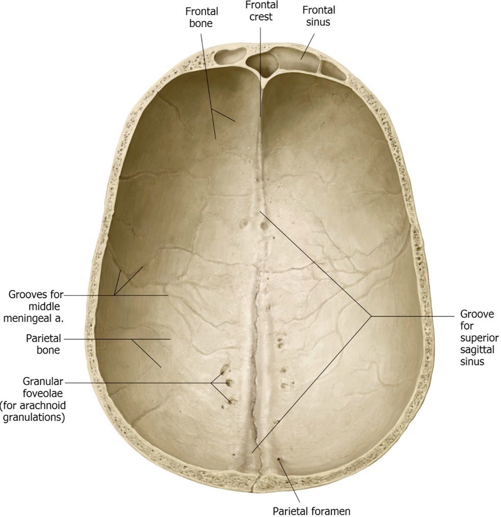
Figure 5.
Question
What are arachnoid granulations, and what is their function?
■Look at the cut surface of the calvaria and notice its construction: an outer table of compact bone, an inner table of compact bone, and spongy bone (called diploe) in the middle.
Tethered to the outside of the dura is the middle meningeal artery (it is sandwiched between the dura and skull bones). The middle meningeal is a branch of the maxillary artery (from the external carotid artery). It is the chief source of blood to the calvaria and its associated dura.
Dural Septa
Examine the dural septa (= infoldings of the meningeal layer of the dura). See Figure 6.
■Falx cerebri
■Contains the superior sagittal sinus and is located within the longitudinal fissure of the cerebrum, separating the left and right cerebral hemispheres. Its inferior free margin contains the inferior sagittal sinus and relates to the brain’s corpus callosum.
■Tentorium cerebelli
■Elevate the brain’s temporal lobes to see the tentorium cerebelli.
■It attaches to the anterior and posterior clinoid processes of the sphenoid bone, the summit of the petrous temporal bones, and to the internal surface of the occipital bone.
■The attached edges of the tentorium contain the superior petrosal and transverse venous sinuses.
■The left and right portions of the tentorium meet in the midline where it contains the straight venous sinus and where it is attached to the falx cerebri. The midline part of the tentorium is higher than the peripherally attached portions (a sloped tent).
■The tentorium has a gap anteromedially called the tentorial notch that contains the brainstem. The tentorium separates the occipital lobes of the brain from the cerebellum.
■Falx cerebelli
■Lies inferior to the tentorium. It attaches to the midline of the occipital bone and to the inferior surface of the tentorium and it separates the left and right cerebellar hemispheres.
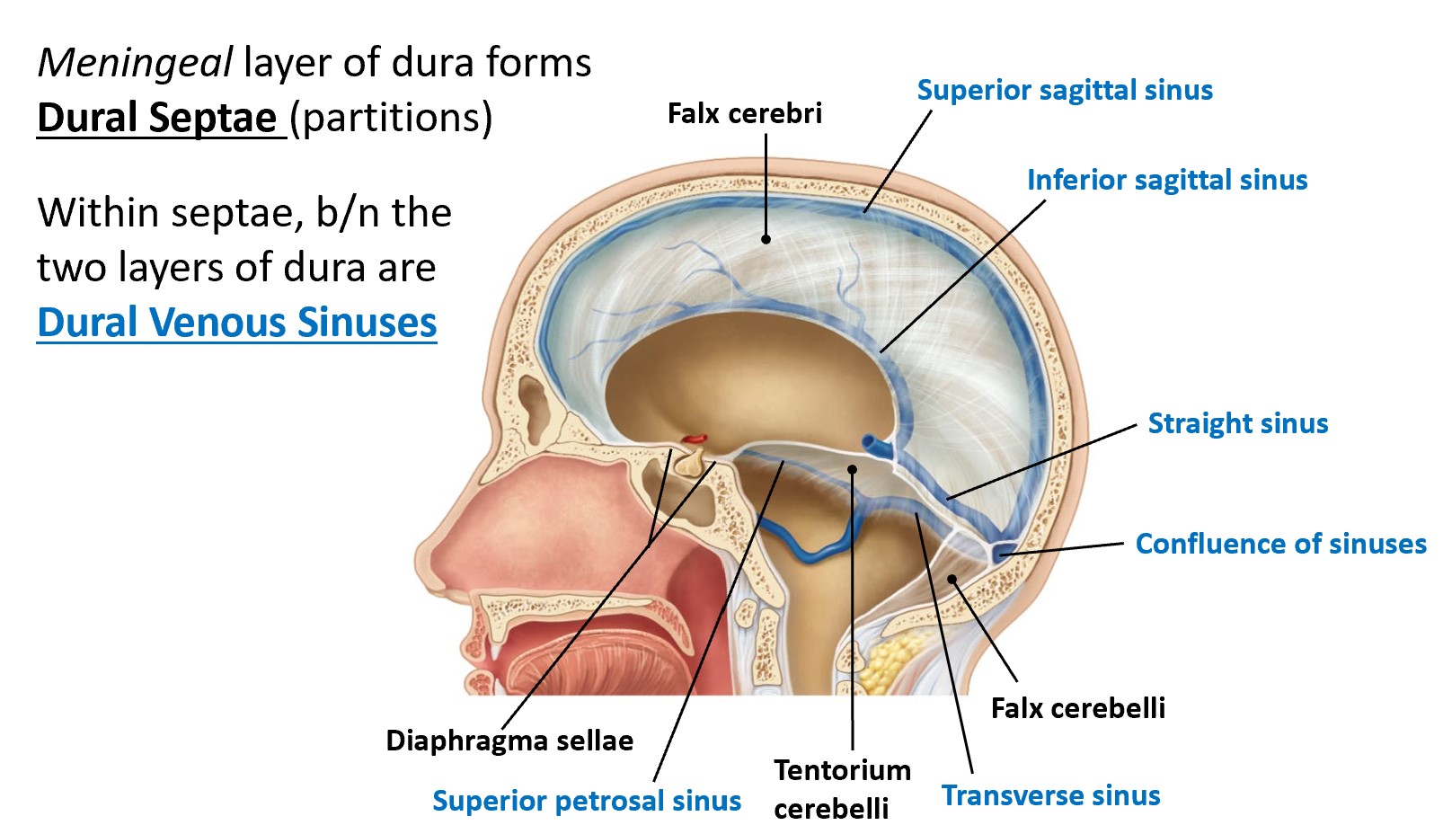
Figure 6.
Remove the entire brain.
Question
This step was already done—what dural folds were cut in order to remove the brain?
Proceed from anterior to posterior; have one member elevate the brain while another cuts.
Question
The 12 cranial nerves were cut—what two sets of major arteries also had to be severed to free the brain?
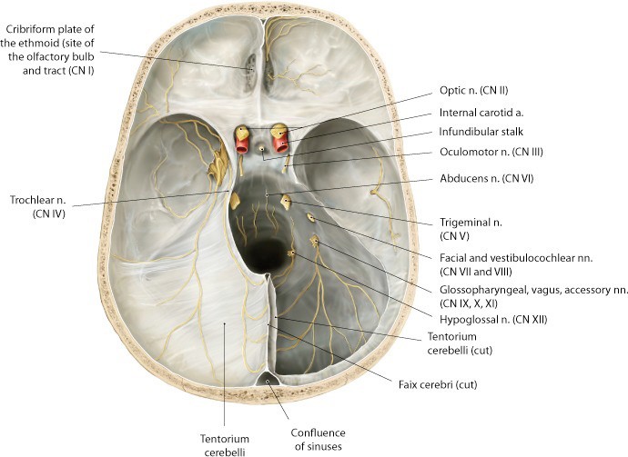
Figure 7.
Brain: Features and Arteries
Identify the following parts or structures on the brain:
■the lobes of the cerebrum: frontal, parietal, temporal, and occipital
■the cerebellum
■the parts of the brainstem: midbrain, pons, and medulla oblongata
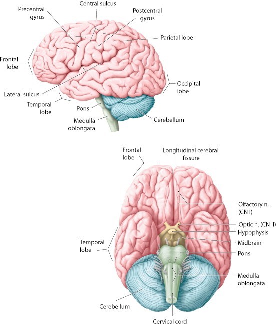
Figure 8. Adapted from Gilroy et al., Atlas of Anatomy, 2nd ed., Figs. 40.6A, 40.6C.
Study the remaining meninges covering the brain (there are now 2).
The arachnoid mater is the outermost shiny layer. It spans across the sulci (invaginations) of the cerebral cortex.
Peel away some of the arachnoid mater to expose the underlying pia mater. This is the most intimate layer of the meninges. It adheres snugly to the cerebrum and follows its contours over the gyri (contours) and down into the sulci.
Question
Examine the meninges on top of the brain along the edges of the longitudinal fissure. Are there any white arachnoid granulations?
Identify the following arteries on the inferior surface of the brain:
■Vertebral artery
■Basilar artery
■Components of the cerebral arterial circle (of Willis)
■posterior cerebral artery
■posterior communicating artery
■internal carotid artery
■anterior cerebral artery
■anterior communicating artery
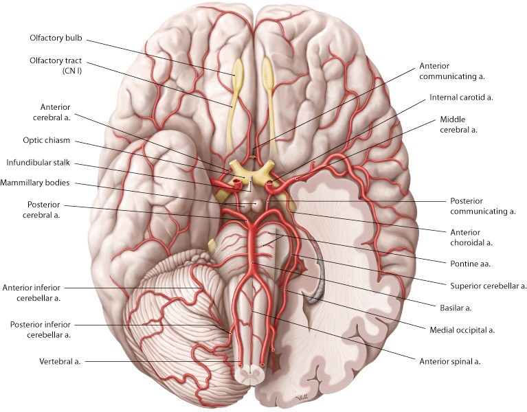
Figure 9. Adapted from Gilroy et al., Atlas of Anatomy, 2nd ed., fig. 41.6.
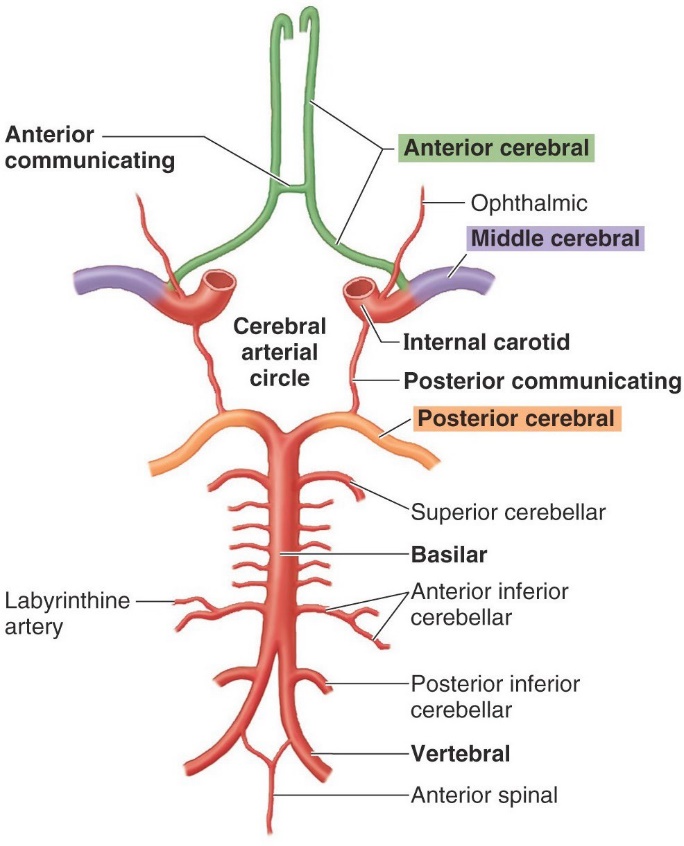
Figure 10. Clinically Oriented Anatomy, Figure 7.43.
Try to identify a few of the cranial nerves attached to the brainstem.
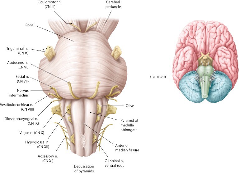
Figure 11. Adapted from Gilroy et al., Atlas of Anatomy, 2nd ed., Figs. 40.19b, 40.6b.
After you're done with the brain,
put it into the plastic Ziplock bag. Store the brain inside the body bag with your donor.
Internal Base of the Skull
Bony Features and Cranial Nerves
Study the bony features of the cranial base in a dried skull. Use a pipe cleaner to identify foramina and other passageways. Correlate the bony features in the skull with what you see in the cranial cavity of your donor.
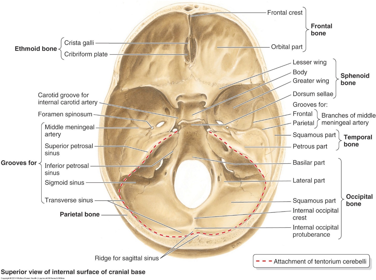
Figure 12. Clinically Oriented Anatomy, Figure 7.30.
Identify the following parts of the skull in the base of the skull:
■Anterior cranial fossa
■Orbital plates of frontal bone
■Cribriform plate of ethmoid bone (CN I pass through the cribriform foramina)
■Lesser wings of sphenoid bone
■Anterior clinoid processes of sphenoid bone
■Crista galli of ethmoid bone
■Contents of anterior cranial fossa: frontal lobes of brain, olfactory bulbs, olfactory tracts
■Middle cranial fossa
■Greater wings of sphenoid bone
■Squamous and petrous parts of temporal bones
■Optic canals (contain CN II, ophthalmic artery)
■Superior orbital fissures (contain CN IV, III, VI, V1)
■Foramina in middle cranial fossa: foramen rotundum (transmits V2), foramen ovale (transmits V3), spinosum (transmits middle meningeal artery), and foramen lacerum (transmits internal carotid artery)
■Sella turcica and its named parts: hypophysial fossa and dorsum sellae
■Posterior clinioid processes of sphenoid
■Grooves for middle meningeal arteries
■Contents of middle cranial fossa: temporal lobes of brain and hypophysis (pituitary). The pituitary rests in the hypophysial fossa of the sella turcica.
■Posterior cranial fossa
■Clivus (made from sphenoid and occipital bones)
■Internal acoustic meatuses (transmits CN VII, VIII)
■Jugular foramina (transmits CN IX, X, XI; internal jugular vein)
■Foramen magnum (contains spinal cord, CN XI, vertebral arteries)
■Hypoglossal canals (transmits CN XII)
■Grooves for transverse, and sigmoid sinuses
■Contents of posterior cranial fossa: midbrain, pons, medulla oblongata, cerebellum, occipital lobes of brain
Question
In the skull base of your donor, can you locate the stumps of all the cranial nerves (I to XII) anterior to posterior? Name their foramina, too!
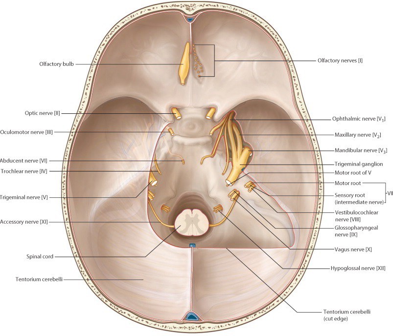
Figure 13. Gray’s Atlas of Anatomy.
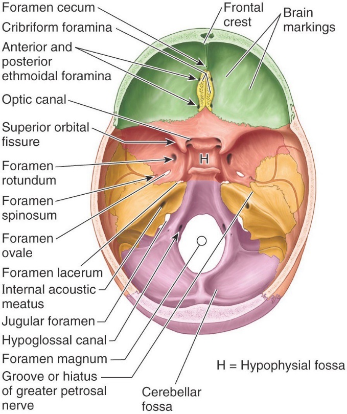
Figure 14. Clinically Oriented Anatomy, Figure 7.11.
Dural Venous Sinuses
Locate and name the dural venous sinuses. See Figure 15. These are comparable to veins (except they have no middle layer of smooth muscle in their walls) and are created between the meningeal and endosteal layers of the dura.
Examine the skull base in your donor and the models:
■Superior sagittal sinus: In upper margin of falx cerebri
■Inferior sagittal sinus: In lower margin of falx cerebri above corpus callosum
■Straight sinus: Formed by the union of the inferior sagittal sinus and the great cerebral vein (vein of Galen).
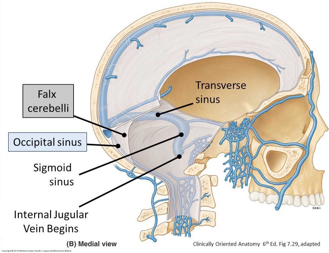
Figure 15.
■Confluence of sinuses
■R & L transverse sinuses: Along margins of tentorium cerebelli
■R & L sigmoid sinuses: Direct continuations of the transverse sinuses; terminate at jugular foramina
■R & L cavernous sinuses: In the middle cranial fossa, just lateral to the sella turcica
■R & L superior petrosal sinuses: Course from anterior to posterior along the tops of the petrous temporal bones, connecting the cavernous sinuses to the sigmoid sinuses
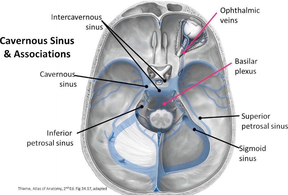
Figure 16.
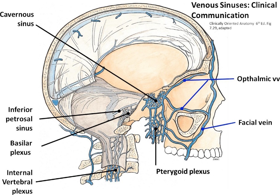
Figure 17.
Question
A donut-shaped shelf of dura called the sellar diaphragm covers the hypophysial fossa in the sella turcica—the hole in the donut is for the passage of the infundibulum (stalk of the pituitary). Where is the pituitary gland? (Can you see it?)
Trigeminal Nerve
Identify the stump of the trigeminal nerve as it passes over the petrous ridge of the temporal bone. Pass a probe next to the nerve and advance it forward to demonstrate the pocket of dura that surrounds the nerve = this is called the trigeminal (Meckel’s) cave. This outpocketing of dura surrounds CN V and the trigeminal ganglion.
Attempt to strip the dura from the floor of the middle cranial fossa on one side of the cranial cavity. (on the side not done already)
Note
Use foreceps or a curved clamp to grasp the dura. It's a tough mother!
■Strip the dura from one side to identify the middle meningeal artery and to observe V1 (ophthalmic division), V2 (maxillary division), and V3 (mandibular division) branching from the trigeminal ganglion
■Trace V1 along the lateral wall of the cavernous sinus into the superior orbital fissure.
■Trace V2 into foramen rotundum.
■Trace V3 into foramen ovale.
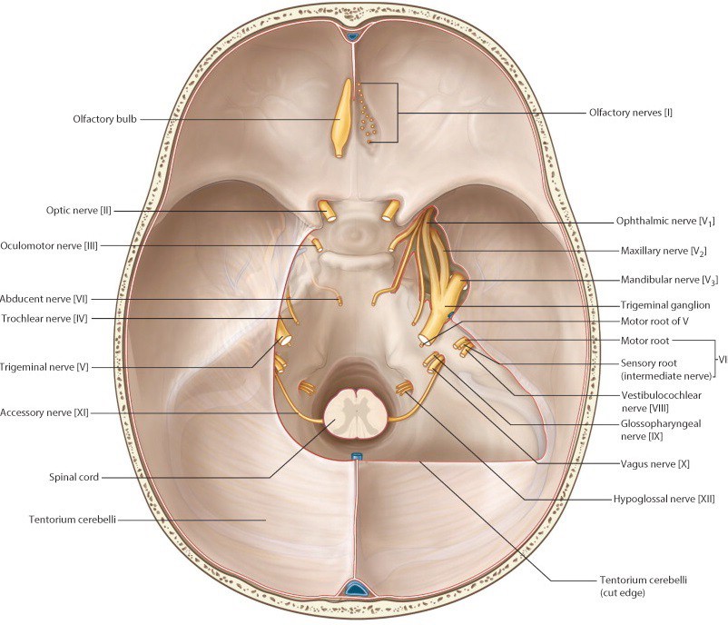
Figure 18. Gray’s Atlas of Anatomy.
Cavernous Sinuses
Open the other cavernous sinus (this was completed on one side—now do the other!)
Trace the oculomotor nerve (CN III) forward and note that it lies in the lateral wall of the cavernous sinus.
Question
What other structures lie within the cavernous sinus and its walls? See Figure 20.
Note that the Internal carotid artery passes through the center of the sinus. Contemplate this: arterial blood in the ICA is bathed in venous blood. Crazy!
Question
Which cranial nerve is most closely associated with the internal carotid artery within the cavernous sinus?
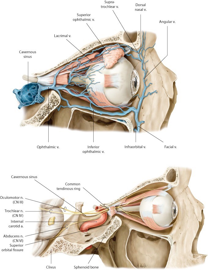
Figure 19. Adapted from Gilroy et al., Atlas of Anatomy, 2nd ed., Figs. 35.6, 35.5.
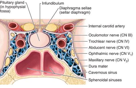
Figure 20. Posterior view of coronal section of cavernous sinus.
Checklist, Lab #22
Review and make sure you have identified each of the structures below.
Scalp
Skin
Connective tissue (superficial fascia)
Epicranial aponeurosis
Loose areolar CT layer
Pericranium
Cranial Cavity/Cranial Base
Dura mater
Falx cerebri
Falx cerebelli
Tentorium cerebelli
Tentorial notch
Anterior cranial fossa
Crista galli
Middle cranial fossa
Petrous part of temporal bone
Infundibulum of hypophysis
Hypophysis (pituitary gland)
Optic nerve (CN II)
Oculomotor nerve (CN III)
Trochlear nerve (CN IV)
Trigeminal nerve (CN V)
Ophthalmic nerve (V1)
Maxillary nerve (V2)
Mandibular nerve (V3)
Abducens nerve (CN VI)
Internal carotid artery
Posterior cranial fossa
Facial nerve (CN VII)
Vestibulocochlear nerve (CN VIII)
Glossopharyngeal nerve (CN IX)
Vagus nerve (CN X)
Spinal accessory nerve (CN XI)
Hypoglossal nerve (CN XII)
Vertebral arteries
Dural venous sinuses—identify in donor and models
Superior sagittal
Straight sinus
Confluence of sinuses
Transverse sinuses
Sigmoid sinuses
Superior petrosal sinuses
Cavernous sinuses
Brain
Arachnoid mater
Arachnoid granulations
Pia mater
Brainstem: medulla, pons, and midbrain
Cerebellum
Olfactory bulb and tracts
Optic tracts, optic chiasma, and optic nerves
Circle of Willis
Basilar artery
Posterior cerebral arteries
Posterior communicating arteries
Anterior cerebral arteries
Anterior communicating artery
Skull: Bones of Cranial Vault
Frontal
Ethmoid
Sphenoid
Temporal
Occipital
Parietal
Coronal suture
Sagittal suture
Lambdoidal suture
Squamous suture
Skull: Skull Base
Cribriform plate with Olfactory (cribriform) foramina
Crista galli
Optic canals
Anterior and posterior clinoid processes
Greater and lesser wings of sphenoid bone
Superior orbital fissures
Sella turcica: Includes the hypophysial fossa + dorsum sellae
Foramen rotundum
Foramen ovale
Foramen spinosum
Foramen lacerum
Groove for middle meningeal artery
Clivus
Internal acoustic meatuses
Jugular foramina
Hypoglossal canals
Grooves for transverse sinuses and sigmoid sinuses