Lab 13: Dissection: Posterior Abdominal Wall (PAW) and Kidneys

  1. Demonstrate the fascias surrounding the kidneys.
  2. Clean the kidneys and identify features of their external and internal anatomy.
  3. Identify the ureters and trace them into the pelvis.
  4. Identify the suprarenal glands and name their parts.
  5. Identify the parts of the diaphragm.
  6. Clean and identify the muscles of the posterior abdominal wall.
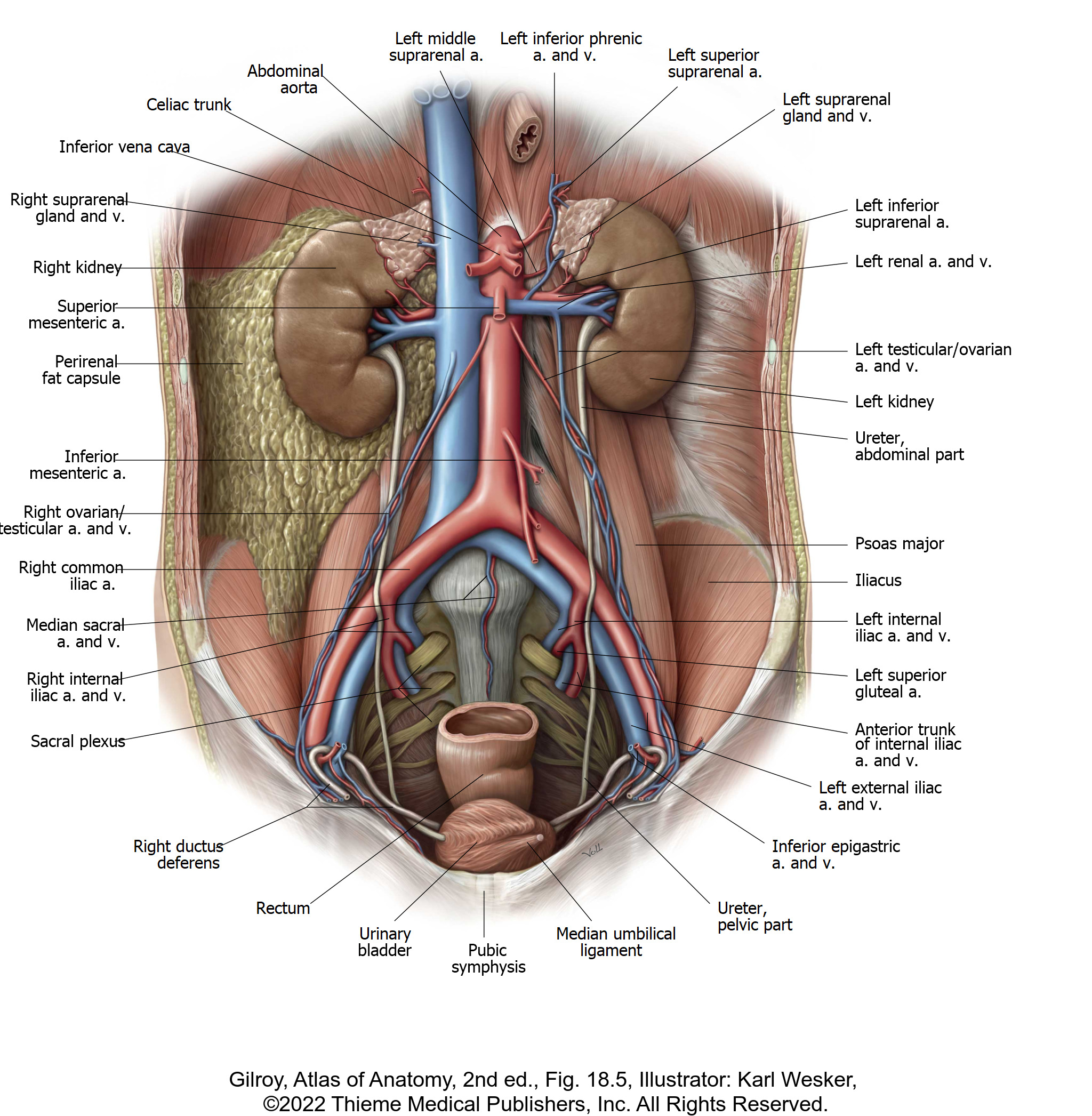
  7. Identify major branches of the abdominal aorta and tributaries of the IVC.
  8. Identify the branches of the lumbar plexus.
  9. Find the lumbar sympathetic trunks and identify the aortic plexus and celiac (prevertebral) ganglia.
  10. Identify structures that leave the PAW and enter the pelvic cavity through the pelvic inlet.

This lab will focus on the posterior abdominal wall (PAW) and the organs associated with it. Clinicians refer to the PAW as the “retroperitoneum.”


To access the PAW, you will need to work around the gastrointestinal tract. Start by finding and reviewing the major parts of the GI tract: esophagus, stomach, small intestine (duodenum, jejunum, ileum) and large intestine (cecum, ascending colon, transverse colon, descending colon, rectum). 

before you begin

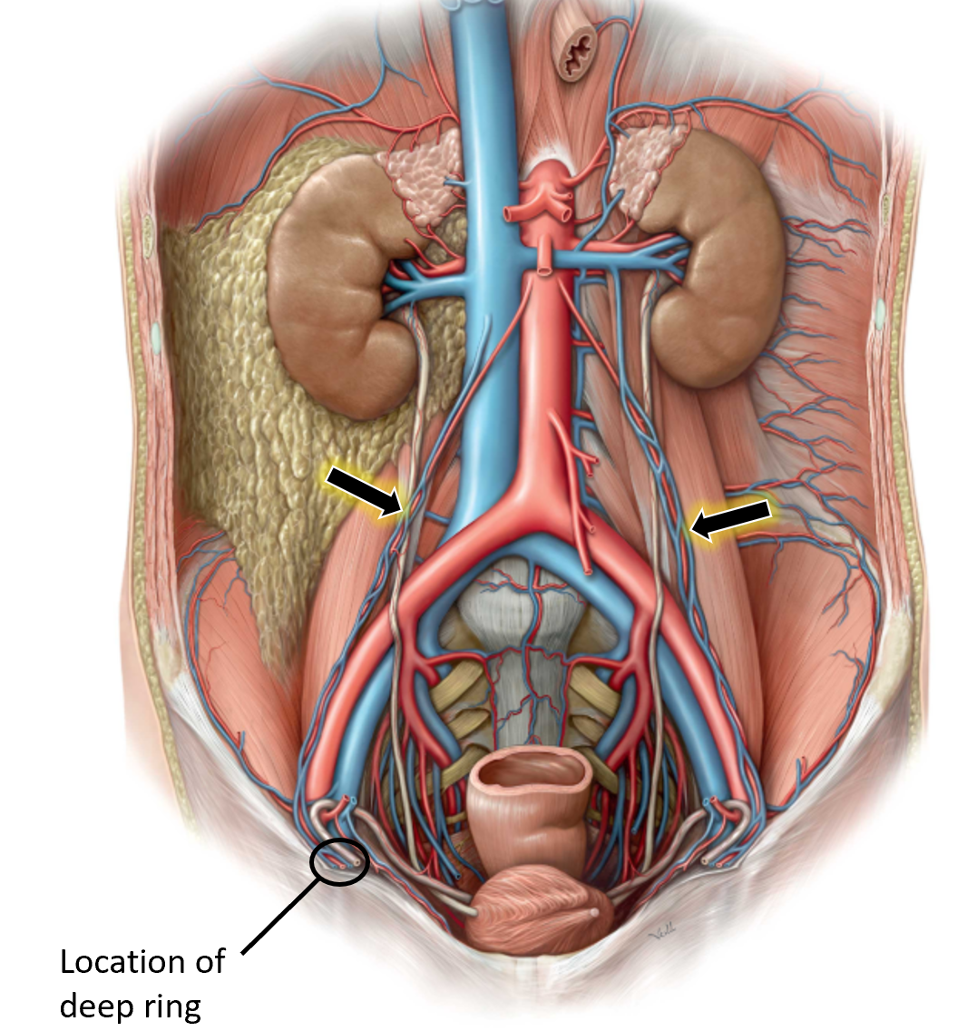
Find and protect the gonadal vessels: they are very thin and vulnerable vessels, so having an idea where they are is important before you dissect!

Figure 1.

Locate the gonadal (testicular or ovarian) vessels. In the male, start at the deep inguinal ring and trace back to the aorta and IVC (consult an atlas). In the female, trace back from the ovaries. Follow the left gonadal (testicular or ovarian) vein upwards and note that it flows into the left renal vein.
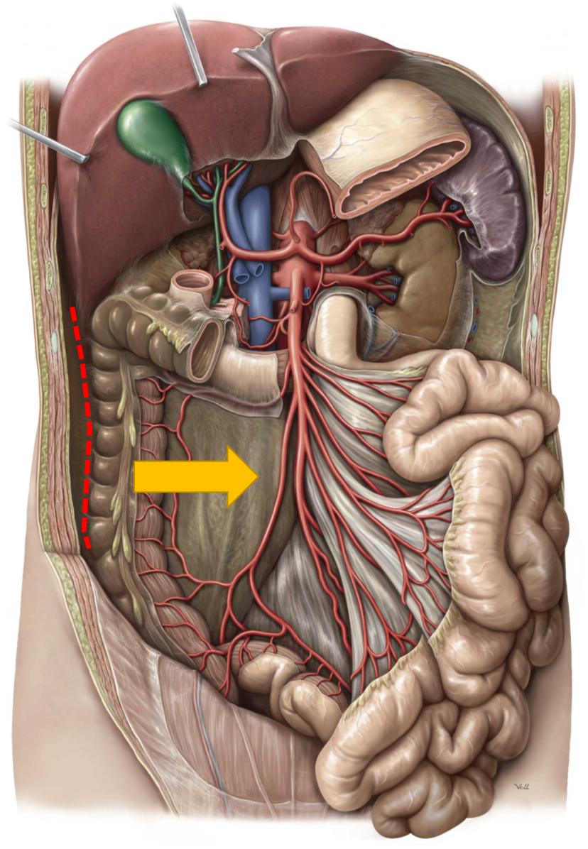
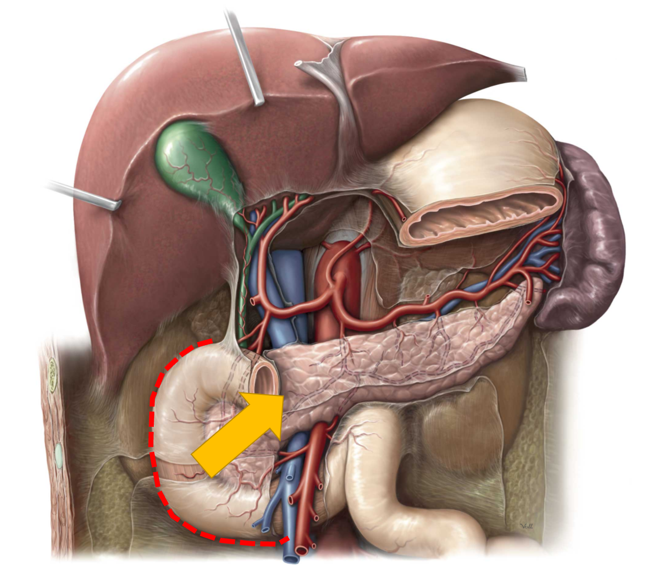
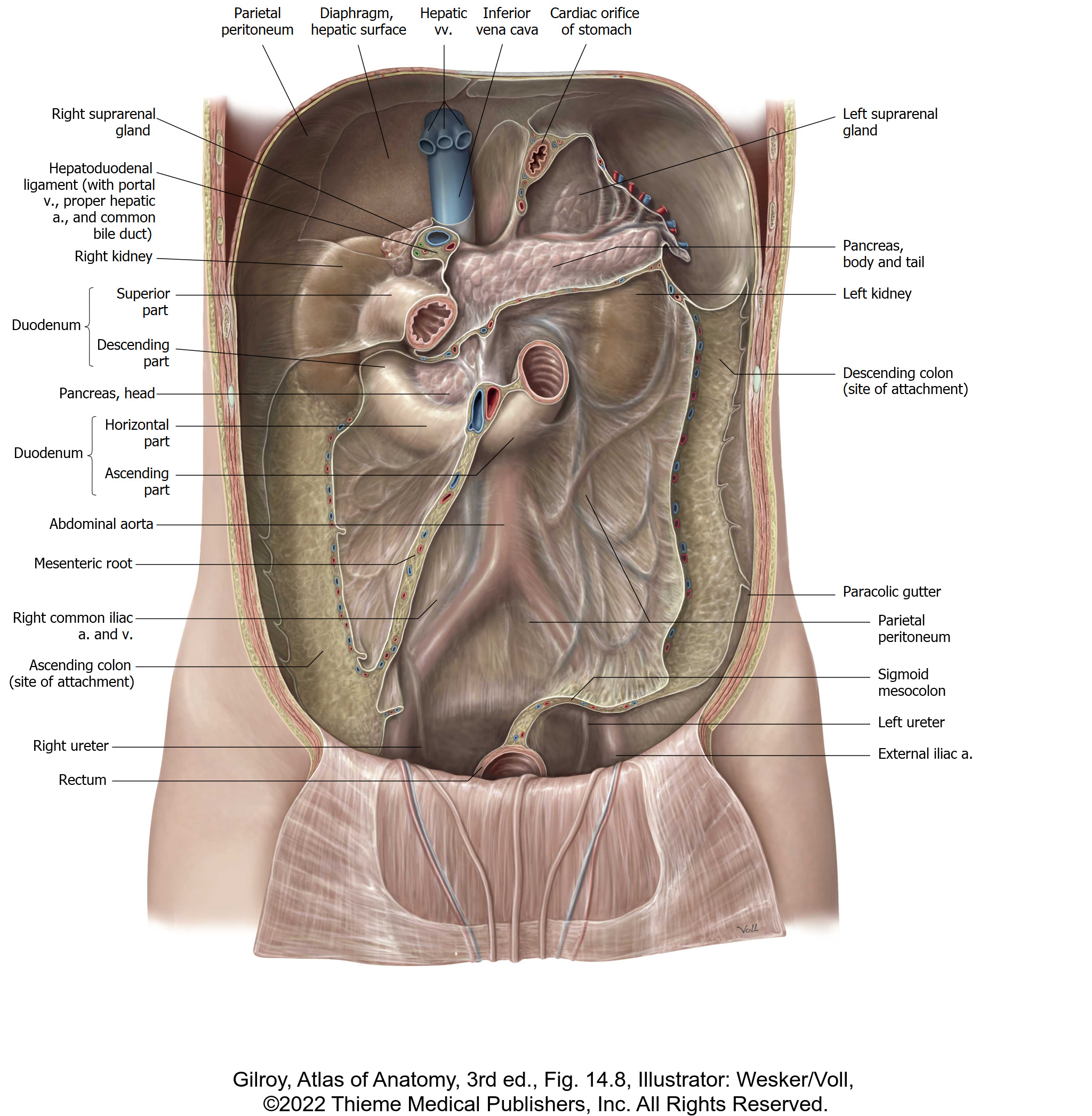
Free retroperitoneal GI structures from the PAW.

In order to free the GI tract, and gain better access to the PAW, you will want to mobilize parts of the GI tract that are retroperitoneal (do you remember what these are?).

1Locate the paracolic gutters, lateral to the ascending and descending colon, between the colon and the lateral body wall. If the colons are still adhered to the posterior abdominal wall, incise the peritoneum in the gutters. Using blunt dissection, pry away the ascending and descending colon from the posterior body wall. 

Figure 2.

2Do the same with the duodenum and pancreas—incise the peritoneum adjacent to the retroperitoneal parts of the duodenum and pancreas. Pry the duodenum and pancreas away from the retroperitoneum. 

Figure 3.

Cut through the rectum in the transverse plane.

1Use plastic locking strips (cable ties) provided in lab to tie off the rectum.

Apply two plastic ties to the rectum as far down in the pelvic cavity as possible.

If the rectum is full of feces, move up to the sigmoid colon. The rectum has no mesentery, so you will have to pry it loose from the connective tissue anterior to the sacrum.

2Cut the rectum between the ties with scissors or a scalpel and lift the upper portion of the rectum out of the pelvic cavity.

Get the lay of the land in the PAW and PALPATE.

With the GI tract mobilized you should be able move it side to side as your work around it to access the PAW – do your best to preserve structures you identified in the supra & infracolic regions! 


The peritoneum may have been partially removed; the peritoneum on the PAW that is still intact should be thin enough to peer/palpate through. Do your best to look and palpate for:

Diaphragm

Abdominal aorta w/bifurcation into common iliac arteries

IVC

Kidneys, surrounded by copious amounts of extraperitoneal fat

Psoas major muscles

Vertebral column

Iliac crest

Pelvic brim

Bladder

Uterus w/Fallopian tubes and ovaries (if present!)

Figure 6.

Question

Note the rigidity of the PAW. How does this compare with the anterior abdominal wall?

Remove the peritoneum from the PAW with forceps.

Gently peel the remaining peritoneum off the PAW. Note the large amounts of extraperitoneal fascia external to the peritoneum that is loaded with fat. This may become liquefied during the course of the lab, so have paper towels on hand, and take care to clean up spills on the floor.

Fascial Layers of the Kidneys

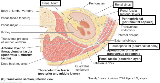
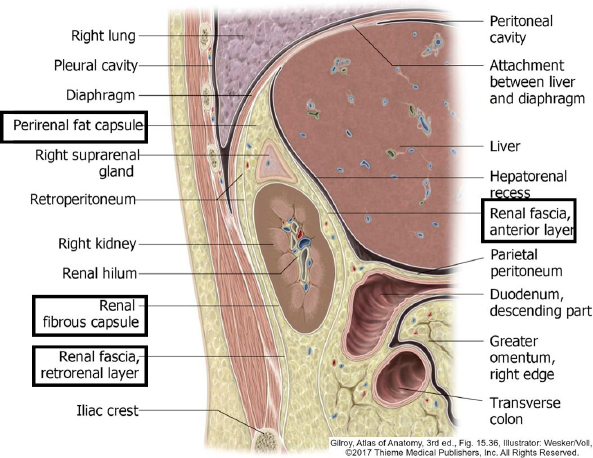
Two categories of fatty tissue are associated with the kidneys.

Paranephric (pararenal) fat is the copious extraperitoneal tissue of the PAW that is mainly posterior and lateral to the kidneys. It is the padding that protects the kidneys.

Perinephric (perirenal) fat is a fairly thin layer that surrounds the kidney on all sides and is continued into the renal sinus of the kidney. Perinephric fat is deep to a layer of abdominopelvic fascia called renal fascia (Gerota’s fascia).

Renal fascia is between paranephric and perinephric layers of fat.

1With blunt dissection and forceps, clean away the massive amounts of paranephric fat adjacent to the kidneys. This will mobilize the kidneys.

2Locate the renal fascia, a thin layer of fibrous tissue deep to the paranephric fat. Deep to the renal fascia is the perinephric fat—also known as the adipose capsule of the kidney. The renal fascia is not always easy to identify!

Figure 7.
Figure 8.

Suprarenal Glands

Identify and carefully clean the suprarenal glands.

Careful!

They are fragile.

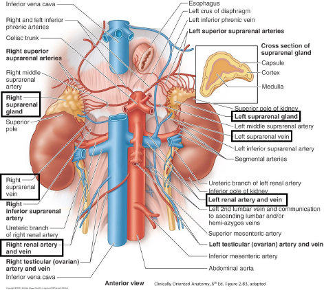
The left suprarenal gland is semilunar in shape and snuggled up against the left crus of the diaphragm. The right suprarenal gland is triangle shaped and partially hidden behind the IVC.

1Clean the left and right renal veins and trace them to the IVC. They are anterior to the renal arteries. Which vein is longer?

2Clean the left and right renal arteries and trace them to the aorta.

3Clean the suprarenal veins—the left one is a tributary of the renal vein while the right flows to the IVC.

Figure 9.

Question

The suprarenal glands are also called the adrenal glands. Why is “suprarenal” a better term?

Question

Read up on the suprarenal glands. They are small but mighty organs! Functionally, they are “two organs in one.” What are the functions of the two subparts?

Kidneys

Clean the remaining paranephric fat from the kidneys.

Rotate the kidneys toward the midline of the body using the renal vessels as hinges.

Clean away all the paranephric fat behind the kidneys. Then return the kidneys to their proper locations.

Question

Between which vertebral levels are the kidneys located?

How are they oriented (in which directions are their “poles” oriented)?

Which kidney is located more superior?

With which bony and muscular structures do they relate?

Clean and identify the renal pelvis and then follow the ureter inferiorly.

Carefully clean the hilum of each kidney and locate the pelvis of the ureter, a.k.a. the renal pelvis (the wide part of the ureter that leaves the kidney).

The renal pelvis is the most posterior structure in the kidney hilum. From anterior to posterior the structures in the hilum are: renal vein, renal artery, and renal pelvis. Clean and trace the ureters down to the pelvic brim. Take care—the ureters are crossed by the delicate gonadal vessels.

Figure 10.

Question

The ureter is described as having three spots of real or potential narrowing, that is, three places where the diameter of the ureter’s lumen actually narrows or where the ureter could be compressed by other nearby organs. Where are these? Why would these spots have clinical relevance?

Remove and section one kidney.

Remove and section one kidney (right kidney if possible). Cut the renal artery, renal vein, and ureter of the kidney near its hilum and remove it from the body.

Examine the internal features and find:
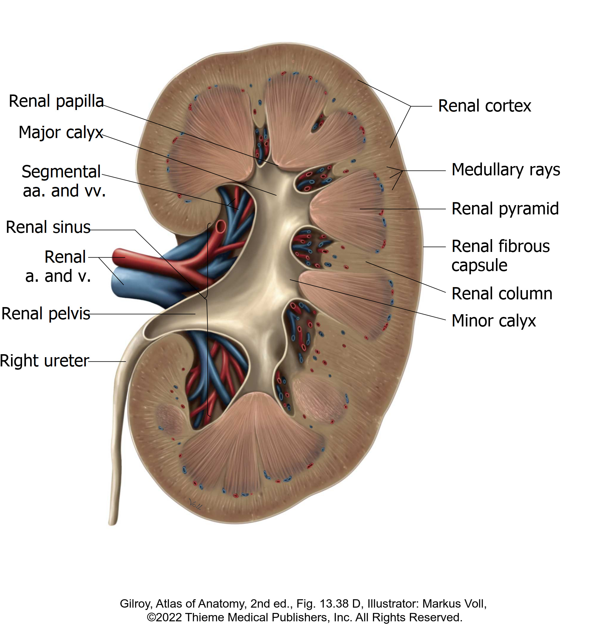
Figure 11.

Renal cortex is the most external layer—it dips down between the renal pyramids as the renal columns

All the renal pyramids together make the renal medulla. The tips of the renal pyramids are the renal papillae. The renal papillae project into the minor calyces.

Minor calyces fuse to form 2 or 3 major calyces. Major calyces fuse to form the renal pelvis. The renal pelvis narrows to become the ureter.
Identify the renal sinus, the central cavity in the kidney that contains the calyces, renal pelvis, and renal vessels. The renal sinus usually contains quite a bit of fat.

Identify the renal hilum = the indented part of the medial border of the kidney that is the doorway to the sinus.

Muscles of the PAW

Clean away the remaining extraperitoneal fat from the PAW.

PALPATE the 12th rib and the iliac crest in preparation for studying muscles.

CLEAN muscles of the PAW. IDENTIFY:

Transversus abdominis

Quadratus lumborum

Psoas major

Psoas minor (40% of folks don’t have one!)

Iliacus

For each muscle—describe attachments. The psoas major and iliacus muscles fuse below the inguinal ligament to form the iliopsoas muscle. 

Question

What is its function?

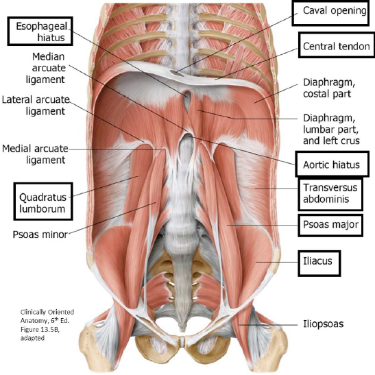
EXAMINE the inferior surface of the diaphragm and clean the two crura of the diaphragm = the “legs” of the diaphragm that attach it to the vertebral column.

Figure 12.

IDENTIFY:

Central tendon of diaphragm

Opening for inferior vena cava (T-8)

Esophageal hiatus (at T-10) = located left of the midline

Aortic hiatus (at T-12)

A MNEMONIC for remembering the vertebral levels of the 3 major apertures in the diaphragm:

I Ate 10 Eggs At Twelve
I Ate = Opening for Inferior Vena Cava is at the level of T-8
10 Eggs = Esophageal hiatus is at the level of T-10
At 12 = Aortic hiatus is at the level of T-12

Question

Besides the esophagus, what else is transmitted through the esophageal hiatus?

[Hint: The esophagus is at T-10; Cranial Nerve 10 accompanies it.]

Question

Besides the aorta, what else passes through the aortic hiatus?

Neurovasculature of the PAW

Examine and clean the surface of the abdominal aorta and IVC.
Figure 13.

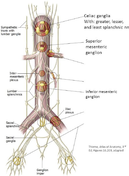
A tough network of autonomic nerve fibers (aortic plexus) covers the entire length of the abdominal aorta. Like the cardiac plexus in the thorax, the aortic plexus contains a mixture of:

Sympathetic, Parasympathetic, and Visceral Afferent nerve fibers

Each time a branch of the aorta is given off, part of the plexus follows the artery. The concept is that autonomic nerves follow vessels to their target organs.

In order to clean the vessels, you will need to use scissors to remove the tough nerves.

Within the aortic plexus of nerves, identify the celiac ganglia. These are the largest of the prevertebral ganglia. They are about the size of a dime and are flat. The celiac ganglia can be mistaken as lymph nodes. There are usually 2 celiac ganglia, one on either side of the celiac trunk.

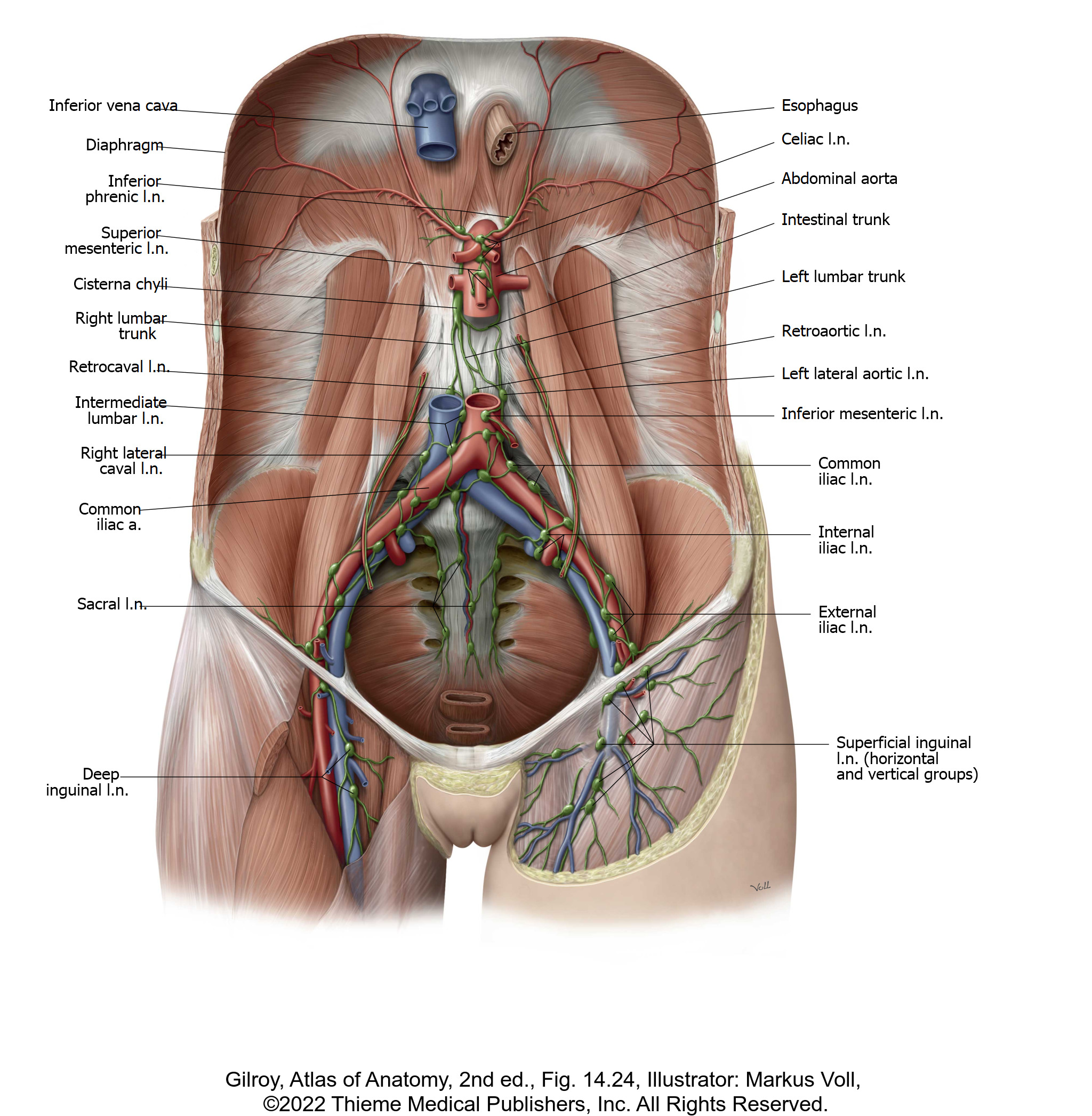
As you clean the aorta and IVC, look for lymph nodes. In general, these are called lumbar nodes.

Clinicians have schemes for naming the subgroups of lumbar nodes. You may hear the terms para-aortic nodes for the lumbar nodes on the left side, and paracaval nodes for the lumbar nodes on the right side.

Question

What is the difference between PARA-aortic (lumbar) nodes and PRE-aortic nodes? (Consider which areas/structures each drain.)

Abdominal Aorta

Find these branches of the abdominal aorta:

Unpaired visceral branches:
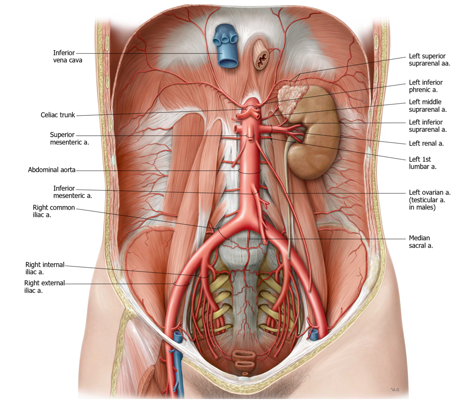
Figure 14.

Celiac trunk, superior mesenteric artery, and inferior mesenteric artery

Paired visceral branches:

Middle suprarenal arteries (very difficult to find)

Renal arteries

Testicular/Ovarian arteries (long, thin, and fragile)

Branches to body wall:

Inferior phrenic arteries (to diaphragm)

Lumbar arteries (lift the aorta—they originate from the posterior side).
The terminal branches of the abdominal aorta are the common iliac arteries. The aortic bifurcation is at L-4—about the level of the umbilicus.

Inferior Vena Cava

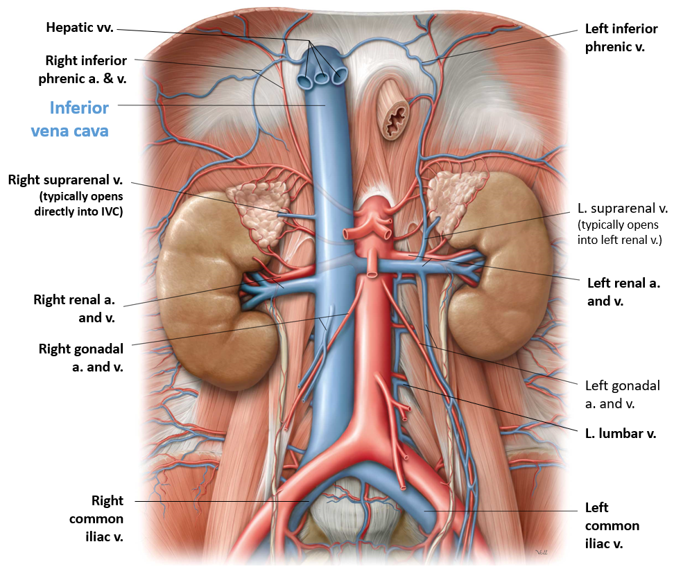
Find these tributaries of the IVC:

Figure 15.

Hepatic veins (2–3; may be with the liver)—if the liver has been removed, you will see these with the segment of IVC attached to the liver. If the liver is still intact, you won’t see these flowing into the IVC

Renal veins

Right suprarenal vein

Right ovarian/testicular vein

Lumbar veins (enter on posterior side)

The left suprarenal vein and left testicular/ovarian vein are tributaries of the left renal vein.

Chalk Talk

Diagram and discuss the branches of the abdominal aorta and tributaries of the IVC.

Cisterna Chyli

Figure 16.

Back to Lymph for a second:

For a thrill, widen the aortic hiatus with scissors by cutting away parts of the crura.

Displace the aorta and look for the cisterna chyli behind it. This variably-shaped sac (sometimes it resembles a plexus of vessels rather than a sac) is the starting point of the thoracic duct.

The cisterna chyli is formed by the union of the two lumbar lymph trunks and the single intestinal lymph trunk. This item is a prize!
Lumbar Plexus and Sympathetic Trunk

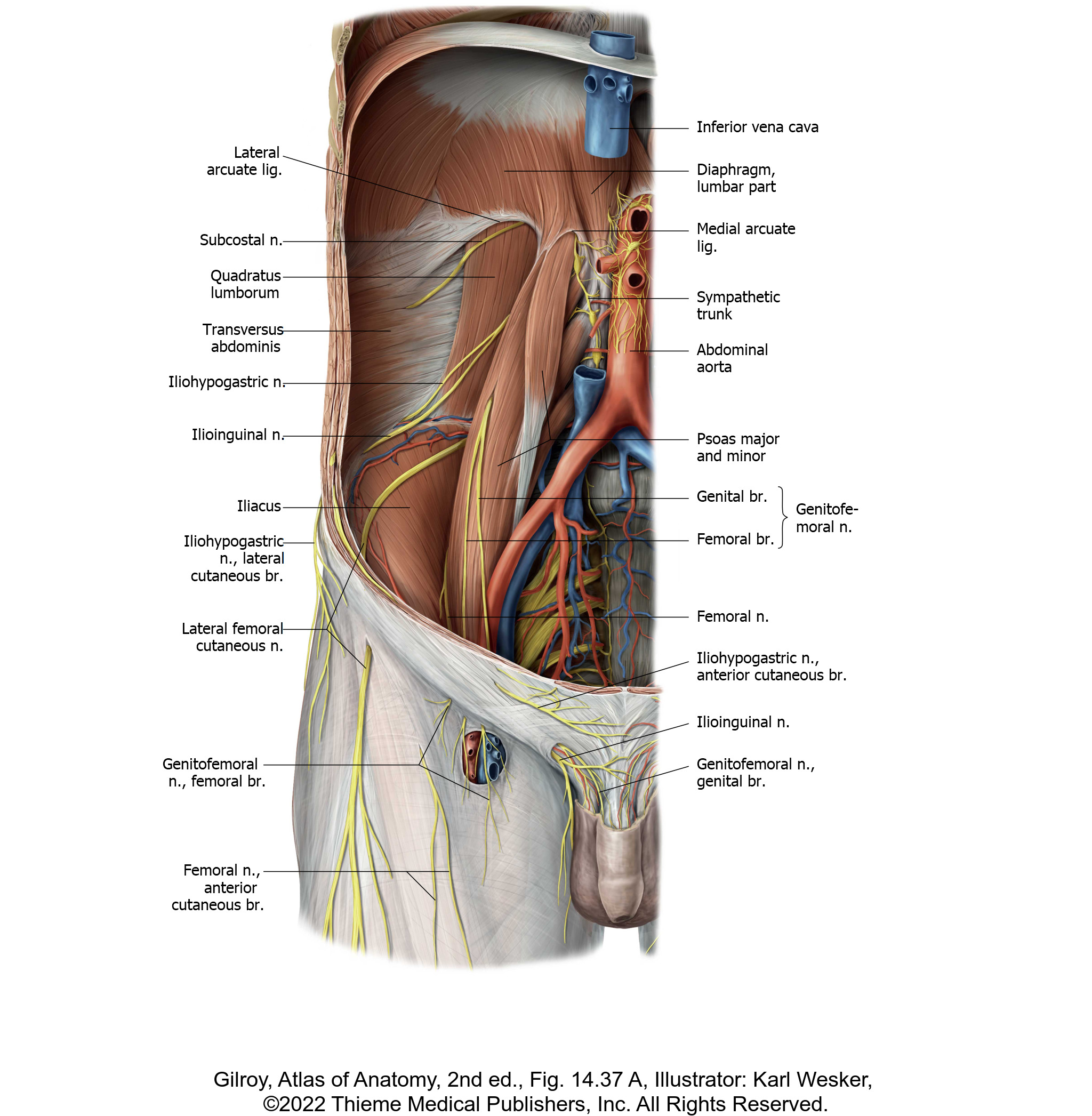
Both sides: Dissect and identify the branches of the lumbar plexus.

The psoas major muscle is a landmark we can use to organize the branches of the lumbar plexus:

Most branches are lateral to the psoas

A few branches are medial to the psoas

One branch is on the anterior surface of the psoas.

Figure 17.

1Remove fatty tissue and fascia from the quadratus lumborum and psoas major. Palpate and identify the 12th rib. Start here by finding the thick subcostal nerve below the rib.

2Work down to locate branches of the lumbar plexus along the lateral border of the psoas major.

Carefully use blunt dissection, scissors and forceps to clean the iliohypogastric and ilioinguinal nerves (both from L-1 spinal nerve). These two nerves head out toward the iliac crest to enter the neurovascular plane of the body wall.

Next, find the lateral cutaneous nerve of the thigh (L-2 and L-3). It passes across the iliacus muscle as it heads toward the ASIS to pass under the inguinal ligament to the lateral thigh.
Note

A common L1 nerve is often present in the PAW, which later divides into the iliohypogastric and ilioinguinal nerves within the body wall. Don’t despair if you only see one L-1 nerve in your dissection.

Clean the deep groove between the psoas major and iliacus muscles to find Big Daddy = the femoral nerve (L-2, L-3, and L-4). It passes through the subinguinal space into the thigh. Note that it gives muscular branches to innervate the iliacus muscle.

3Carefully clean along the anterior surface of the psoas major to find the genitofemoral nerve (L-1 and L-2).

4Medial to the psoas major, in the lateral wall of the pelvic cavity just below the pelvic brim, find:

The obturator nerve (L-2, L-3, and L-4). It passes out of the pelvic cavity through the obturator canal to the medial thigh.

Look for the lumbosacral trunk (L-4 and L-5 rami) passing into the pelvic cavity anterior to the sacro-iliac joint. This nerve is better seen in the pelvis—you will see this again when we study the sacral plexus in the pelvis.

DISCUSS with your team the spinal nerve segments in each of these nerves (e.g. iliohypogastric is L-1) and their functions.

These nerves serve the anterior abdominal wall, pelvis, and lower limb, all areas you studied in 501, so now is a good time to review these innervations.

Figure 18.

DISCUSS with your team the spinal nerve segments in each of these nerves (e.g. iliohypogastric is L-1) and their functions.

These nerves serve the anterior abdominal wall, pelvis, and lower limb – some we have discussed, while others we will continue to follow in your up coming studies.

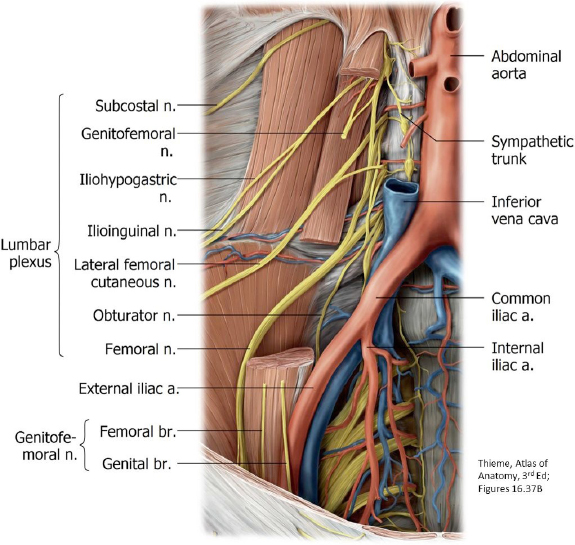
FOR THE ADVENTUROUS DISSECTOR: pick a side and carefully remove the psoas major in a piece meal fashion (pulling muscle fascicles away from the underlying nerves) to expose the formation of the lumbar plexus nerves as shown in the diagram


Chalk Talk

Draw and discuss the formation and branches of the lumbar plexus. Show the relationship of its branches to the psoas major muscle.

Clean and identify the sympathetic trunk in the PAW.

Clean along the attachment of the psaos major muscle to the vertebral column to find the sympathetic trunk.

The ganglia here are hard to see and there is not a one-to-one ratio of ganglia and vertebrae.

The sympathetic trunk enters the abdominal cavity by passing behind the diaphragm. Trace it down over the pelvic brim onto the pelvic surface of the sacrum.

See any tiny nerves joining the sympathetic trunk to the anterior rami of lumbar spinal nerves? These would be gray rami communicantes.

Checklist, Lab #13

Review and make sure you have identified each of the structures below.

Para- and perinephric fat

Renal fascia—you are a superstar if you can find it

Hilum of kidney

Kidneys: internal anatomy

Cortex and medulla

Renal columns and pyramids

Major and minor calyces

Renal papillae

Renal sinus

Renal pelvis (of ureter)

Suprarenal glands (L and R)

Abdominal aorta and its branches

Unpaired visceral arteries

Celiac trunk; superior mesenteric and inferior mesenteric arteries

paired visceral arteries

Renal arteries

Gonadal arteries (ovarian/testicular)—thin and fragile. If you find them, give a “shout out.”

Arterial branches to the body wall

Inferior phrenic arteries

Lumbar arteries (lift the aorta—they originate from the posterior side)

Bifurcation of aorta into common iliac arteries

External and internal iliac arteries

Veins

IVC

Renal veins

Right suprarenal vein and right gonadal (testicular/ovarian) veins To IVC

Left suprarenal vein and left gonadal (testicular/ovarian) veins To left renal vein

Common iliac and external iliac veins

Muscles

Diaphragm—crura, central tendon, opening of IVC, esophageal hiatus, aortic hiatus

Psoas major muscle

Psoas minor muscle—if present

Transversus abdominis muscle

Quadratus lumborum muscle

Iliacus muscle and iliac crest above it.

Nerves

Subcostal nerve

Lumbar plexus

Iliohypogastric nerve

Ilioinguinal nerve

Lateral cutaneous nerve of the thigh

Femoral nerve

Genitofemoral nerve

Obturator nerve

Lumbar sympathetic trunk and ganglia

Celiac ganglia (these are the largest of the prevertebral ganglia)

Other

Ureter

Cisterna chyli (and thoracic duct)—another superstar item!

Table of Contents
Headshot of David Conley, PhD · Professor, Department of Translational Medicine & Physiology
David Conley
PhD · Professor, Department of Translational Medicine & Physiology
Office: PBS 41A
envelope icon
Headshot of Shannon Helbling, PhD · Clinical Assistant Professor, Department of Translational Medicine & Physiology
Shannon Helbling
PhD · Clinical Assistant Professor, Department of Translational Medicine & Physiology
Office: PBS 41C
envelope icon

Dear students,
We would love to hear from you about your learning materials.

Page/Content Feedback Form