
Dissector instructions
Tap the Dissector Instructions menu on the left side of your screen to open the steps of your dissection. It's sticky, so it will scroll with you as you progress through the lab.
Need to log in to view the dissector?
Tap here for instructions on how to show the keyboard on the displays in the anatomy lab
- Goals
- Identify the parts of the distal humerus and proximal radius and ulna associated with the elbow region.
- Identify the borders and contents of the cubital fossa.
- Identify the muscles, nerves, and vessels in the anterior compartment of the forearm.
- Identify and describe bones and ligaments of the elbow joint.
Bony Anatomy Relevant to This Area
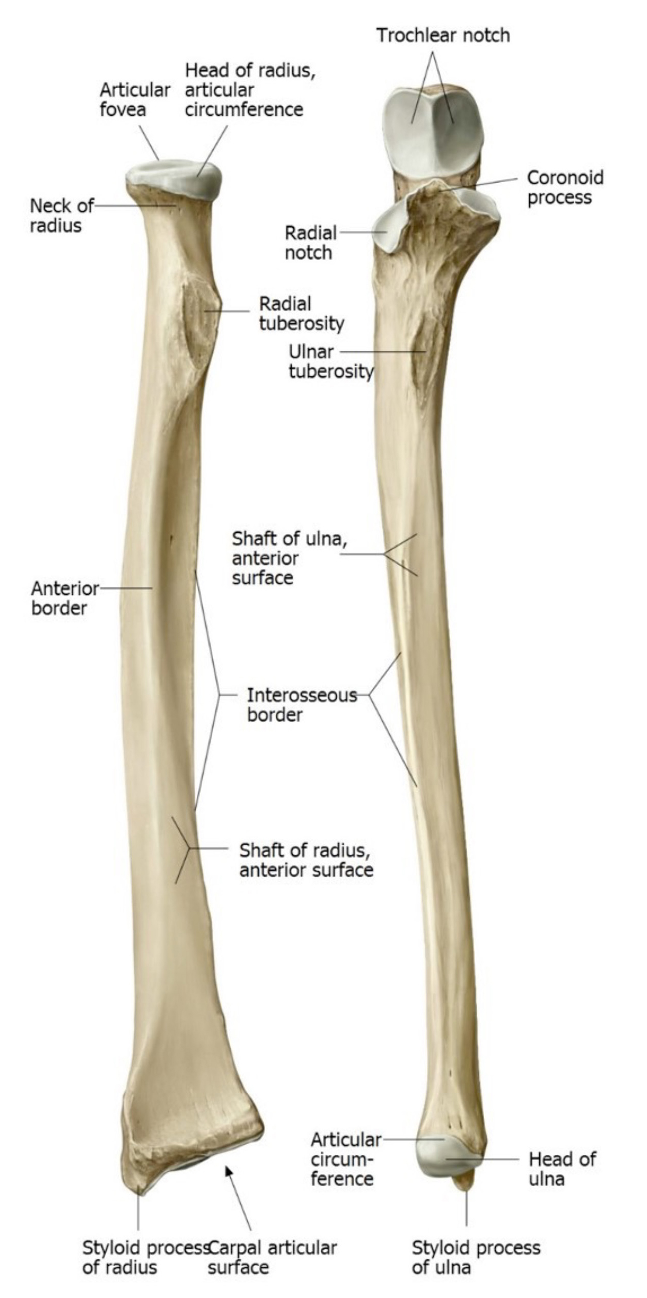
Bony landmarks of the elbow
Identify the following parts on bone specimens:
Radius
■Head
■Neck
■Radial tuberosity. Which muscle attaches here?
Ulna
■Olecranon. Which muscle attaches here?
■Coronoid process. Which muscle attaches here?
■Trochlear notch
■Radial notch (the radial head fits here at the proximal radio-ulnar joint)
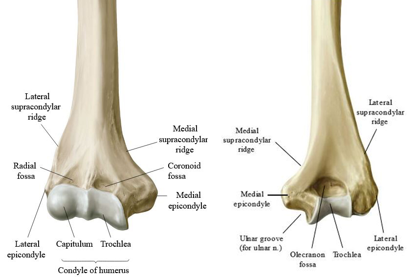
Distal Humerus
■Medial and lateral supracondylar ridges
■Medial and lateral epicondyles. Which muscle groups attach to the epicondyles?
■Capitulum (also known as the capitellum)
■Trochlea
Cubital Fossa
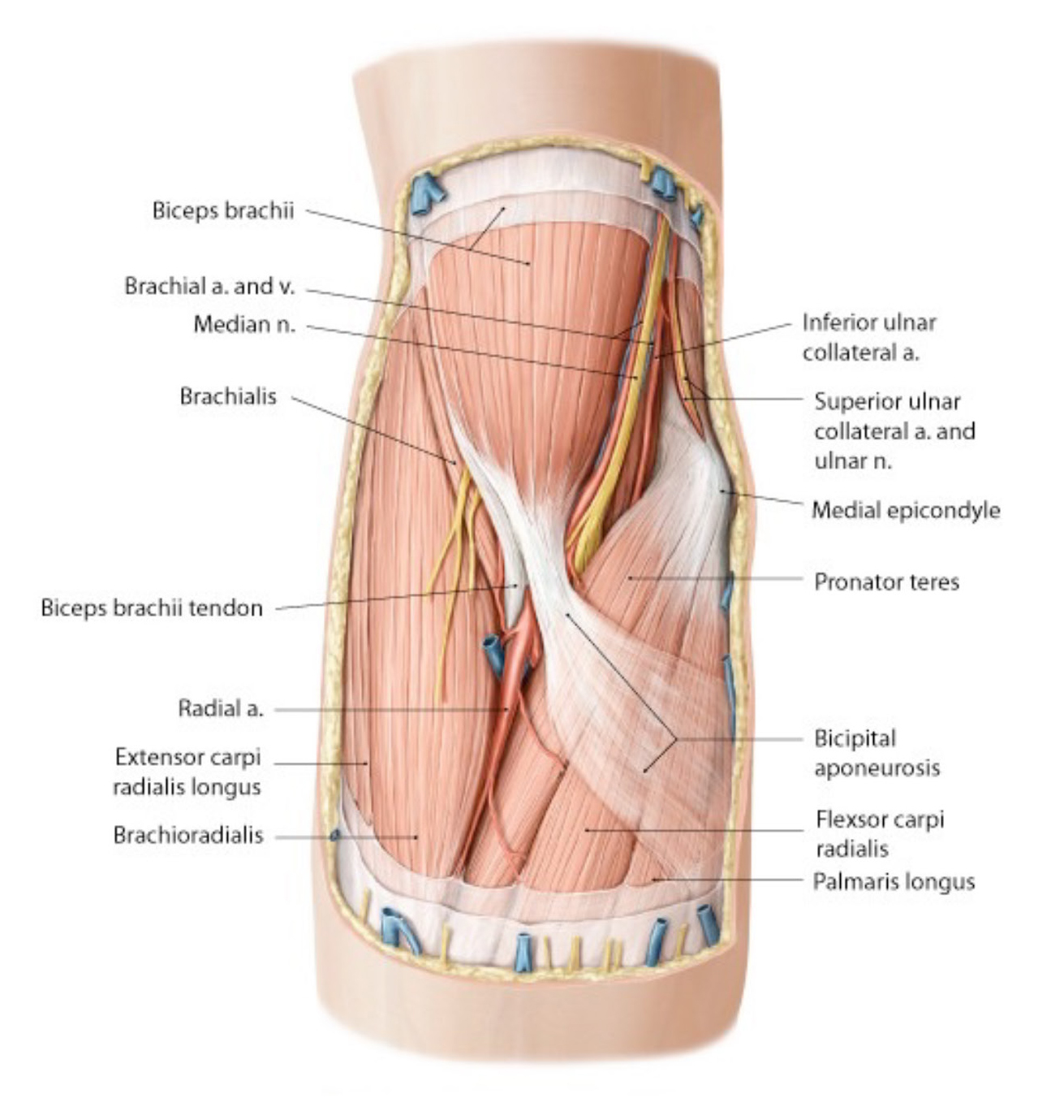
Before you cut, review the boundaries and contents of the cubital fossa. See Figure 3.
Chalk Talk
A chalk talk on the whiteboard might be in order here!
Boundaries
The cubital fossa is an inverted triangle:
■Base: transverse line through medial and lateral epicondyles of humerus
■Lateral side: brachioradialis muscle
■Medial side: pronator teres muscle
■Floor: brachialis and supinator muscles
■Roof: bicipital aponeurosis (an extension of the biceps brachii tendon) and the deep fascia of the forearm
Cubital Fossa Contents: From Medial to Lateral
■Median nerve
■Brachial artery and veins (paired)
■Tendon of biceps brachii
■Radial nerve dividing into deep and superficial branches (peek under the brachioradialis to see these)
Skin Incisions for Cubital Fossa and Forearm (make the incisions in both limbs)
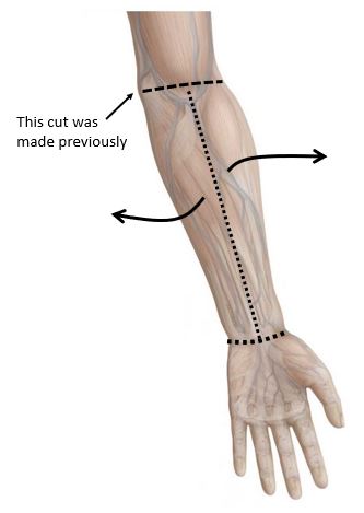
Make a shallow skin incision from the center of the cubital fossa along the midline of the anterior forearm to the center of the wrist.
Then make an incision in the transverse plane around the wrist. A similar incision was made proximally when you dissected the arm in the previous lab.
Reflect the two shallow flaps of skin medially and laterally to expose the anterior forearm. See Figure 4.
Choose one side for a superficial dissection and the other for a deep dissection.
You will leave the veins intact on one side and remove them on the other.
Superficial Dissection of the Cubital Fossa
Superficial structures of the cubital fossa
Identify the following structures within the superficial fascia external to the cubital fossa:
■Lateral cutaneous nerve of the forearm: emerges between the biceps and brachioradialis at the lateral margin of the cubital fossa (comes from the musculocutaneous n.)
■Medial cutaneous nerve of the forearm: accompanies the basilic vein from the arm (comes from the medial cord of the brachial plexus)
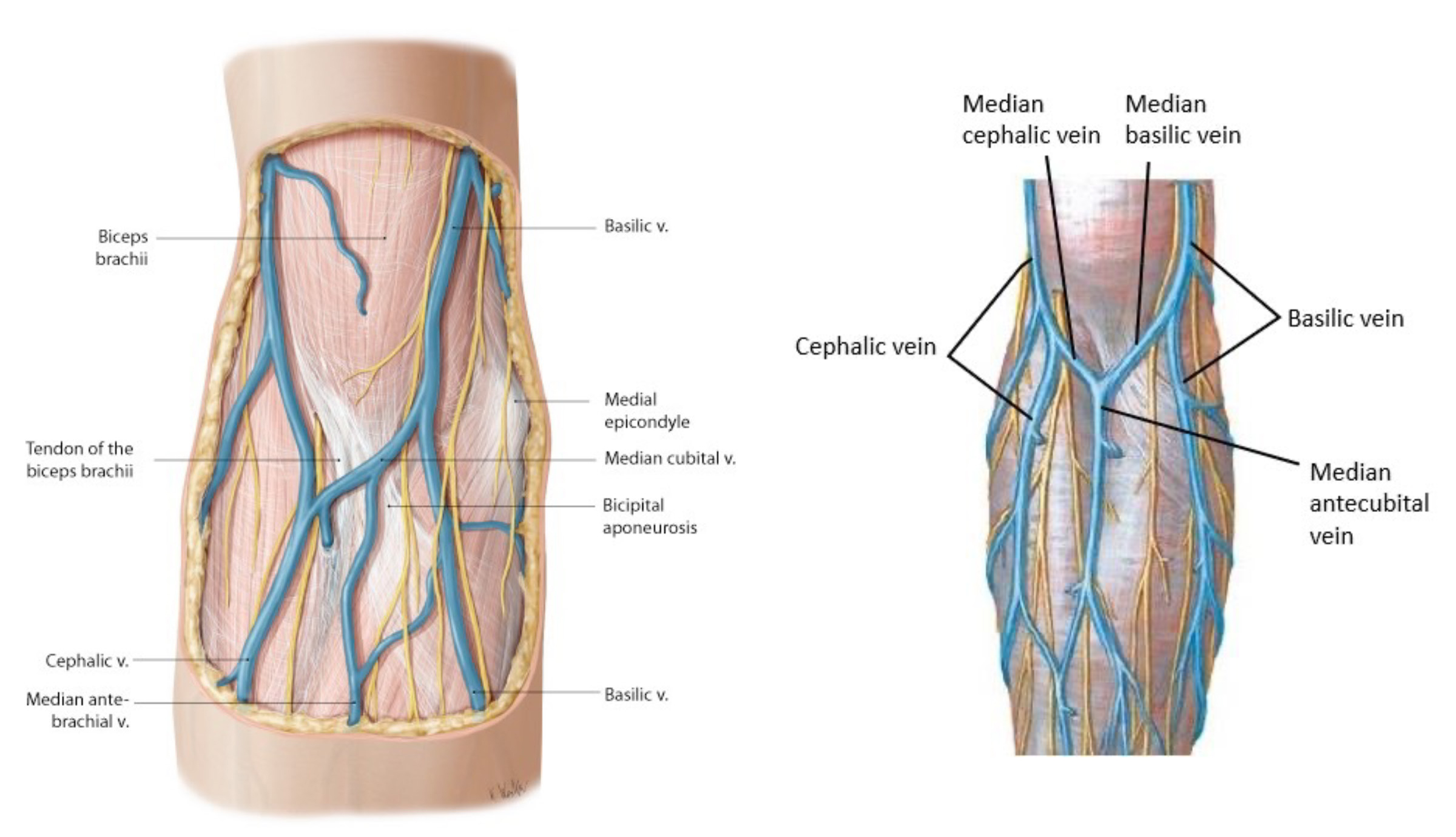
■Cephalic, basilic and median cubital veins
Deep to the median cubital vein is the bicipital aponeurosis.
Clinical correlation
The veins superficial to the cubital fossa have a variable arrangement (see Fig. 5.5). Normally they follow one of two patterns: an “H” or “M” pattern. In the “H” pattern, the crossbar of the “H” is called the median cubital vein. In the “M” pattern, two short veins cross over the cubital fossa and fuse to form a vein in the midline of the anterior forearm (median antecubital vein).
Regardless of the configuration, veins over the cubital fossa (such as the median cubital vein) are common sites for venipuncture.
Deep Dissection of the Cubital Fossa
Cubital fossa contents
Deep dissection: Remove the median cubital vein/superficial veins over the fossa.
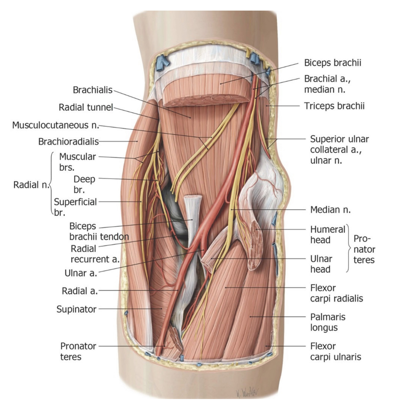
Cut the bicipital aponeurosis and reflect it inferiorly.
Cut the distal attachment of the pronator teres muscle (where it attaches to the radius) and reflect it superiorly. This will expose the contents of the cubital fossa more clearly.
Identify from medial to lateral in the fossa:
■median neve
■brachial artery
■biceps tendon
The pronator teres muscle has two proximal attachments: ulnar and humeral.
Clinical correlation
Entrapment of the median nerve between the two proximal attachments of the pronator teres can produce what is called pronator syndrome.
Locate the brachialis muscle in the floor of the cubital fossa.
Deep dissection: Remove the brachial veins and locate the terminal bifurcation of the brachial artery into the ulnar artery and radial artery.
Just distal to the bifurcation, you may be able to identify the short common interosseous artery, a branch of the ulnar artery.
Deep dissection: Retract the brachioradialis laterally and look under it to find the radial nerve.
Complete anatomy
Deep contents of the cubital fossa
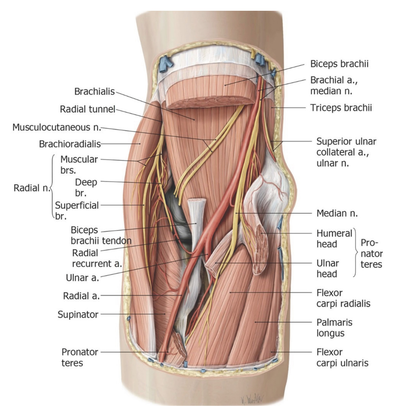
Here, the radial nerve divides into a deep branch and a superficial branch.
The deep radial nerve is motor to the posterior compartment of the forearm, where the muscles that produce extension of the wrist and fingers are located.
The deep radial nerve passes through the supinator muscle, dividing the muscle into superficial and deep parts. See Figure 7.
Clinical correlation
The superficial (proximal) part of the supinator muscle arches over the deep radial nerve, producing what clinicians call the arcade of Frohse or supinator arch = a possible site for entrapment of the deep radial nerve.
The deep radial nerve curls around the lateral side of the radius under the supinator. After piercing the supinator, it then courses deep in the posterior forearm, just external to the interosseous membrane. Here the deep radial nerve changes its name to the posterior interosseous nerve (don’t sweat this now—we will cover this again soon).
The superficial branch of the radial nerve continues distally under cover of the brachioradialis muscle. It is a sensory nerve only, to the dorsum of the hand.
Anterior Compartment of Forearm
Identify the deep fascia of the forearm, a continuous sleeve that invests the muscles of the forearm.
■The deep fascia, intermuscular septa, radius, ulna, and interosseous membrane combine to separate the forearm into anterior and posterior compartments.
To help us divide and conquer, the muscles in the anterior compartment can be sorted into three layers: superficial, intermediate, and deep.
Both sides: Clean the fascia and fat from the anterior compartments in both limbs. Using blunt dissection, separate the muscles.
Perform a superficial dissection on one side and deep on the other:
Do these on the same sides as the superficial and deep cubital fossa dissections.
Note that there are specific instructions for superficial and deep dissections.
Superficial Dissection Side
Superficial muscles of the forearm
Leave all the muscles in the anterior forearm intact.
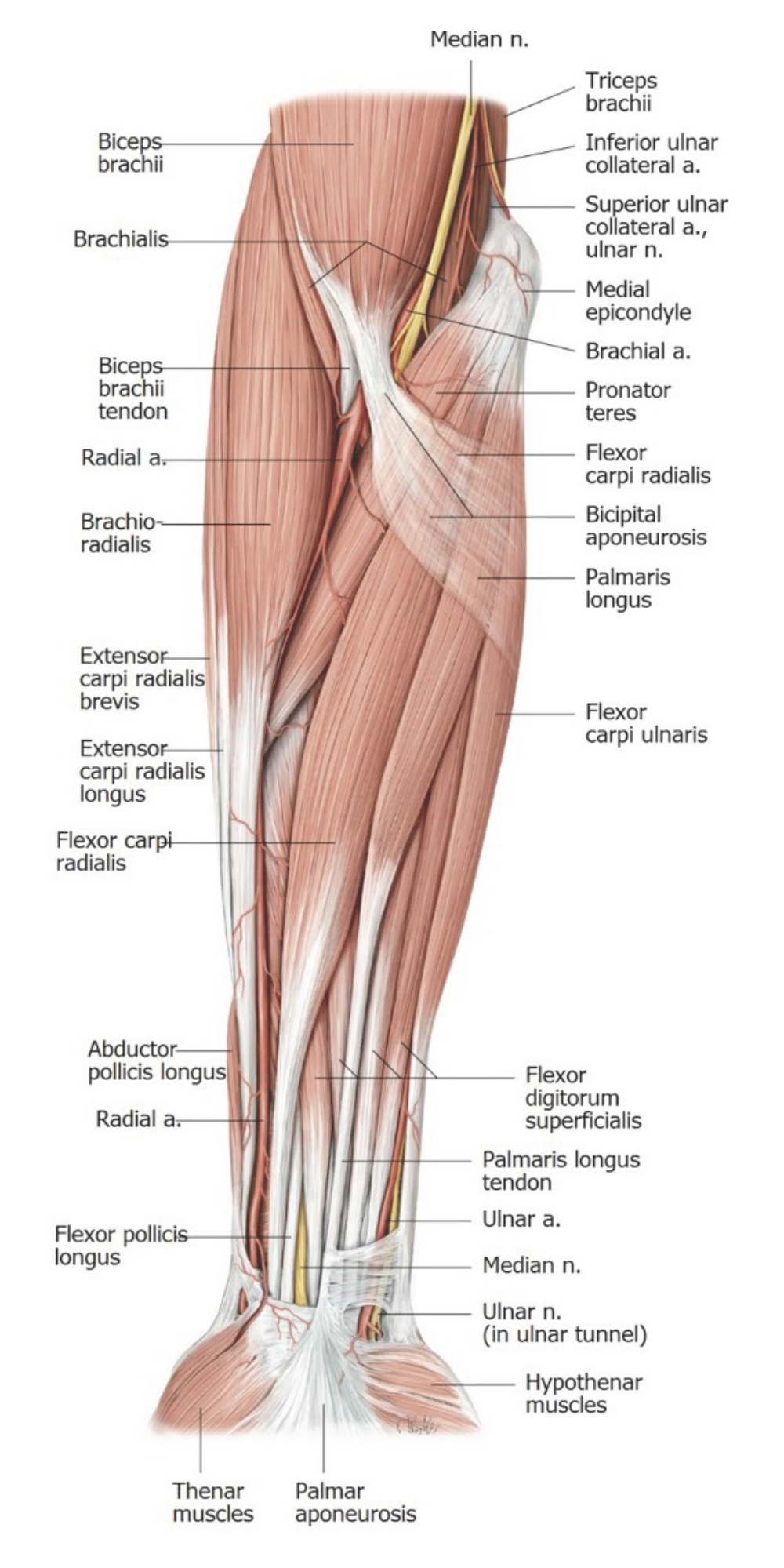
Superficial layer of muscles—lateral to medial (Figure 8):
1Pronator teres (median n. innervation)
2Flexor carpi radialis (median n.)
3Palmaris longus (median n.)
4Flexor carpi ulnaris (ulnar n.)
The four muscles in the superficial layer share a common proximal attachment, the medial epicondyle of the humerus via a common flexor tendon.
Deep Dissection Side (Same Side as Deep Cubital Fossa Dissection)
Complete anatomy
Intermediate structures of the forearm
Deep dissection: Cut across the flexor carpi radialis and palmaris longus muscles approximately 5" below the medial epicondyle.
Reflect the cut ends distally to reveal the flexor digitorum superficialis (FDS).
The FDS forms the intermediate layer of muscles in the anterior compartment of the forearm. The FDS is innervated by the median nerve.
Follow the flexor digitorum superficialis muscle distally and find its four tendons. These vanish at the wrist as they pass into the hand via the carpal tunnel. The thick band of deep fascia over the carpal tunnel is the flexor retinaculum.
Question
Distally the tendons of FDS insert on the middle phalanges of the medial four digits. What is their action? Pull on the tendons to verify your answer.
Deep dissection: Separate the humeral and ulnar attachments of the pronator teres and see if you can find the median nerve coursing between.
Trace the median nerve distally from the cubital fossa. There is an arched gap between the proximal attachments of the FDS (these are called humeral, ulnar, and radial attachments)—the median nerve and ulnar artery dive under this gap. See Figure 9.
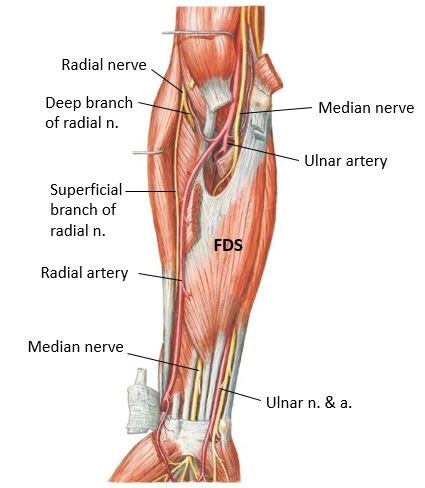
Identify these neurovascular structures (see Figure 10):
■Superficial to FDS: radial artery
■Deep to the FDS: median nerve, ulnar nerve, and ulnar artery
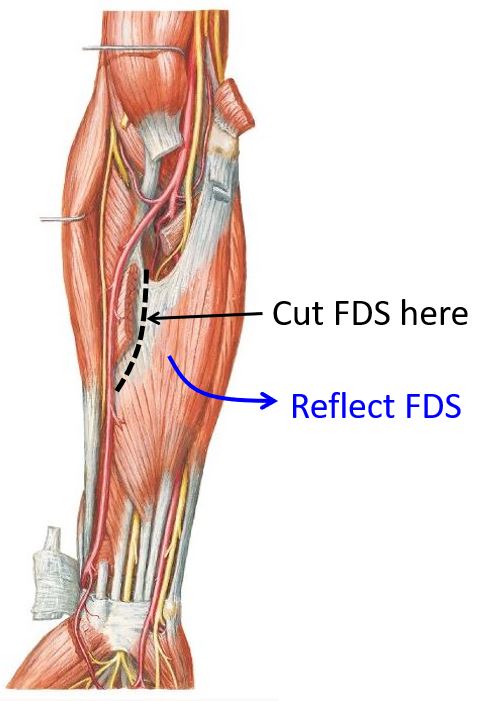
Deep dissection: Cut through the radial attachment of the flexor digitorum superficialis and reflect it away from the radius.
Reflect the FDS medially away from the radius to see the median nerve beneath the muscle. It may be tricky to see since it is tethered to the deep surface of the FDS, sandwiched between the FDS and the deep layer of muscles in the anterior forearm. See Figure 10.
Deep Side (Continued)
Complete anatomy
Deep muscles of the forearm
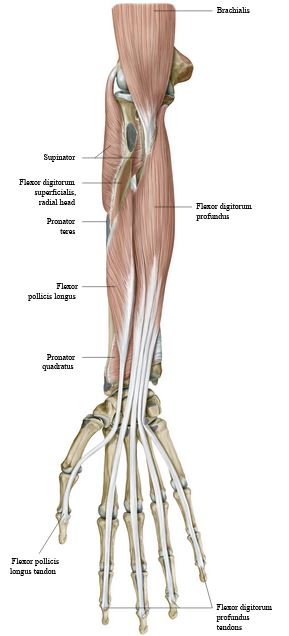
Cut transversely through the entire muscle belly of the FDS halfway down the forearm, while protecting the median nerve. Reflect the distal part of the cut FDS toward the hand. This will expose the Deep Muscle Layer = the 3rd layer of muscles in the anterior forearm.
Deep layer of muscles (Figure 11):
1Flexor digitorum profundus (FDP) (has a dual innervation = the medial half of the muscle is supplied by the ulnar nerve; the lateral half of the muscle is supplied by the median nerve). Like the FDS, the FDP splits into four tendons distally and these pass through the carpal tunnel. The tendons of FDP insert distally on the distal phalanges of the medial four digits. What is the function of the FDP?
2Flexor pollicis longus—inserts on the distal phalanx of the thumb. Function?
3Pronator quadratus. Function?
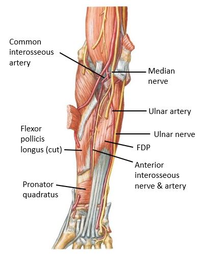
The median nerve gives off an important branch in the proximal deep forearm: the anterior interosseous nerve. After arising from the median nerve, it runs distally with the anterior interosseous artery along the interosseous membrane in the groove between the flexor digitorum profundus and flexor pollicis longus muscles. See Figure 12. The anterior interosseous nerve innervates the muscles in the deep layer of the anterior forearm (except for the medial half of the FDP).
Locate the ulnar nerve in the arm, proximal to the elbow. Trace it distally and note that it disappears behind the elbow. Here the ulnar nerve runs in a bony groove between the olecranon and the medial epicondyle. This groove is covered over by fascia to produce the cubital tunnel. The ulnar nerve passes through the cubital tunnel—it is subcutaneous here and vulnerable to injury. Compression of the nerve in this vicinity is “hitting your funny bone”—but it’s not a laughing matter! Trace the ulnar nerve distally in the forearm, beneath the flexor carpi ulnaris muscle (Figure 12).
Clinical correlation
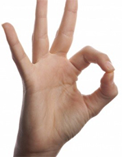
The integrity of the anterior interosseous nerve can be checked by the having the patient make the “o.k. sign”. Which muscles are used to perform this gesture?
Question
Review the muscles in the anterior compartment. Identify which are primary wrist flexors and which act on the digits?
Prosection: The Elbow Joint
The elbow joint
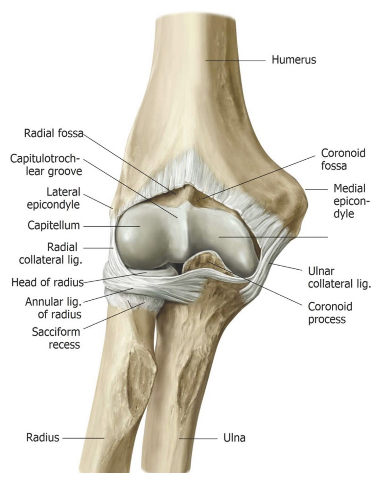
Review the bony anatomy of the elbow joint (Figure 14).
The radius and ulna articulate with the humerus at the elbow joint. There are two articulations:
■Humero–ulnar articulation. Which bony parts come together here?
■Humero–radial articulation. Which bony parts come together here?
Question
The elbow is a hinge joint. Which movements can occur?
Like all synovial joints, the bones of the elbow joint are enclosed by a joint capsule made of dense connective tissue.
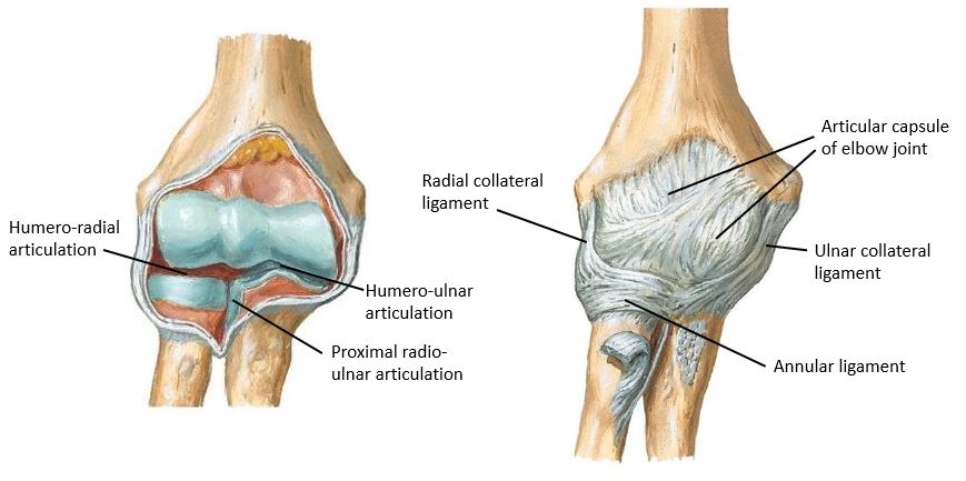
A third articulation is structurally within the joint capsule, but functions separately from the elbow joint. This is the proximal radio-ulnar joint (Figure 15). Rotation occurs at this joint so that the radius can pivot around the ulna. Which movements of the forearm can occur thanks to the radio-ulnar joints?
Prosection: Identify the three ligaments of the elbow joint.
Ligaments reinforce the joint capsule and provide stability to the joint (Figure 15). They are named based on their locations and fiber orientation.
■Radial collateral ligament
■Ulnar collateral ligament
Question
What are the functions of the elbow collateral ligaments?
■Annular ligament = this ligament is important to the function of the proximal radio-ulnar joint: It keeps the radial head in place (preventing it from drifting distally when the forearm is pulled distally) so that it can spin, allowing the radius to pivot during the motions of supination and pronation.
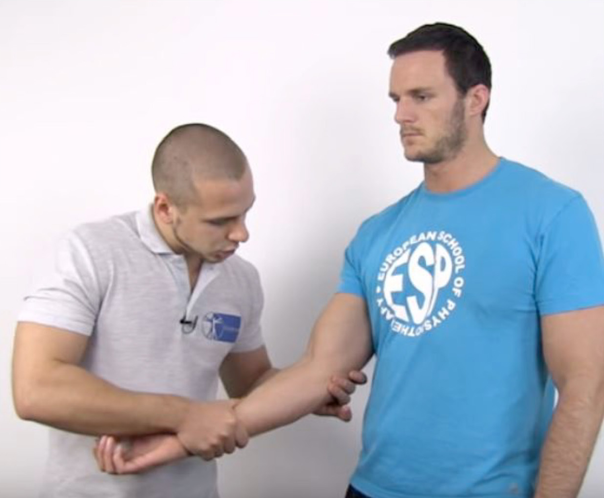
VARUS and VALGUS. These terms refer to movements at the distal end of a limb segment, with the proximal end of the limb segment fixed and not moving. Take the forearm for example. If the distal forearm and hand are deviated medially, this is a varus movement that exerts a varus force at the elbow joint. If the distal forearm and hand are deviated laterally, this a valgus movement and exerts a valgus force at the elbow joint.
Hint
The word valgus has the letter l in it. L = Lateral deviation of the distal part of the limb segment.
■Varus stress test: Move the distal forearm medially with one hand, while cradling the elbow with the other hand with the fingers of that hand palpating the lateral side of the elbow joint (see Figure 16). This puts stress on the lateral (radial) side of the elbow joint, thus testing the radial collateral ligament. You should feel the radial side of the joint tense up.
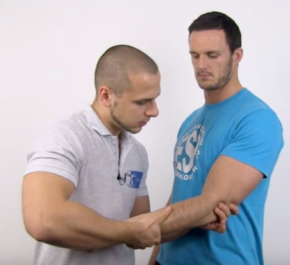
■Valgus stress test: Move the distal forearm laterally with one hand, while cradling the elbow with the other hand and the fingers of that hand palpating the medial side of the elbow joint (see Figure 17). This puts stress on the medial (ulnar) side of the elbow joint, thus testing the ulnar collateral ligament. You should feel this side of the elbow become tense.
Checklist, Lab #5
Review and make sure you have identified each of the structures below.
Bones
Radius
Head and neck of radius
Radial tuberosity (what muscle attaches here?)
Ulna
Olecranon process (which muscle attaches here?)
Coronoid process (which muscle attaches here?)
Trochlear notch
Radial notch (part of the proximal radio-ulnar joint)
Distal Humerus
Medial and lateral supracondylar ridges
Medial and lateral epicondyles
Capitulum (also known as the capitellum)
Trochlea
Olecranon fossa
Cubital fossa and forearm
Cephalic and basilic veins [and median cubital vein if present]
Borders of cubital fossa
Bicipital aponeurosis
Tendon of biceps brachii muscle
Brachial artery and veins
Radial artery
Ulnar artery
Common interosseous artery
Anterior interosseous artery (Wish List item—get a gold star for finding this!)
Median nerve
Anterior interosseous nerve (from median n.)
Ulnar nerve
Radial nerve (with its deep and superficial branches)
Lateral cutaneous nerve of forearm
Medial cutaneous nerve of forearm (if present—may have been cut in the arm)
Muscles
You should know the actions and innervations of all these muscles. Know specific bony attachment points if you have learned them when studying the bones (example = radial tuberosity, coronoid process, olecranon). Otherwise, have a general idea of origins and insertions of the muscle and the joints they cross.
Pronator teres
Flexor carpi radialis
Palmaris longus
Flexor carpi ulnaris
Flexor digitorum superficialis
Flexor digitorum profundus
Flexor pollicis longus
Pronator quadratus
Elbow Joint (Prosection)
Humero-radial and humero-ulnar articulations
Proximal radio-ulnar joint
Ulnar collateral ligament
Radial collateral ligament
Annular ligament