
Lab 10: Dissection: Anterior Abdominal Wall (AAW) and Inguinal Region

  1. Reflect the skin from the anterior and lateral abdominal wall.
  2. Identify the two layers of the superficial fascia and reflect them.
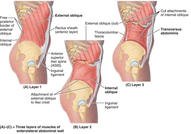
  3. Clean and identify the external oblique, internal oblique, and transversus abdominis muscles. 
  4. Identify and open the rectus sheath on one side and identify the rectus abdominis muscle.
  5. Identify nerves and blood vessels supplying the anterior abdominal wall.
  6. Identify the inguinal ligament, inguinal canal, and deep and superficial inguinal rings.
  7. Open the scrotum in male cadavers and identify the spermatic cord and its contents.
  8. Identify structures in the subinguinal space.
  9. Discuss the clinical anatomy of inguinal hernias.

Surface Anatomy: Inspect and Palpate

Anterior superior iliac spine and iliac crest

Pubic tubercle

Inguinal fold (skin fold in the “groin,” where the thigh meets the abdomen)

Umbilicus

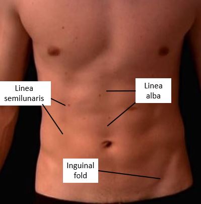
Linea alba and linea semilunaris (in lean donors)

Note

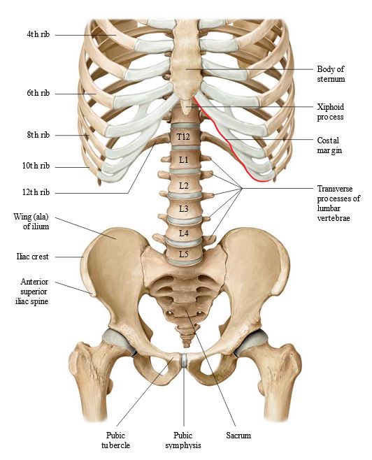
Consult Figures 10.1 and 10.2.

Figure 10.1. Bony Framework of Abdomen: Thieme Atlas of Anatomy, 4th ed., Figure 13.1.
Figure 10.2. Surface anatomy of abdomen: Wikiradiography.net.
Reflect the skin from the abdominal wall.

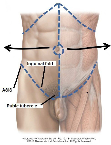
Make incisions as shown in Figure 10.3. Make the incisions deep enough to carry through the skin AND superficial fascia. Reflect them together.

The upper incisions along the costal margin were made in a previous lab.

The lower incisions stretch from the anterior superior iliac spines laterally to the pubic tubercles medially. Make the incisions just below the inguinal folds (the skin crease between thigh and abdomen).

Make the final incision vertically in the midline. Circumscribe the umbilicus.

Figure 10.3. Skin incisions.

Reflect the skin and superficial fascia with a scalpel and blunt dissection as far lateral as possible. Leave the skin flaps attached to the body! The superficial fascia may have a very thick layer of adipose tissue.

Identify the two layers of the superficial fascia:

Fatty layer of superficial fascia (Camper’s fascia)

Membranous layer of superficial fascia (Scarpa’s fascia)—may be difficult to discern, but you should see a pale layer with parallel fiber bundles internal to Camper’s fascia, adjacent to the deep fascia on the muscles. Scarpa’s fascia is the equivalent of the retromammary fascia of the female breast. You should be able to pass your fingers into the plane between Scarpa’s fascia and the muscles of the abdominal wall and separate them.

Now, identify the white rectus sheath covering the rectus abdominis muscle in the midline of the AAW. The rectus sheath is an aponeurosis. We will learn more about the rectus sheath, shortly.

It may be possible to identify a few nerves at this point:

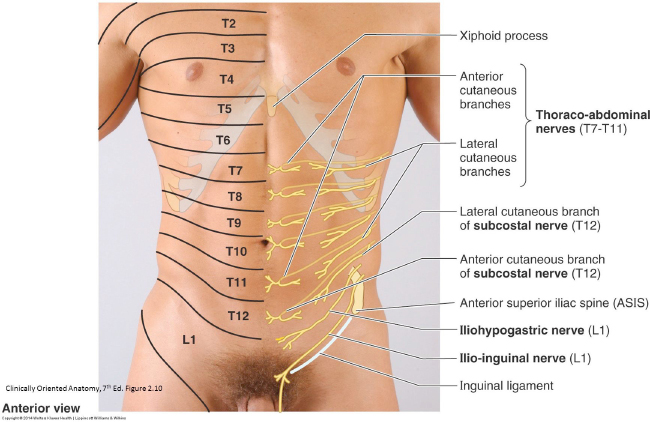
Anterior cutaneous branches of thoraco-abdominal nerves penetrate the rectus sheath about an inch to either side of the linea alba. What are thoraco-abdominal nerves?

Lateral cutaneous branches of thoraco-abdominal nerves pass into the superficial fascia along the mid-axillary line.

The T-10 dermatome crosses the umbilicus. Appendicitis pain often refers here.

Figure 10.4. Cutaneous nerves and dermatomes of abdominal wall.

Muscles of Anterolateral Abdominal Wall

Complete anatomy

Abdominal wall muscles and structures

Figure 10.5. Overview of AAW muscles. Clinically Oriented Anatomy. 8th ed., Figure 5.5.
Both sides: Clean the external oblique muscle, its aponeurosis, and the rectus sheath.

Use a sharp scalpel blade and scissors to remove fascia from the surface of the external oblique muscle and its aponeurosis. Don’t worry if it’s not super clean.

Question

What are aponeuroses? Why do muscles of the anterior abdominal wall have them?

Question

Where is the origin of the external oblique? See Figure 10.5.

Question

How would you describe the direction of the muscle fascicles in the external oblique?

Question

The posterior-most fleshy part of the external oblique is nearly vertical in orientation and inserts onto the iliac crest. The aponeurotic part is directed obliquely and inserts into the linea alba and on the pubic tubercle (Figure 10.5). What is the linea alba?

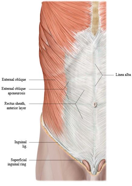
Both sides: Identify the superficial inguinal rings.

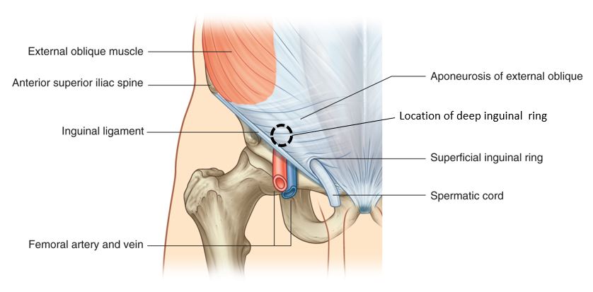
The superficial inguinal rings are bilateral openings in the external oblique aponeuroses near the pubic tubercles. The spermatic cord traverses the superficial inguinal ring in males. The round ligament of the uterus passes through the superficial ring in females. See Figure 10.6.

Figure 10.6. External oblique muscle and superficial inguinal ring. Thieme Atlas of Anatomy, 4th ed., Figure 13.4A.
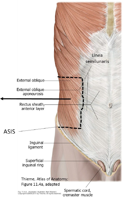
Side one: Cut and reflect a portion of the external oblique as shown in Figure 10.7.

alert

Do this procedure on one side of the donor.

Palpate the anterior superior iliac spine (ASIS). Use scissors to make a shallow horizontal cut in the external oblique at the level of the ASIS from lateral to medial, stopping at the linea semilunaris. Lift the external oblique as you cut it to avoid damaging the underlying internal oblique muscle.

Separate the two muscles with your fingers. Once you have a plane established between the muscles, use scissors to continue cutting: (1) make a vertical cut parallel to the linea semilunaris, (2) then a horizontal cut in a lateral direction to the costal margin. See the dashed lines in the Figure 10.7.

Reflect the external oblique laterally as shown.

Figure 10.7. Cuts and reflection of the external oblique.
Side one: Now you can inspect the internal oblique muscle.

question

Where is the origin of the internal oblique? See Figure 10.5.

Question

How would you describe the orientation of the muscle fascicles in the external oblique (fan shaped, eh?). Compare to the external oblique.

Question

The internal oblique inserts on the lower ribs, linea alba, and the pubic bone of the pelvic skeleton. See Figure 10.5.

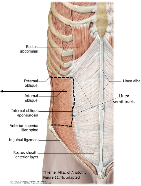
Side one: Cut and reflect the internal oblique as shown in Figure 10.8.

alert

Do this procedure on the same side where you previously made a window in the external oblique.

Use the same scissor technique as before: make a horizontal cut in the internal oblique from lateral to medial, ending at the linea semilunaris. Separate it from the underlying transversus abdominis.

Complete the incisions as shown in Figure 10.8 to form a flap in the internal oblique, then reflect the flap laterally.

Use blunt dissection to separate the internal oblique and transversus abdominis. Fusion of these muscle layers sometimes makes this difficult—do the best you can to reveal as much of the transversus abdominis as possible.

You are in the neurovascular plane of the abdominal wall. The thoraco-abdominal nerves and vessels are in this plane.

Figure 10.8. Cuts and reflection of internal oblique.

Note

This is the same plane in the body wall that you demonstrated in the thorax dissection—between the 2nd and 3rd muscle layers.

Complete anatomy

Nerves and structures of anterior abdominal wall

Side one: Now you can inspect the transversus abdominis.

Question

Where does the transversus abdominis originate? See Figure 10.5.

Question

In which direction are the muscle fascicles of the transversus abdominis oriented? (Not a trick question!)

Question

The transversus obdominis inserts into the linea alba and pubic bone.

Note

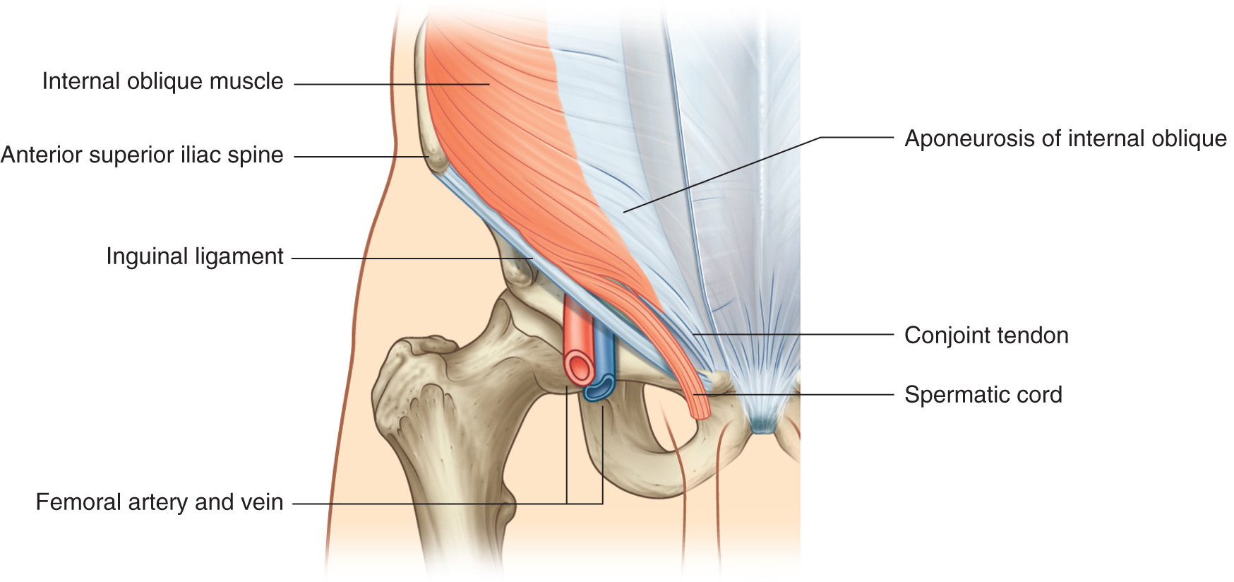
The aponeuroses of the internal oblique and transversus obdominis fuse as they attach to the pubic bone—this fused portion is called the conjoint tendon (see Figure 10.9).

Figure 10.9. Conjoint tendon. Gray’s Anatomy for Students, 4th ed., Figure 4.45.
Side one: Open the rectus sheath; cut and reflect the rectus abdominis.

alert

Do this procedure on the same side where you cut windows in the oblique muscles. Leave the rectus sheath intact on the other side.

Points to Ponder

The rectus sheath that surrounds the rectus abdominis is formed by the fused aponeuroses of the external oblique, internal oblique, and transversus abdominis. It has two layers = anterior and posterior—these form an envelope around the rectus abdominis that prevents bowstringing when the trunk is flexed.

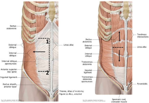
Study Figure 10.10. Use a scalpel to make a shallow incision in the rectus sheath just off the midline; from the xiphoid process above to a point parallel to the ASIS below (points 1 to 2 in Figure 10.10). Make scissors cuts laterally from points 1 and 2, ending the cuts at the lateral edge of the rectus abdominis (at the linea semilunaris). This will create a flap in the anterior layer of the rectus sheath.

Use scalpel, forceps, and scissors to reflect the anterior layer of the rectus sheath laterally toward the linea semilunaris. This will expose the rectus abdominis inside the two layers of the rectus sheath.

Question

Where does the rectus abdominus arise and insert? What are its actions?

Once the rectus sheath is opened, horizontally transect the rectus abdominis above the umbilicus and reflect the inferior portion down and the superior portion up (see Figure 10.10). This exposes the posterior layer of the rectus sheath behind the muscle.

Figure 10.10. Open the anterior layer of the rectus sheath; cut through and reflect the upper and lower portions of the rectus abdominis.

Identify:

Tendinous intersections of the rectus abdominis (these separate the muscles into segments, forming the “six-pack”)

Anterior and posterior layers of the rectus sheath

Linea alba and linea semilunaris

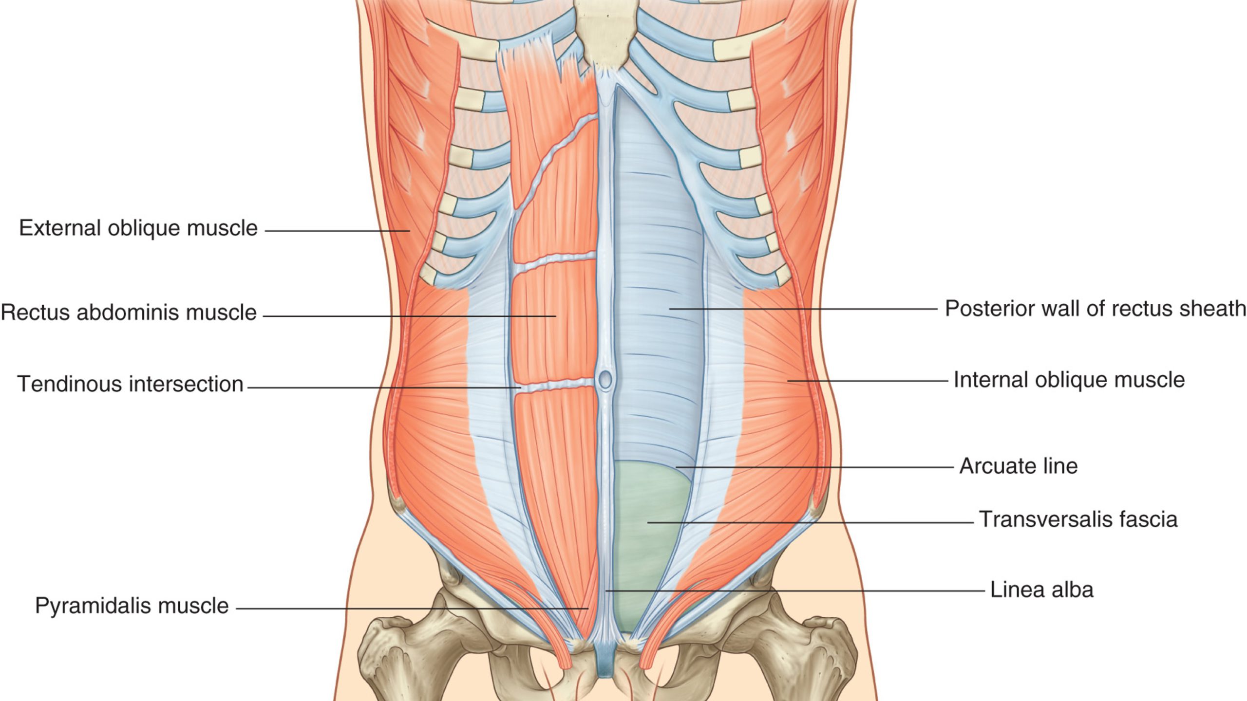
Arcuate line (about halfway between umbilicus and pubic tubercles—Figure 10.11): a curved line formed by the sudden forward transition of aponeuroses—below the arcuate line all 3 layers of aponeuroses pass anterior to the rectus abdominis (discuss this in the Chalk Talk below).

Transversalis fascia—there is no rectus sheath behind the rectus abdominis below the arcuate line—instead, the transversalis fascia is directly adjacent to the posterior surface of the muscle, and deep to the fascia is the parietal peritoneum. Transversalis fascia is a layer of internal fascia, deep to the abdominal wall muscles, between the muscles and the parietal peritoneum.

Inferior epigastric vessels—these pass under the arcuate line and enter the rectus sheath. They ascend behind the rectus abdominis.

Figure 10.11. Arcuate line. Gray’s Anatomy for Students, 4th ed., Figure 4.32.

Question

Where do the inferior epigastric arteries arise?

Question

The inferior epigastric arteries anastomose with arteries that descend behind the rectus abdominis from above. Which arteries? Where do they arise?

Complete anatomy

Posterior view of anterior abdominal wall

Chalk Talk

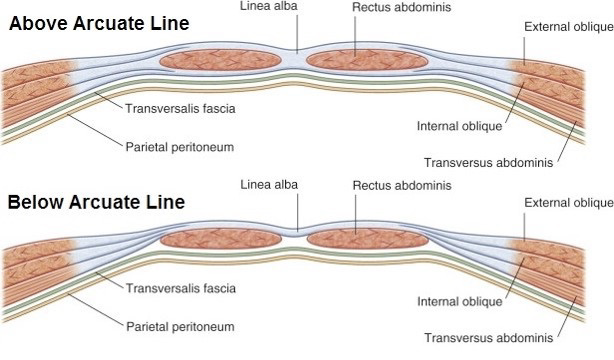
Draw and discuss the layers of the abdominal wall and the composition of the rectus sheath. Understand the concept of the arcuate line. Draw transverse sections of the abdominal wall above and below the arcuate line to show how the rectus sheath is formed by the aponeuroses (see Figure 10.12).

Above the arcuate line, the anterior and posterior layers of the rectus sheath have perfect symmetry – each is formed from 1 and ½ aponeuroses. Below the arcuate line, all three aponeuroses pass in front of the rectus abdominis to form the anterior layer of the rectus sheath—therefore, there is no posterior layer of rectus sheath below the arcuate line.

Figure 10.12. Composition of the rectus sheath layers. Gray’s Anatomy for Students, 4th ed., Figure 4.33.

Inguinal Region

Complete anatomy

Inguinal canal and spermatic cord

Locate:

Anterior superior iliac spine

Pubic tubercle

Inguinal ligament

Question

Where are the bony attachments of the inguinal ligament? Recognize that the ligament is the free inferior edge of the external oblique aponeurosis, rolled inward like the letter 'J'.

Locate the midpoint of the inguinal ligament and place a finger there. This is the surface projection of the deep inguinal ring. Note the location of the superficial ring (near the pubic tubercle). The rings are the internal and external openings of the inguinal canal. Appreciate that the inguinal canal has an oblique orientation and is only a few inches long. See Figure 10.13.

Figure 10.13. Inguinal canal and inguinal rings. Gray’s Anatomy for Students, 4th ed., Figure 4.44.
Side two: Expose the inguinal canal.

Open the inguinal canal on ONE side of the donor only.

Do this procedure on the side opposite from where you cut windows in the muscles earlier. See Figure 10.14.

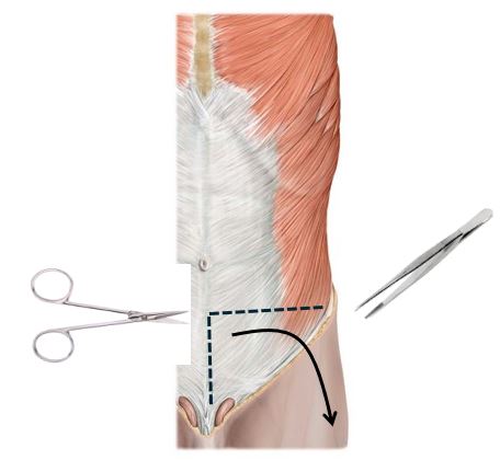
Make a shallow horizontal incision in the external oblique and its aponeurosis beginning at the ASIS and carry it medially, almost to the midline.

Then, make a vertical incision in the aponeurosis near the midline, as shown in Figure 10.14, and carry the incision downward just medial to the pubic tubercle.

Figure 10.14. Exposing the inguinal canal. Thieme Atlas of Anatomy, 4th ed., Figure 13.4A.

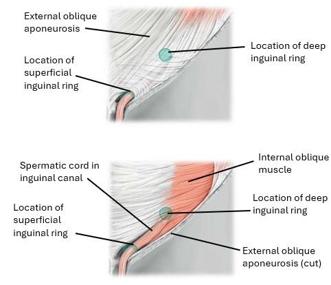
Use scissors and forceps to carefully peel down the aponeurosis toward the thigh, using the inguinal ligament as a hinge. Reflect only the external oblique aponeurosis. Since this layer forms the anterior wall of the inguinal canal, the inguinal canal and deep inguinal ring should now be exposed. See Figure 10.15.

Figure 10.15. Inguinal canal exposed.

Identify the following (see Figure 10.15):

Deep and superficial inguinal rings

The fibers of the internal oblique and transversus abdominis muscles arching over the inguinal canal. These are in the roof of the canal.

The conjoint tendon (fused internal oblique and transversus abdominis aponeuroses) attaching to the pubic bone. The conjoint tendon is the medial-most part of the posterior wall of the canal.

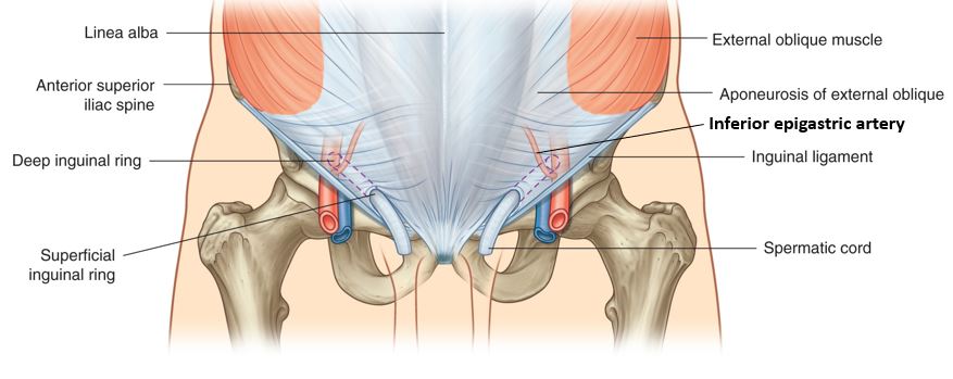
See if you can spot the inferior epigastric vessels ascending on the abdominal wall, medial to the deep inguinal ring (Figure 10.16). They are important landmarks for hernias (discussed later).

Figure 10.16. Deep ring and inferior epigastric artery. Gray’s Anatomy for Students, 4th ed., Figure 4.42.

Chalk Talk

Draw and discuss the inguinal canal, noting the structures that form its walls, floor, and roof, and the locations of the inguinal rings.

BOUNDARIES of the inguinal canal:

Lateral:

Medial:

Superior:

Inferior:

Anterior:

Posterior:

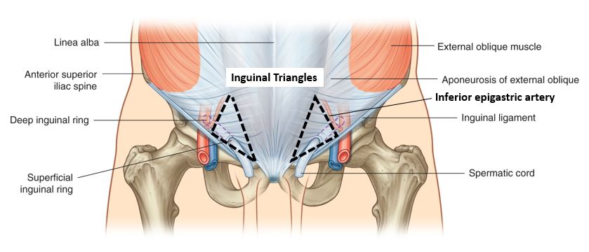
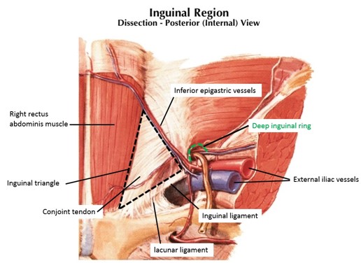
An important region of the anterior body wall just medial to the deep inguinal ring is called the inguinal triangle (Hesselbach’s triangle). See Figures 10.17 and 10.18. 

Figure 10.17. Structures bordering the inguinal canal. Gray’s Anatomy for Students, 4th ed., Figure 13.14B.
Figure 10.18. Inguinal (Hesselbach’s) triangles. Gray’s Anatomy for Students, 4th ed., Figure 4.42.

The region of the abdominal wall within the inguinal triangle is reinforced only by the conjoint tendon (fused aponeuroses of the internal oblique and transversus abdominis) and the transversalis fascia (se Figure 10.19). Aging and/or illness can weaken the muscles and tissues and make this area less stable.

Figure 10.19. Inguinal triangle—internal view looking at posterior side of the anterior abdominal wall. Netter, Atlas of Human Anatomy, Plate 262.

Chalk Talk

Sketch and discuss the boundaries of the inguinal triangle.
•  Medial: Lateral border of rectus abdominus
•  Lateral: Inferior epigastric vessels
•  Inferior: Inguinal ligament

Clinical correlation

What is the definition of a hernia in general? What are inguinal hernias? Spend some time in lab discussing the applied anatomy of inguinal hernias = DIRECT vs. INDIRECT.

Direct inguinal hernias bulge into the inguinal triangle, medial to the inferior epigastric vessels. Indirect inguinal hernias enter the deep ring lateral to the inferior epigastric vessels. Which type of hernia would enter the spermatic cord?

Complete anatomy

Inguinal triangle

Spermatic Cord

In male donors, open the scrotum on one side.

Note

If you have a female donor, skip down a section, but be sure you visit a male dissection and study the anatomy.

Using a scalpel or scissors, make a vertical skin incision starting at the superficial inguinal ring and extending to the inferior pole of the scrotum.

Use blunt dissection to separate the skin and superficial fascia of the scrotum (called the dartos) from the spermatic cord and swing the entire cord with testes superiorly out of the scrotum. The testes hang from the body wall via the spermatic cord like a pendulum. Why are the testes outside of the body cavity?

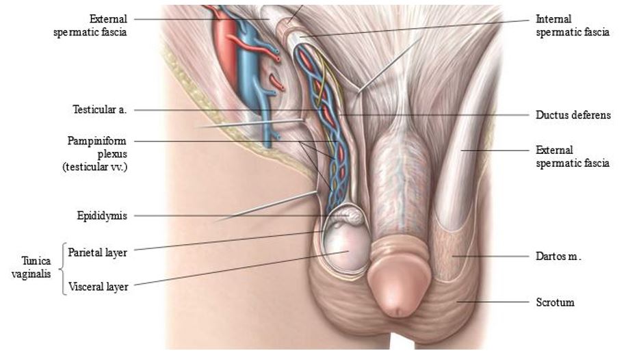
Use scissors to open the fascial layers of the spermatic cord and to tease out the contents of the cord = you should find the pampiniform plexus of veins and the ductus deferens with ease.

Figure 10.20. Fascia and contents of spermatic cord. Thieme Atlas of Anatomy, 4th ed., Figure 13.17B.

Identify the following in males:

The external spermatic fascia is the outermost layer of the spermatic cord. Developmentally, it is derived from the fascia of the external oblique muscle.

Deep to the external spermatic fascia is the cremasteric fascia containing the cremaster muscle. Illuminate the spermatic cord with a light and note the loops of skeletal muscle in the fascia = cremaster muscle. What is the function of the cremaster? From which layer of the abdominal wall is the cremaster derived developmentally?

Pampiniform plexus of veins (translation = “tendril” or “vine-like”). What is the function of the pampiniform plexus?

Ductus deferens = palpate its wire-like texture! What is the function of the ductus (vas) deferens?

The testicular artery is challenging to find—it is surrounded by the twisted veins of the pampiniform plexus—it is less tortuous and paler in color than the veins. Where does the testicular artery arise?

Coverings of the Testes and Epididymis

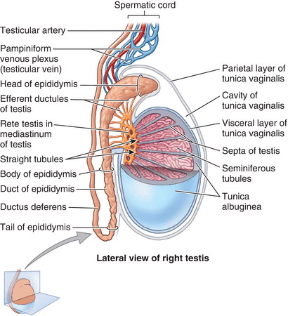
Figure 10.21. Testis and epididymis. Clinically Oriented Anatomy, 8th ed., Figure 5.21.

Each testis and epididymis are surrounded by the same three layers of fascia that surround the spermatic cord.

Deep to the fascias of the spermatic cord there is a serous sac on the anterior surface of the testis. This is the tunica vaginalis. Use scissors to make a vertical cut to open the tunica vaginalis so its two parts can be examined.

The layer of serous membrane facing the testis (on the deep surface of the spermatic cord fascia) is the parietal layer of the tunica vaginalis. The shiny layer of serous membrane directly adhered to the testis itself is the visceral layer of tunica vaginalis.

The thick capsule of the testis is the tunica albuginea (“white coat”). The visceral tunica vaginalis is firmly attached to the tunica albuginea. Use scissors to cut open the testis—inside are the many tiny seminiferous tubules.

Identify the epididymis. The ductus deferens attaches to the tail of the epididymis (Figure 10.21). The epididymis is a storage unit where sperm mature.

Note

The tunica vaginalis is a small serous sac developmentally derived from a diverticulum of peritoneum from the abdomen = the processus vaginalis. Like all good serous sacs, it has two continuous serous layers (parietal and visceral) with a potential space (serous cavity) between them.

Note

Review these clinical terms: Hydrocele and Varicocele.

In female donors, incise the skin of a labium majus.

The inguinal canal in females contains the round ligament of the uterus. Proximally, the round ligament attaches to the uterus in the pelvic cavity. It leaves the pelvic cavity through the deep inguinal ring and enters the inguinal canal. Distally, it emerges from the superficial inguinal ring and blends with the fibrous tissue of the labium majus.

Make a vertical incision, starting at the superficial inguinal ring and extending downward into the labium majus. Probe around in the fascia and try to find the round ligament of the uterus as it emerges from the superficial inguinal ring into the labium majus. It may be difficult to locate.

Note

Embryology: A processus vaginalis (diverticulum of the peritoneal cavity) also develops in females, pushing into the body wall and carving out the inguinal canal. The processes vaginalis in females is known as the “Canal of Nuck.” It usually closes completely and obliterates—thus there is no tunica vaginalis counterpart in females. The inguinal canal contains the round ligament of the uterus, wrapped in the same fascial layers as those that surround the spermatic cord structures in males. Do you know which embryonic structure gives rise to the round ligament of the uterus?

Subinguinal Space

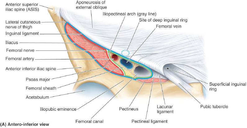
This is the passageway beneath the inguinal ligament, connecting the abdominopelvic cavity to the proximal thigh.

Reflect the skin from the proximal thigh, about an inch or two below the inguinal ligament. Do this on one side only. Use scissors and blunt dissection to clean the femoral artery and vein.

Clinical correlation

The mid-inguinal point (midpoint of the inguinal ligament) is an important landmark. Earlier we located the deep inguinal ring superior to it—the femoral artery is inferior to the mid-inguinal point. Its pulsations can be felt here. If you can locate the femoral artery—you know that the femoral vein is medial to it!

Figure 10.22. Subinguinal space. Clinically Oriented Anatomy, 8th ed., Figure 5.14.

Question

What are the names of the femoral artery and vein proximal to the inguinal ligament (within the abdominopelvic cavity)? Realize that these vessels are one and the same—only the names have changed.

Lateral to the artery, locate the femoral nerve.

Medial to the femoral vein, use blunt dissection to locate the femoral canal. The femoral canal is a potential space, filled with loose connective tissue and a few lymph nodes and vessels. Its function is to provide space for expansion of the femoral vein when venous return from the lower limb increases.

Place a finger in the femoral canal and rotate it medially to palpate the sharp edge of the lacunar ligament. The lacunar ligament is the part of the inguinal ligament that is reflected downward on to the pubic bone.

Note

A handy mnemonic for recalling structures in the subinguinal space, from lateral to medial = NAVEL. Can you recall which structures the letters refer to?

Clinical correlation

The femoral canal is a potentially weak area where a viscus from the abdominal cavity could protrude into the thigh, below the inguinal ligament = a femoral hernia. A femoral hernia can strangulate if compressed against the sharp edge of the lacunar ligament.

Checklist, Lab #10

Review and make sure you have identified each of the structures below.

Anterior abdominal wall and inguinal region

Fatty layer of superficial fascia (Camper’s)

Membranous layer of superficial fascia (Scarpa’s) 

External oblique muscle & aponeurosis

Internal oblique muscle & aponeurosis

Transversus abdominis muscle & aponeurosis 

Rectus abdominis muscles

Tendinous intersections in rectus abdominis

Rectus sheath (Anterior and posterior layers)

Arcuate line

Transversalis fascia (can be seen behind the rectus abdominis, inferior to the arcuate line)

Linea alba and linea semilunaris

Thoracoabdominal nerves (= lower intercostal nerves in the neurovascular plane of AAW) 

Inferior epigastric vessels

Inguinal ligament

Lacunar ligament (can be felt)

Superficial and deep inguinal rings

Inguinal canal

Inguinal (Hesselbach’s) triangle 

Dartos fascia of scrotum (= the superficial fascia of the scrotum)

External spermatic fascia of spermatic cord 

Cremasteric fascia w/ cremaster muscle

Ductus deferens

Pampiniform plexus

Testicular artery (hard to separate from pampiniform plexus, but understand its location in the spermatic cord) 

Testes

Tunica vaginalis (visceral and parietal layers) 

Epididymis

Round ligament of the uterus—EXTRA BONUS POINT (seen leaving the superficial inguinal ring in female donor)

Femoral artery and vein 

Femoral nerve

Femoral canal

Table of Contents
Headshot of David Conley, PhD · Professor, Department of Translational Medicine & Physiology
David Conley
PhD · Professor, Department of Translational Medicine & Physiology
Office: PBS 41A
envelope icon
Headshot of Shannon Helbling, PhD · Clinical Assistant Professor, Department of Translational Medicine & Physiology
Shannon Helbling
PhD · Clinical Assistant Professor, Department of Translational Medicine & Physiology
Office: PBS 41C
envelope icon

Dear students,
We would love to hear from you about your learning materials.

Page/Content Feedback Form