

Station 1. Meninges and ventricles
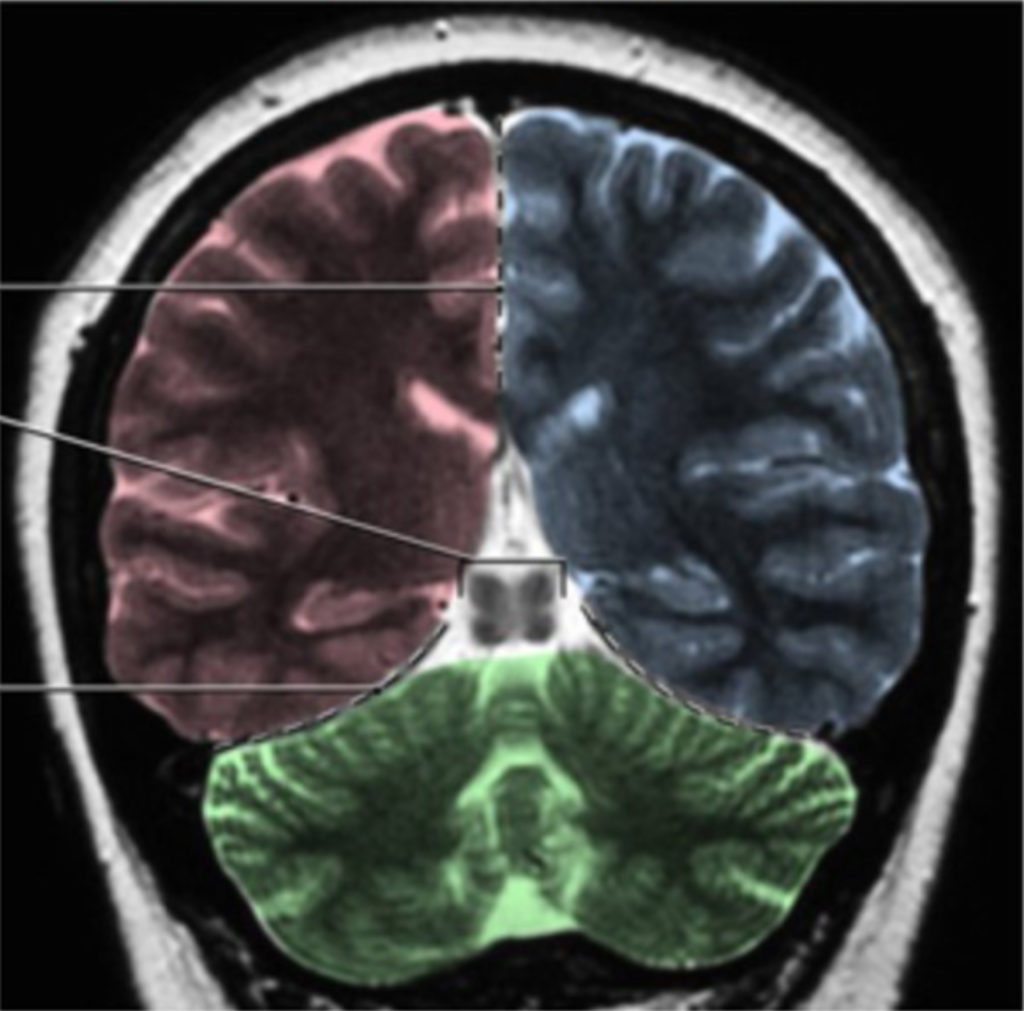
Neuroanatomy lab navigation Identify the parts of the ventricular system on a mid-sagittal brain, coronal sections, and model. The formation of the ventricular system, as it relates to development, was discussed in the Neuroembryology session. Review the clinical significance of the flow and blockage of CSF For further reference Figure 1. Figure 2. Lateral ventricles […]
Station 5. Blood supply and circulation
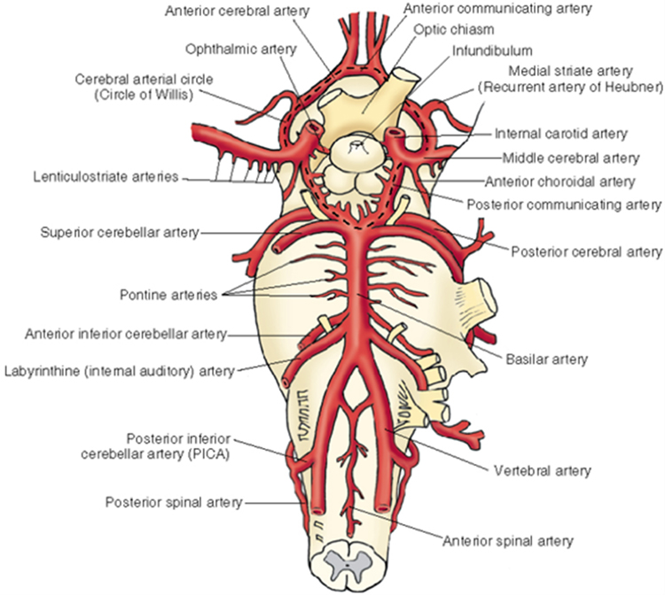
Neuroanatomy lab navigation Arterial blood supply Review and identify the arterial supply. Vertebral arteries supplying the brainstem and cerebellum Anterior and posterior spinal Posterior inferior cerebellar Basilar (unpaired)—multiple pontine branches also come directly off the basilar artery Anterior inferior cerebellar Superior cerebellar Posterior cerebral Posterior communicating Internal carotid supplying the midbrain, thalamus, hypothalamus, and cerebrum […]
Station 3. Diencephalon
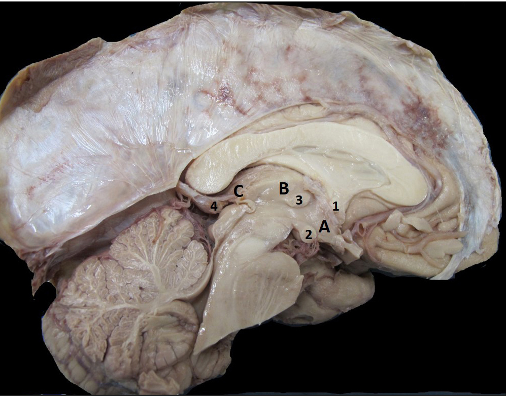
Neuroanatomy lab navigation The diencephalon is composed of the thalamus, hypothalamus, and epithalamus. Thalamus The thalamus is key structure that relays information to the cortex associated with motor and sensory information. There are also thalamus nuclei that form the connections in the memory and emotion circuit from the hippocampal formation. This portion of the thalamus […]
Station 2. Cerebrum, sulci, and gyri

Neuroanatomy lab navigation Introduction to neuroanatomy Figure 1. Anatomic directional terms. From Neuroanatomy: A Laboratory Guide (2e); Jansen and Lampa. Introduction to the Central Nervous System Watch this excellent Introduction to the Central Nervous System from University of British Columbia Neuroanatomy. Cerebrum, sulci, and gyri Lobes of the cerebrum Recall that the surfaces of the […]
Station 4. The brainstem
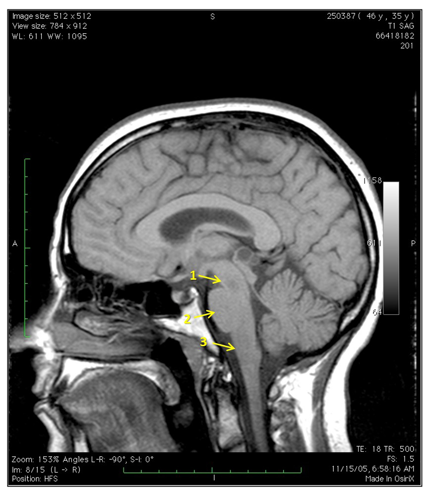
Neuroanatomy lab navigation Parts of the brainstem Midbrain Mesencephalon Pons Metencephalon Medulla Myelencephalon The basal subdivision of the brainstem is the most anterior and contains mostly the descending fiber tracts. The tegmentum is the location of the cranial nerve nuclei, ascending fiber tracts, and the reticular formation. In the medulla, it also contains important neurons […]
Lab 16: Anterior and Medial Compartments of Thigh; Hip Joint
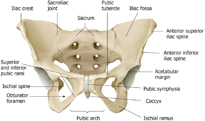
Download this lab as a PDF Goals Identify the parts of the hip bones, femur, patella, and tibia that are associated with the anterior and medial thigh. Identify the femoral triangle, its boundaries, and contents, including the structures passing through the subinguinal space. Identify the adductor canal and its contents. Identify the muscles, nerves, and […]
Lab 15: Dissection: Pelvic Viscera and Pelvic Vessels (Hemisection)
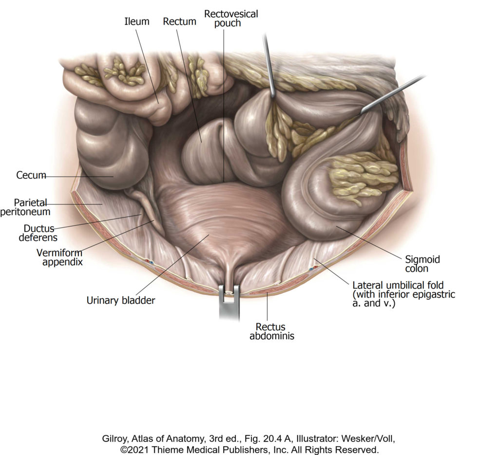
Download this lab as a PDF Goals Understand the general arrangement of pelvic organs in the male and female and identify those organs that can be approached through the pelvic inlet Identify pelvic organs and their subparts Identify muscles in the walls and floor of the pelvic cavity Clean and identify the internal iliac vessels […]
Lab 14: Pelvic Hemisection
Lab 14 navigation Pelvic Hemisection This page is for the half of the class whare are working in their groups to complete the hemisection in preparation for the next dissection lab. Before you begin the hemi-section, partner up with another group to review the male and female external genitalia. External Female Genitalia Figure 29. External […]
Lab 14: Prosection lab: Pelvic Skeleton, Pelvic Cavity, and Pelvic Vessels; Perineum
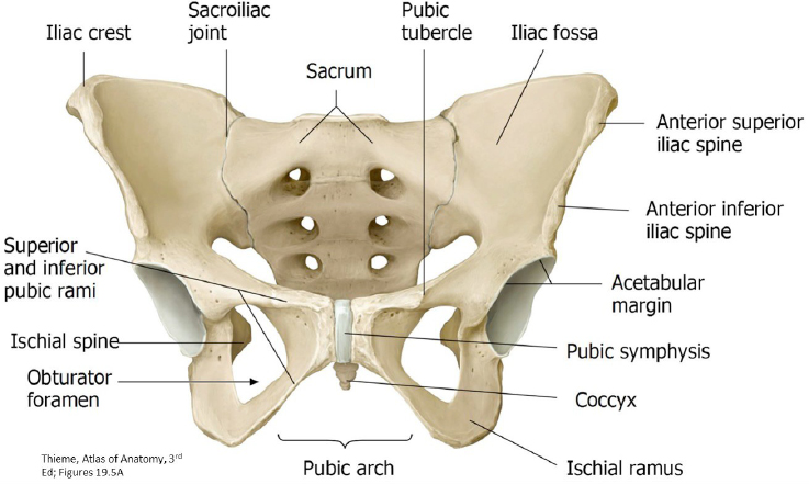
Students: This lab will include 2 parts: Half of the time will be spent rotating through prosection stations, and the other half perfoming a pelvic hemisection in preparation for the next lab Those groups in lab 45 (group A) will start by completing the hemi-section. Find your donor table and pull up the instructions on […]
Lab 14, Station 3: Perineum (Urogenital Triangle)
Lab 14 navigation Station 3: Perineum—Urogenital Triangle Question What are the boundaries of the perineum? What are its triangles called? (See Figure 18.) This station covers the male and female urogenital triangle. The anal triangle has a similar construction in both sexes—it is covered at Station 2. Figure 18. Female perineum showing its triangles. The UG […]