Lab 2, Station 4: Spinal Cord

Table of Contents
Headshot of David Conley, PhD · Professor, Department of Translational Medicine & Physiology
David Conley
PhD · Professor, Department of Translational Medicine & Physiology
Office: PBS 41A
envelope icon
Headshot of Shannon Helbling, PhD · Clinical Assistant Professor, Department of Translational Medicine & Physiology
Shannon Helbling
PhD · Clinical Assistant Professor, Department of Translational Medicine & Physiology
Office: PBS 41C
envelope icon

Complete anatomy

Spinal nerves

Identify the following on the spinal cord prosection or isolated spinal cord:

Dorsal and ventral roots—because the cadaver is prone, only the dorsal roots will be easily visible. Rootlets gather together to form the roots.

Dorsal root ganglionwhat does this contain?

Spinal nerves—formed by the union of dorsal and ventral roots

Dorsal rami of spinal nerves (in the cadaver these are loose, because they are detached from the overlying skin and muscles on the posterior side of the body)

Ventral rami of spinal nerves (In the thorax these become the intercostal nerves as they pass between the ribs)

Conus medullaris of spinal cord

Filum terminale (made of pia mater)

Cauda equina—which structures form the strands of the cauda equina?

Question

The inferior-most tip of the spinal cord is at which vertebral level in the adult?

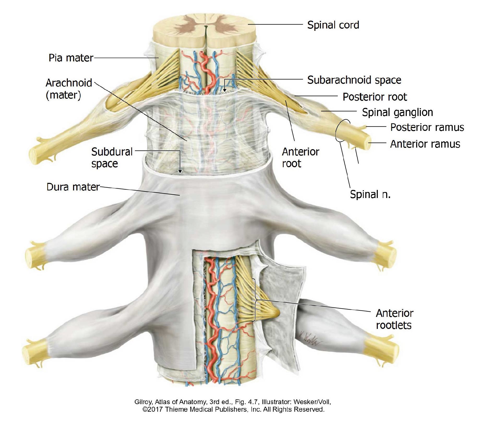
Spinal meninges—these are the protective coverings of the spinal cord

Dura mater

Arachnoid mater

Pia mater


The arachnoid and pia may be difficult to see in the prosection— nonetheless you should understand their locations and relationships.

Figure 46.

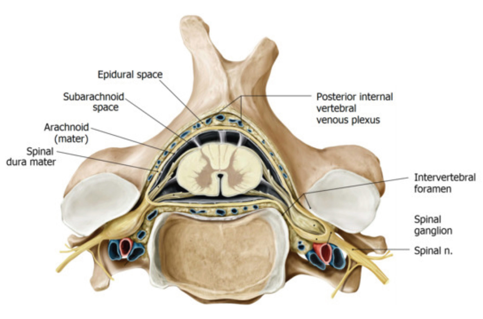
Spaces associated with the meninges:

Epidural space—located within the vertebral canal, between the bone and dura mater. Contains fat and vessels.

Subdural spacea “potential space” between the dura and arachnoid. In a normal individual the space does not exist since the two layers of meninges are touching. The dura and arachnoid are adjacent—so together are called the thecal sac.

Subarachnoid spaceThis space cannot be appreciated in the cadaver because it is collapsed. Why? What does it contain in the living body?

Figure 47. Spinal cord in situ: transverse section. From Anatomy: An Essential Textbook, 2nd ed., ©2018 Thieme Medical Publishers, Inc. All rights reserved.

Discussion: Spinal cord and spinal nerves

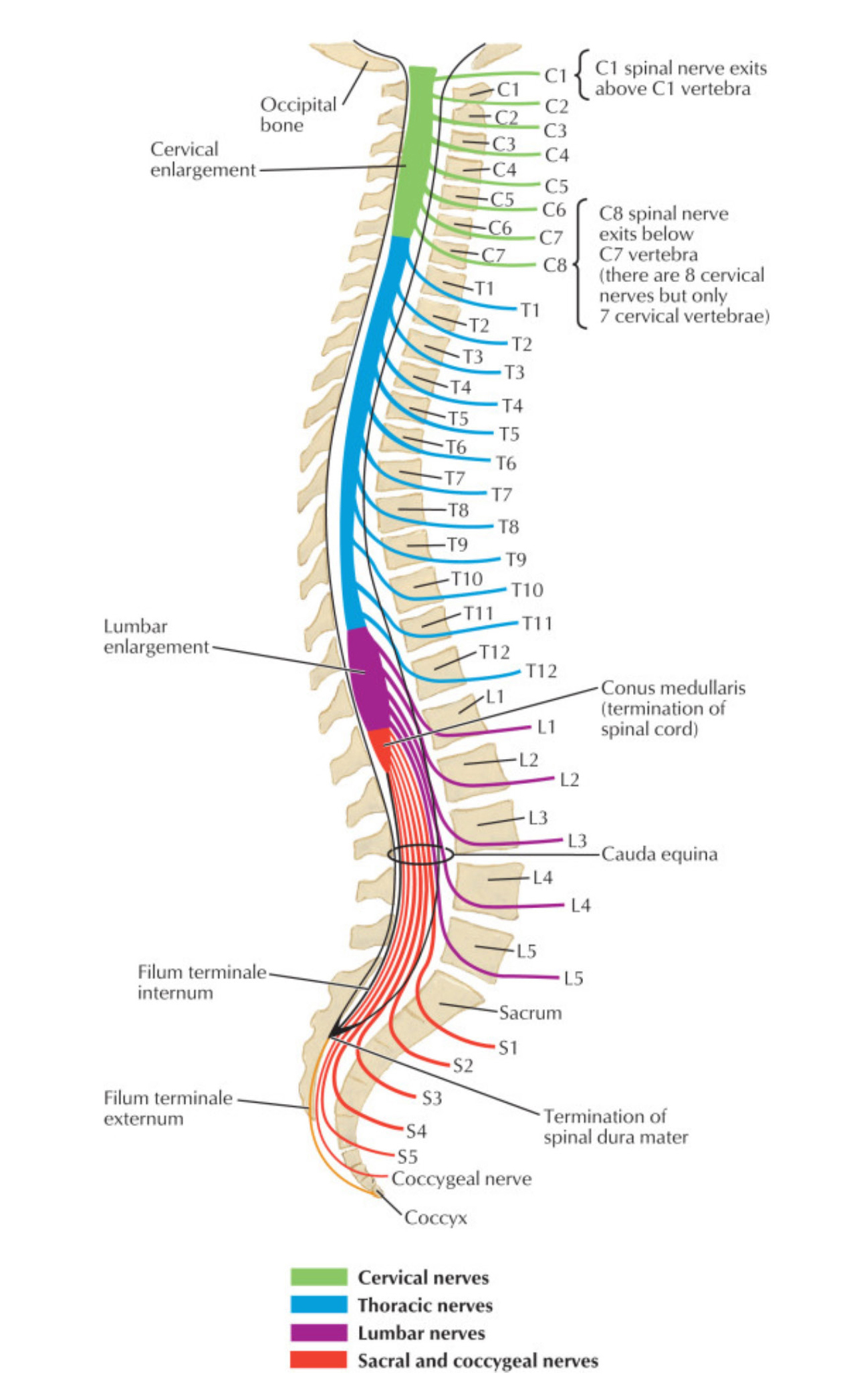
Spinal cord segments are the physical regions of the spinal cord that give rise to the ventral and dorsal roots that form the individual spinal nerves—they are named and numbered the same as the spinal nerve they give rise to (example = L-1 spinal cord segment gives rise to the roots that form L-1 spinal nerve).

The spinal cord contains 31 segments—corresponding to the 31 pairs of spinal nerves. The sacral and coccygeal segments are compressed within the tip of the spinal cord (in the conus medullaris).

  • In Figure 2.48, locate the sacral spinal cord segments (shown in RED). Opposite which vertebrae are they located? (Are they located opposite sacral vertebrae?)
  • Now locate the sacral spinal nerves (also in RED). Where do they exit the vertebral column? Do sacral spinal nerves exit the vertebral column at the same levels as where the sacral spinal cord segments are located?
  • Which structures make up the individual strands of the cauda equina?
  • Where is the safest place to do a lumbar puncture procedure (between which vertebrae)? Why?
  • If a fracture of the body of L-1 vertebra forced a bone fragment directly posterior, what parts of the spinal cord would be affected?
Figure 48. Netter, Atlas of Human Anatomy, 6th ed., Plate 161.

Clinical correlation

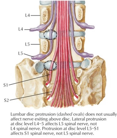
In the upper regions of the spine, a postero-lateral disc herniation affects the spinal nerve passing through the intervertebral foramen at the level of the herniation, because the disc is directly in line with the center of the intervertebral foramen.
However, in the lumbar region (where disc herniations are common) the herniated disc affects the spinal nerve that exits the intervertebral foramen below the herniation (one level down), not the nerve that exits the intervertebral foramen at the level of the disc herniation. This is because IV discs in the lumbar region are located opposite the lower half of the intervertebral foramen, and not the center. Thus, spinal nerves in this region pass through the IV foramina above the disc.

Summary example: A herniated disc between L-4 and L-5 affects the L-5 spinal nerve, and not L-4 (see Figure 2.49).

Figure 49. Netter, Atlas of Human Anatomy, 7th ed., plate 170.