Lab 15: Anterior and Lateral Leg, Knee and Ankle Joints, and Dorsum of Foot
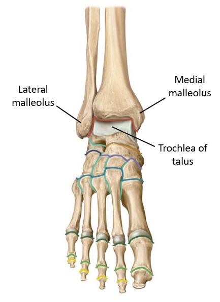
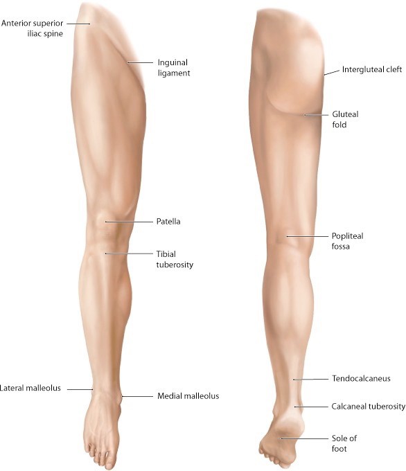
Download this lab as a PDF Goals Identify bony features associated with the knee, leg, and ankle. Identify the muscles in the anterior and lateral compartments of the leg and discuss their actions, innervations, and blood supply. Identify the muscles, nerves, and vessels on the dorsum of the foot. Identify the bones, ligaments, and cartilages […]
Lab 14: Gluteal Region, Posterior Thigh, and Popliteal Fossa
Download this lab as a PDF (Updated 28 October 2024, 8:42 a.m.) Goals Clean and identify the muscles, nerves, and vessels of the gluteal region. Identify the greater sciatic foramen and the structures that traverse it. Clean and identify the muscles, nerve, and vessels of the posterior compartment of the thigh. Identify the boundaries and […]
The knee joint
The knee is a modified hinge joint There are three articulations within the knee joint complex: Lateral and medial tibiofemoral and patellofemoral joints Movements can occur along two axes: The primary movements are flexion and extension Because the radii and lengths of the articular surfaces of the femur and tibia differ, there is a small […]
Gluteal region, posterior thigh, and popliteal fossa
Pertinent osteology Please review the anatomy of the acetabulum, posterior hip bone, sciatic notches, and femur. Figure 24.1 Osteology of the os coxae. Figure 24.2 Sciatic foramina. Figure 24.3 Osteology of the femur. Gluteal region Physically, the gluteal region is part of the trunk, but functionally, it is clearly part of the limb. The gluteal […]
The hip joint
Figure 23.1 The hip joint is the articulation between the round femoral head and the concave acetabulum (“little vinegar cup”). The lunate surface is the articular surface of the acetabulum, forming an arc that fills ¾ of the acetabular cup. It is covered with articular cartilage. The acetabulum is deepened by the acetabular labrum, a […]
Anterior and medial compartments of the thigh
Optional Reading Clinically Oriented Anatomy, 8th ed., Anterior and medial regions of thigh section through Surface anatomy of anterior and medial regions of thigh. Compartmentalization of the thigh The deep fascia, intermuscular septa, and femur together define anterior and posterior compartments in the thigh. The anterior compartment contains muscles that flex the hip and extend […]
Lab 11, Station 4: Muscles and Nerves of the PAW
Lab 11 navigation Muscles of the PAW IDENTIFY these muscles of the PAW: ■Transversus abdominis ■Quadratus lumborum ■Psoas major ■Psoas minor (40% of folks don’t have one!) ■Iliacus For each muscle—describe attachments. The psoas major and iliacus muscles fuse below the inguinal ligament to form the iliopsoas muscle. EXAMINE the inferior surface of the diaphragm and […]
Lab 11, Station 3: Vessels of the PAW
Lab 11 navigation Abdominal Aorta Find these branches of the abdominal aorta: Unpaired visceral branches: ■Celiac trunk, superior mesenteric artery, and inferior mesenteric artery Paired visceral branches: ■Middle suprarenal arteries (very difficult to find) ■Renal arteries ■Testicular/Ovarian arteries (long, thin, and fragile) Branches to body wall: ■Inferior phrenic arteries (to diaphragm) ■Lumbar arteries (lift the […]
Lab 11: Peritoneal Cavity and Overview of GI

Download this lab as a PDF Students: Unlike most lab sessions, which are dissection-based, this session is a “prosection lab.” Assemble in 5 groups, each with 16 students. There are 5 learning stations situated around the labs. Pick a station to start at. Groups will spend about 25 minutes at each station, rotating around the […]
Protected: Lab 11, Station 2: Peritoneum and the Peritoneal Cavity
There is no excerpt because this is a protected post.