Lab 26: Scalp, Cranial Cavity, and Meninges
Download this lab as a PDF Goals Identify the layers of the SCALP. Examine the dura mater and the dural septae. Remove the brain and study: meningeal coverings, gross features, and arterial supply (circle of Willis). Examine the internal aspect of the base of the skull and the cranial fossae. Identify cranial nerves and blood […]
Protected: Lab 17, Station 5: Head and Neck Anatomy—Sagittal View
There is no excerpt because this is a protected post.
Lab 17, Station 4: Blood Vessels
Lab 17 navigation Station 4: Blood Vessels Major Arteries Common carotid artery Carotid bifurcation Carotid body—vascular structure that lies in the “crotch” of the carotid bifurcation—contains chemoreceptors that monitor the status of blood gases (may not be visible in prosection) Internal carotid artery Carotid sinus—a swelling of the proximal internal carotid artery—contains baroreceptors that monitor […]
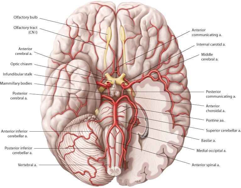
Lab 17, Station 3: Gross Topography of the Brain
Lab 17 navigation Station 3: Gross Topography of Brain Cerebrum Composed of left and right hemispheres. The hemispheres are connected across the midline by a thick tract of nerve fibers called the corpus callosum. Each hemisphere contains a cavity called a lateral ventricle—filled with cerebrospinal fluid.
Lab 17, Station 2: Cranial Base and Cranial Nerves
Lab 17 navigation Station 2: Cranial Base and Cranial Nerves The cranial cavity is the space within the neurocranium. It contains the brain, meninges, blood vessels, and the proximal parts of the cranial nerves. The floor is the cranial base. Foramina in the cranial base transmit cranial nerves and blood vessels. The cranial base has […]
Lab 17, Station 1: The Skull
Lab 17 navigation Station 1: The Skull The skull is divided into two parts: Neurocranium and Viscerocranium. Neurocranium (“brain case”) = Frontal bone, ethmoid, sphenoid, occipital bone, temporal bones (2), and parietal bones (2) The neurocranium has a roof called the calvaria (“skull cap”). Fibrous joints called sutures join the bones. The largest sutures are […]
Lab 17: Introduction to Head and Neck Anatomy
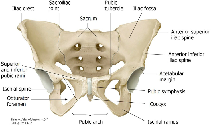
Download this lab as a PDF Goals Identify the bones and ligaments of the pelvic skeleton. Compare and contrast the features of the pelvic skeleton in the male and female. Define the boundaries of the pelvic inlet, pelvic outlet, and pelvic cavity. Identify the muscles in the pelvic walls and pelvic floor and discuss their […]
Cranial nerves appendix
Templates for drawing the pathways of the cranial nerves Cranial nerves templates Previous The mixed cranial nerves
The mixed cranial nerves
Cranial nerve V: Trigeminal nerve Functions Innervates skin of the face, mucous membranes in oral and nasal cavities, external ear, and teeth (general sensory). Innervates muscles of mastication and other muscles in the neck, palate, and middle ear (skeletal motor). Skull opening Ophthalmic branch (V1) = Superior orbital fissure Maxillary branch (V2) = Foramen rotundum […]
The mainly motor cranial nerves
Cranial nerve III: Oculomotor nerve Functions Innervates four of the six extrinsic muscles that move the eye + the muscle that elevates the upper eyelid (skeletal motor). Innervates smooth muscle in the eyeball (parasympathetic). Skull opening Superior orbital fissure. Attachment to brainstem Midbrain. Nucleus of origin/destination in CNS Skeletal motor fibers originate from the oculomotor […]