Protected: Lab 14, Station 3: Perineum (Urogenital Triangle)
There is no excerpt because this is a protected post.
Protected: Lab 14, Station 1: Pelvic Skeleton and Ligaments
There is no excerpt because this is a protected post.
Protected: Lab 14, Station 2: Pelvic Cavity and Anal Triangle of Perineum
There is no excerpt because this is a protected post.
Protected: Lab 13: Dissection: Posterior Abdominal Wall (PAW) and Kidneys
There is no excerpt because this is a protected post.
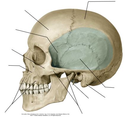
Temporal and infratemporal regions; temporomandibular joint
Optional reading Clinically Oriented Anatomy, 7th ed., Temporomandibular joint section through Arthritis of TMJ. Temporal region Figure 1. Attachments of temporal fascia. GRAY’S ANATOMY FOR STUDENTS, FIGURE 8.138. Figure 2. GRAY’S ANATOMY FOR STUDENTS, FIGURE 8.139. The temporal fossa is the sunken area located on the lateral skull above the zygomatic arch. Boundaries Its floor […]
Protected: Lab 12: Infracolic Region
There is no excerpt because this is a protected post.
Protected: Lab 24: Peritoneal Cavity and Supracolic Region
There is no excerpt because this is a protected post.
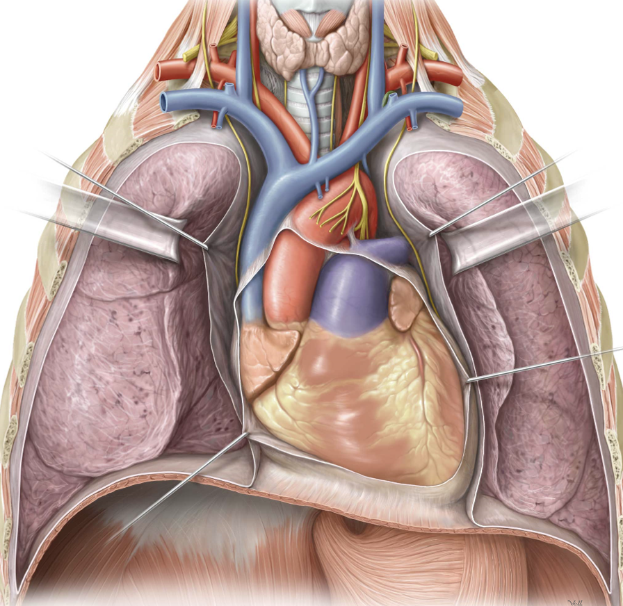
Thoracic cavity
Protected: Lab 25: Infracolic Region
There is no excerpt because this is a protected post.
Protected: Lab 23: Oral Cavity, Pharynx, and Muscles of Mastication
There is no excerpt because this is a protected post.