


Craniocaudal magnification showing malignant breast calcifications. Case courtesy of Melbourne Uni Radiology Masters, Radiopaedia.org, rID: 43928
X-rays are produced by bombarding a dense target (the anode) with high-energy (5–110 Kv) electrons, which produces a beam that radiates outward from a small point source. The beam is shaped with lead shutters. This geometry can lead to significant image magnification, which in mammography is beneficial but in other situations can cause confusion. The farther the X-ray tube is from the imaging plate, the less magnification. Also, the closer the patient is to the imaging plate, and the farther the patient is from the X-ray tube, the less magnification.
In clinical practice
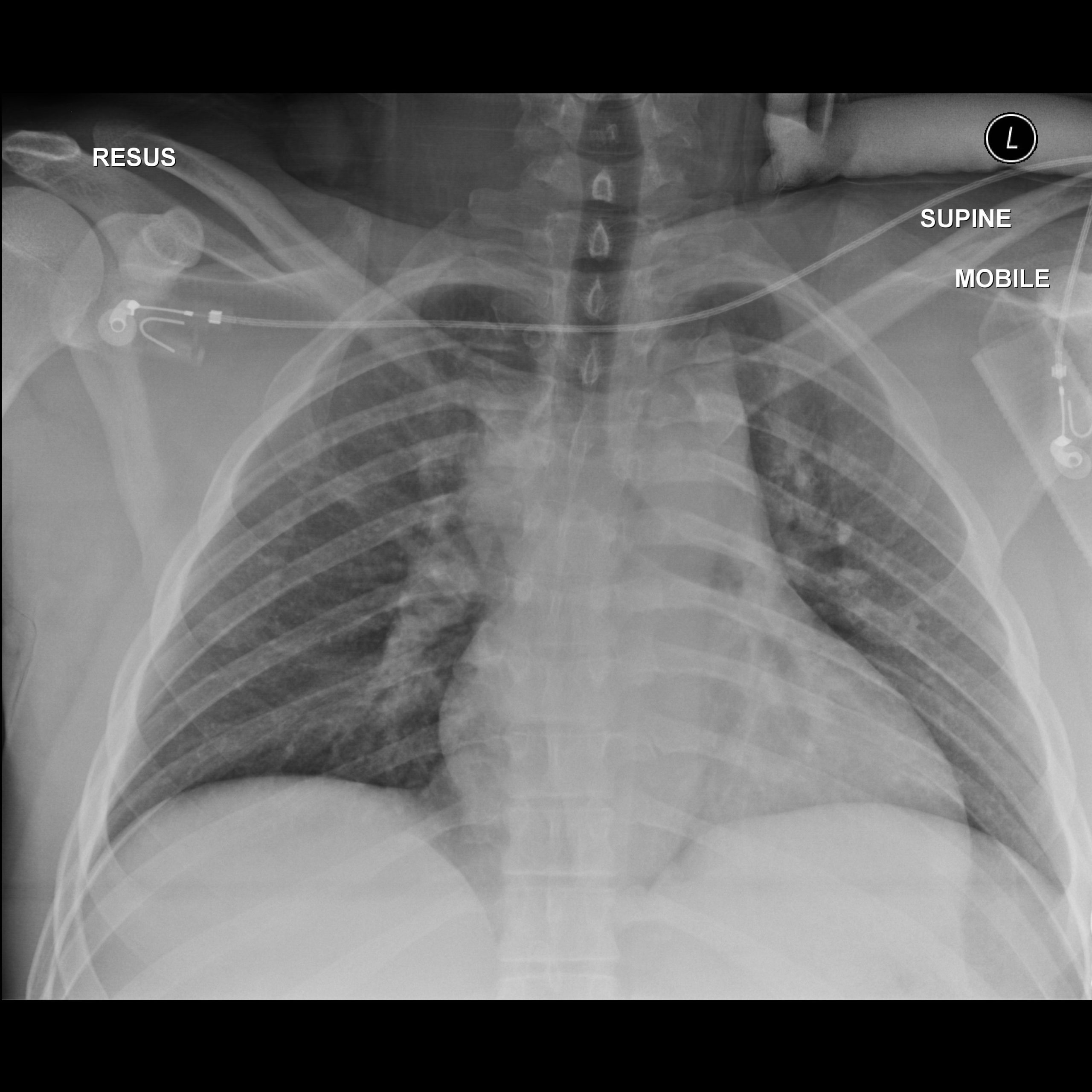
Tap the images to enlarge. PA and AP supine chest X-ray on the same patient. Note the difference in the heart size and central structures called the mediastinum. Case courtesy of Dr Yi-Jin Kuok, Radiopaedia.org, rID: 17910.
Portable films have short tube-to-imaging plate distance, so there is significant magnification on the image (up to 20%). Measurements on a portable film have an often unknowable amount of magnification. Therefore, measurements are approximate!
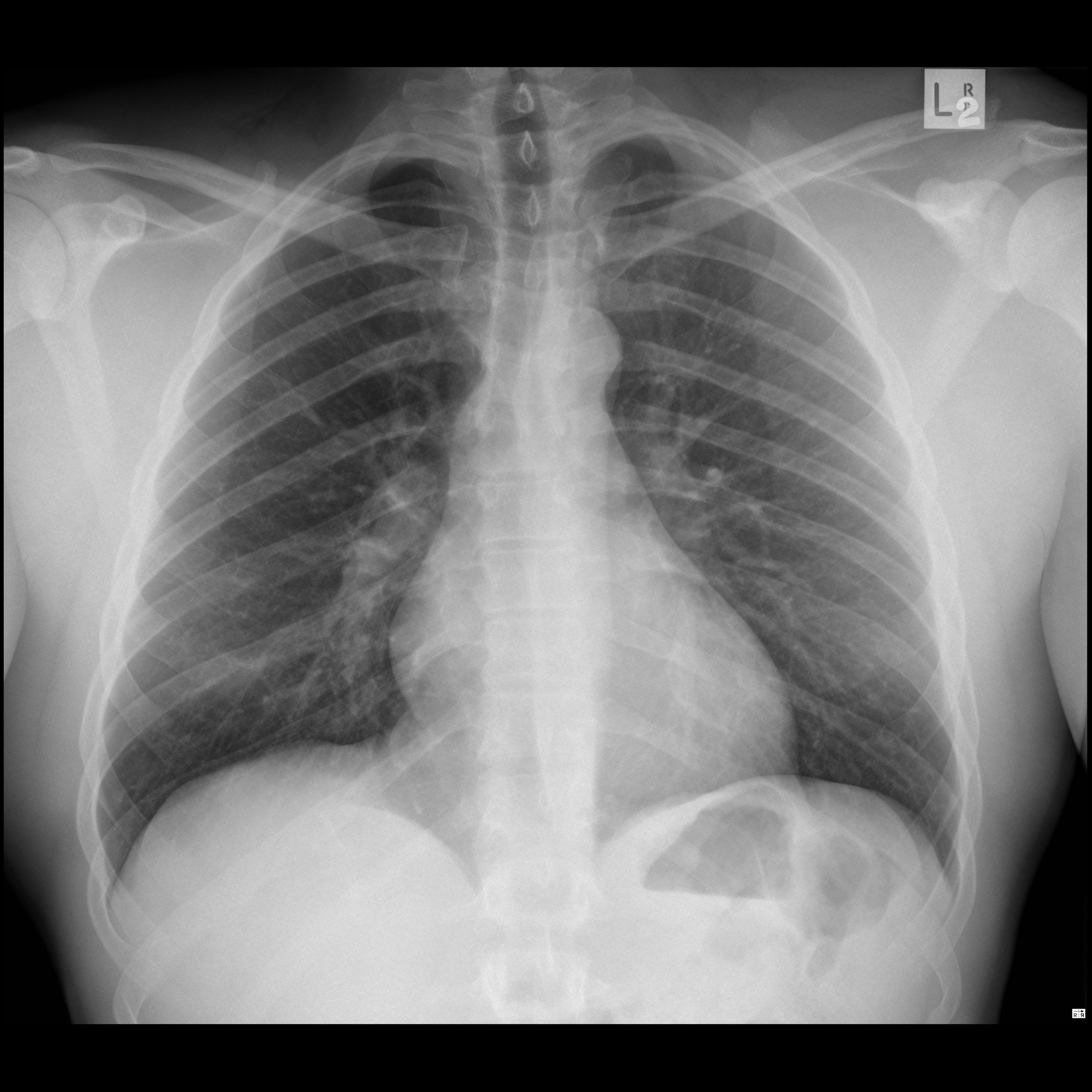
PA upright X-rays, where the heart is as close as possible to the imaging plate and a long (72-inch) tube-to-imaging plate distance gives the least magnification of the heart and is best for cardiac evaluation.
There are five basic X-ray densities, from least dense to highest X-ray density:
- Air
- Fat
- Water
- Bone
- Metal
X-ray density
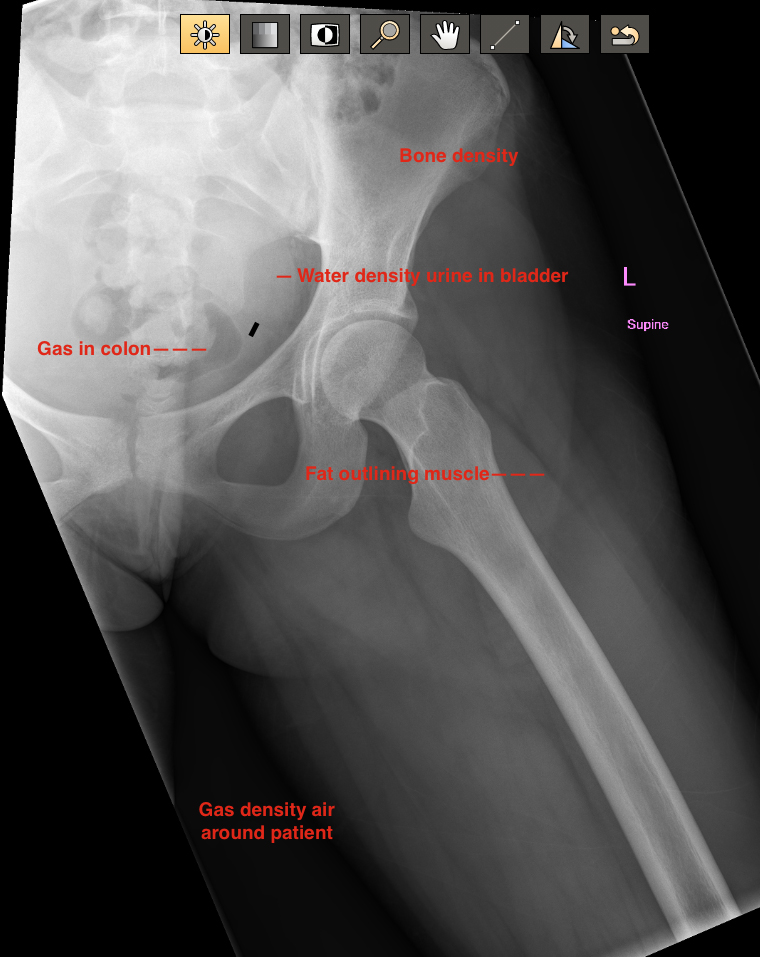
Tap the image to enlarge.
The interplay in the X-ray image gives the contrast necessary for diagnosis— the standard for more than 100 years!
X-rays are ionizing radiation, which result in some degree of tissue damage. However, modern digital equipment used for plane film radiography has greatly reduced X-ray exposures.
With the advent of computers, X-ray beam modeling technology, and advances in mathematics, Computed Tomography Scan (CT) expanded perception of X-ray densities more than 100-fold! Magnetic Resonance Imaging (MRI) built on the mathematics from the CT experience to further enhance imaging capabilities.
First, let’s look at another technology that doesn’t use ionizing radiation.
X-ray terms
Patient's anterior is against the imaging plate.
X-rays enter from posterior.
Patient's posterior is against the imaging plate.
X-rays enter from anterior.
View from the side. Left lateral puts left side against the plate.