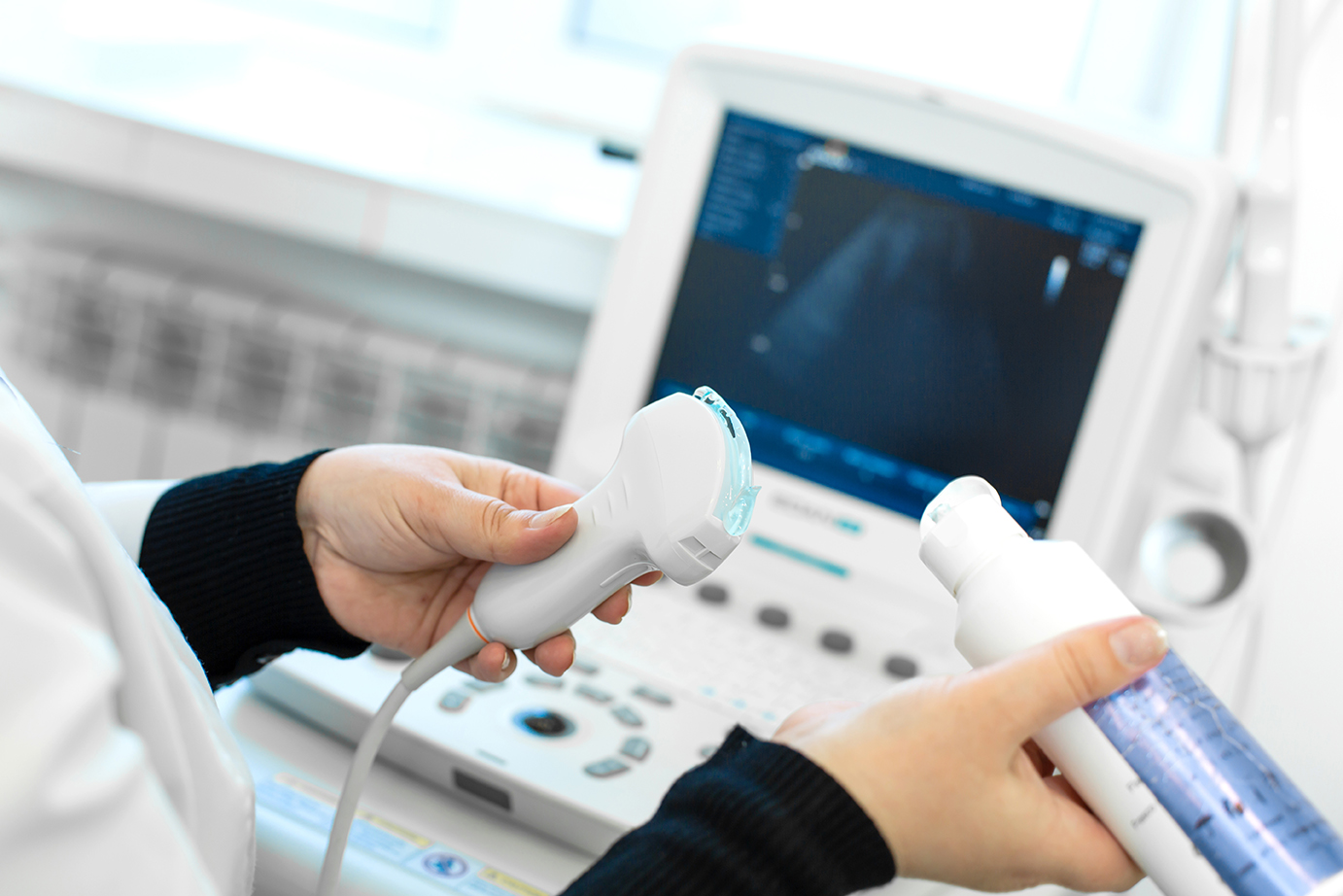
Ultrasound utilizes ultrasonic sound waves in the 2–10 MHz range generated by a handheld piezoelectric crystal, sonically coupled to the skin by gel. A computer controls the process, allowing complex modeling of the sound beam. Higher frequencies have better resolution but poorer depth penetration. Transducer selection is a compromise between the depth of the area of interest and needed resolution.
Technique
The technique is freeform, so image orientation can be challenging. Like a pilot who follows a highway or river, ultrasonographers tend to follow vascular anatomy. When possible, image orientation attempts to follow CT and MRI conventions. Pay attention to annotations on the ultrasound images.
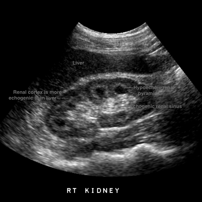
Strongly reflected sound waves are brighter on the image; areas of few returned echos are darker.
These patterns can give clues to the type of tissue in the image.
Sound waves are blocked by metal, air, and bone, although some bone can be imaged. Bowel gas can block large areas of the abdomen, so carbonated beverages and gum chewing should be avoided prior to the imaging session.
Patient preparation
Patients should be NPO for four hours to fill the gallbladder for more accuracy in gallstone detection.
The urinary bladder should be filled to lift the bowel from the pelvis, away from the uterus and ovaries.
Contrast agents are sometimes used in specialized ultrasound exams.
New terminology
New terminology was necessary to describe image characteristics.
Echogenic: Strongly reflected sound waves, typically seen in structurally complex or fatty tissue, bright on the image. Relative to other structures, a finding may be described in degrees of echogenicity:
Less echogenic—hypoechoic.
Equal echogenicity—isoechoic.
No echogenicity—anechoic or sonolucent.
Reverberation: Sound bounced back and forth between a structure and the transducer typically seen from surgical clips or gas, repetitive evenly spaced echos on image.
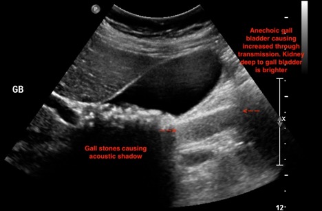
Increased Through Transmission: Tissues deep to an object appear more echogenic (brighter) than normal. This is very useful to diagnose a cyst.
Case courtesy of Associate Professor Natalie Yang, Radiopaedia.org, rID: 9874.
Image of gall bladder with reverberation artifact from tiny calcifications in gall bladder wall in this patient with adenomyomatosis. Ultrasound artifacts are often useful in making a specific diagnosis. Case courtesy of Dr Vikas Shah, Radiopaedia.org, rID: 65505.
Case courtesy of Dr Hani Makky Al Salam, Radiopaedia.org, rID: 14461.
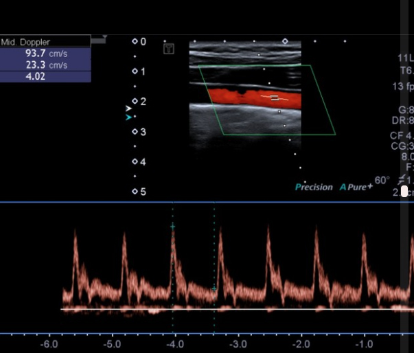
Doppler ultrasound
Moving objects (e.g., blood) within the sound beam shifts (increases or decreases) the frequency of the sound beam. This Doppler shift can be analyzed to give information regarding motion. This is particularly valuable to assess blood flow.
Safety
Ultrasound does not use ionizing radiation.
Color and pulsed doppler interrogation of the carotid artery. Attestation Case courtesy of Dr Bruno Di Muzio, Radiopaedia.org, rID: 54495.
Ultrasound has been used for more than 40 years, with extensive scrutiny. No adverse effects have been found. However, chromosome alterations have been induced in vitro at higher energy levels than are allowed by the FDA for imaging devices. The prudent practitioner will view ultrasound as very safe but limit exams to what is necessary.
Ideal patients are children and non-obese adults (although many severely obese patients image very well). Exams take a variable amount of time depending on the exam, typically 30–120 minutes.