

- Key definitions
Learning is a neural mechanism by which the organism’s behavior changes because of an experience or stimulus.
Memory is the storage mechanism for what is learned.
Normally, once memory information is encoded, it is no longer dependent on the hippocampus for retrieval.
Memory involves distinct brain regions
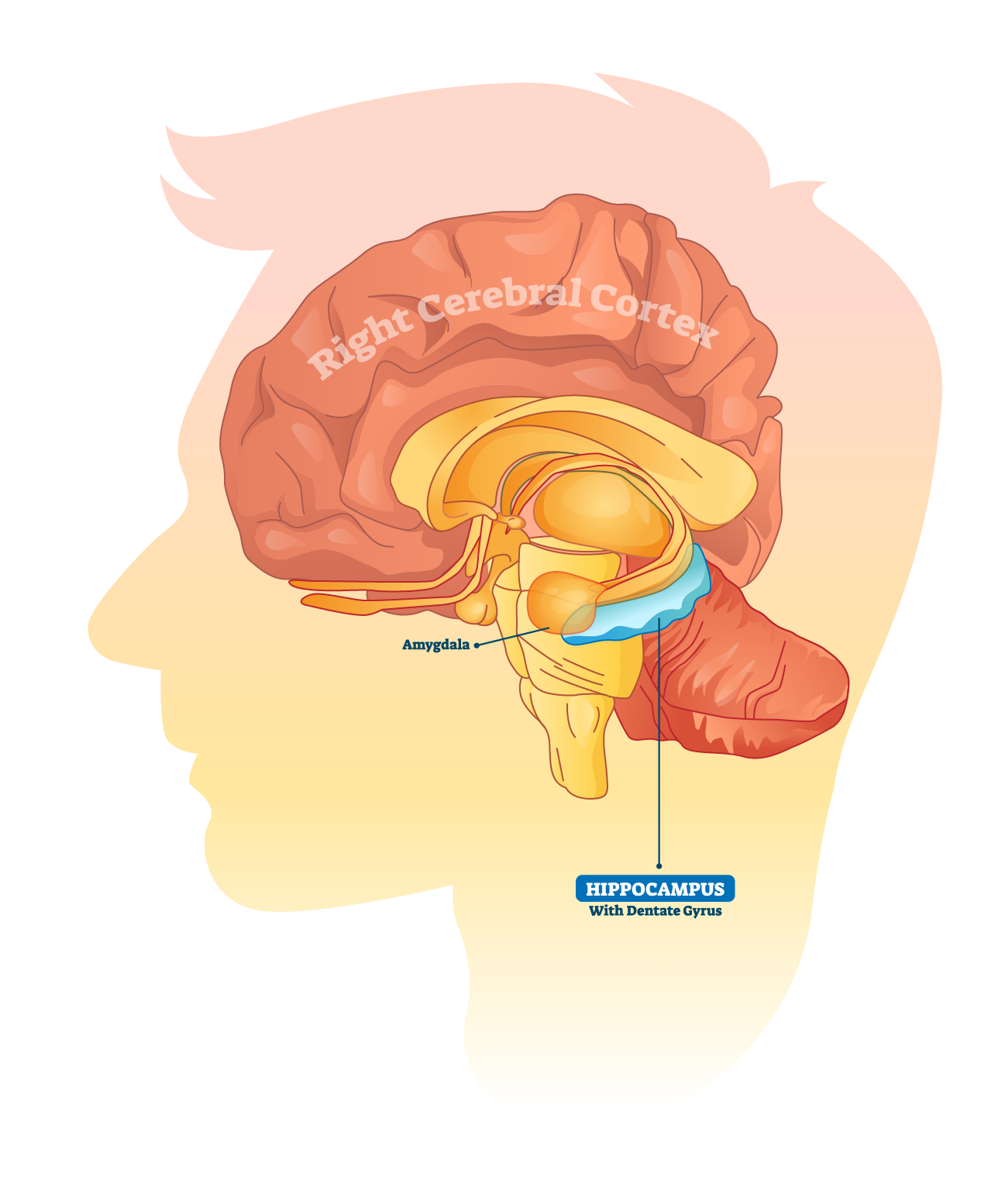
Hippocampus
Critical for encoding and consolidating explicit (declarative) memories such as facts and events.
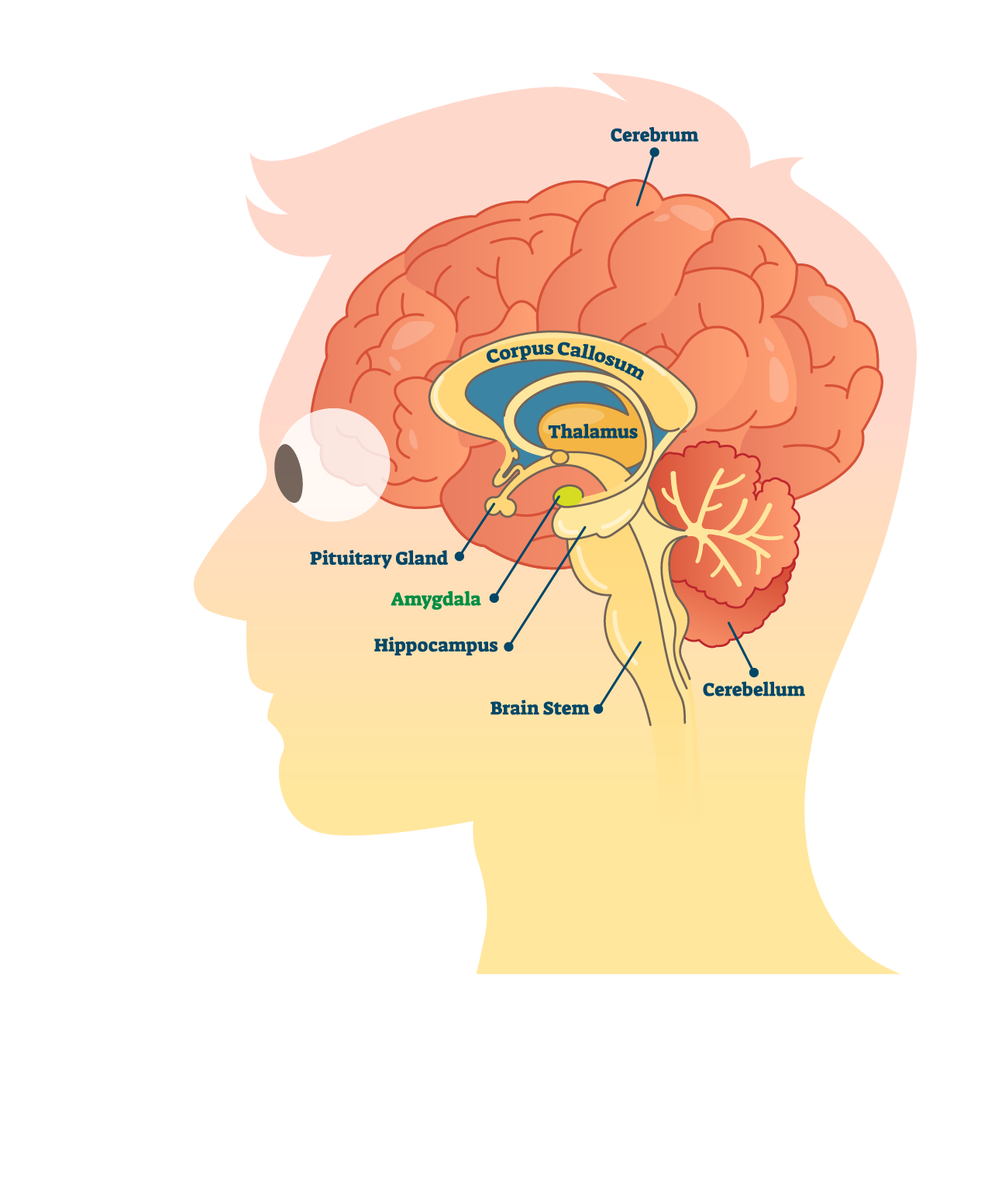
Amygdala
Adds emotional significance to memories, enhancing their strength and likelihood of retention.
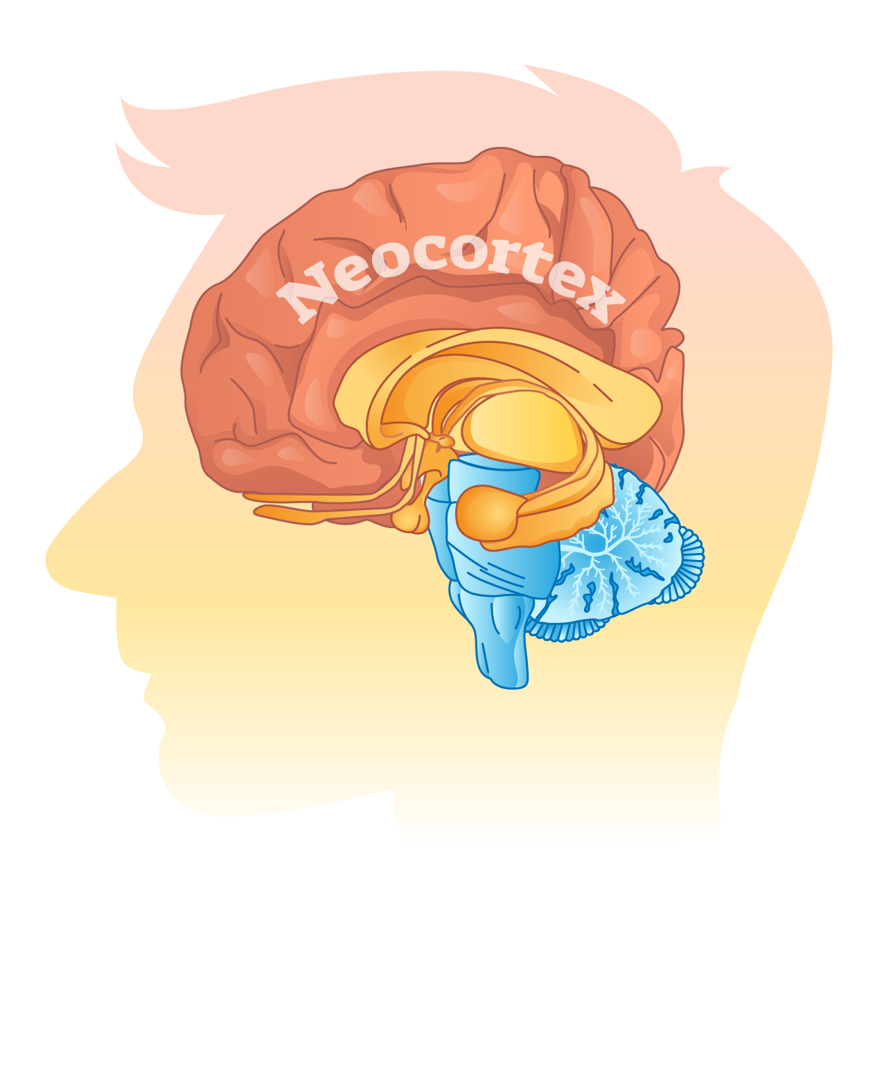
Neocortex
Becomes the primary site for long-term storage after consolidation.
Types of memory and their neural correlates
Working memory
Involves transient storage and manipulation of information.
Supported by prefrontal cortex and hippocampus.
Explicit memory
Includes episodic (personal experiences) and semantic (facts and concepts).
Relies on hippocampus, entorhinal, and parahippocampal cortices.
Implicit memory
Involves learned motor skills and procedures.
Supported by basal ganglia, cerebellum, and motor cortex.
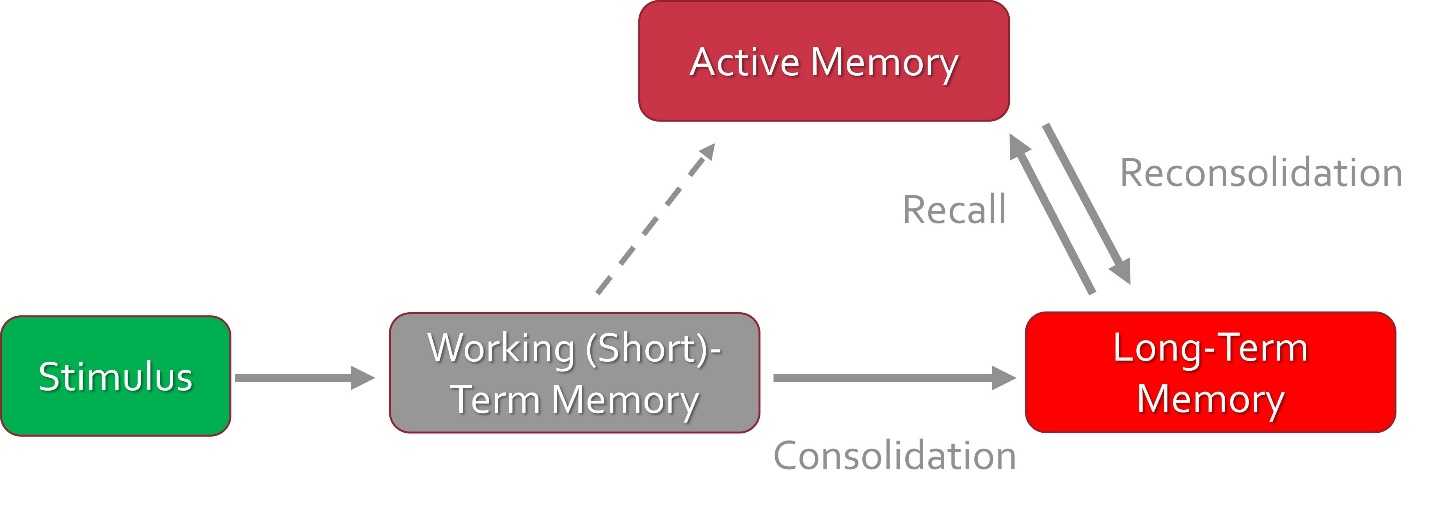
Consolidation transfers memory from hippocampus to cortex
-
Novel information is encoded in the hippocampal formation and then relayed to association areas of the cortex.
-
Over time, retrieval becomes independent of the hippocampus, relying more on neocortical regions.
-
Long-Term Potentiation (LTP) at synapses is the cellular mechanism underlying consolidation.
Functional pathways supporting memory
Table 1. Key Brain Regions in Memory.
|
Brain Region |
Function |
|
Hippocampus |
Encodes and consolidates explicit memory |
|
Amygdala |
Tags emotional significance to memories |
|
Fornix |
Connects hippocampus to mammillary bodies |
|
Mammillary Bodies |
Relay memory signals to thalamus |
|
Thalamus |
Projects to cingulate gyrus for emotional processing |
|
Cingulate Gyrus |
Integrates emotion and behavior |
|
Neocortex |
Long-term memory storage |
|
Basal Ganglia |
Supports procedural memory |
|
Cerebellum |
Coordinates motor memory |
Clinical correlations highlight regional importance
Clinical correlation
Bilateral hippocampal damage leads to anterograde amnesia, affecting the ability to form new declarative memories.
Clinical correlation
Alzheimer’s disease prominently affects episodic memory, due to degeneration in the hippocampus and medial temporal lobe.
Amnesic patient H.M. provided compelling evidence that normal memory function depends on a properly functioning medial temporal lobe.
H.M. had a bilateral medial temporal lobe resection performed for severe epilepsy.
This patient's report was significant because:
-
- It informed neurosurgeons that a bilateral lesion of medial temporal lobe structures placed recent memory functions at risk.
- It suggested that the establishment of memory had a distinct neural substrate memory for new experiences was disturbed, but other cognitive functions and sensory capacities were mostly unimpaired.
- It demonstrated that medial temporal lobe structures are critical for establishing long-term explicit/declarative memory.
This was all conjecture based on surgical reports—it wasn’t until MRI images were taken in the 1990s that showed the extent of the damage for a definitive understanding of medial temporal lobe functional anatomy (see image below).
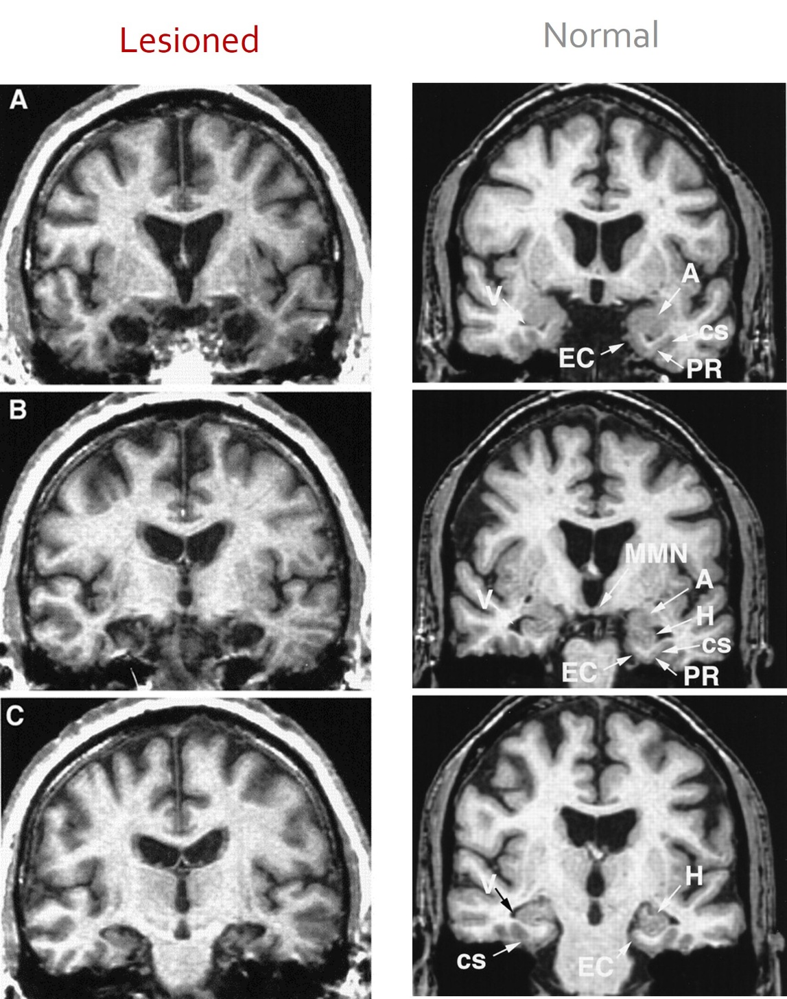
Key anatomic features on coronal MRI: (left) H.M. and (right) a 66-year-old control patient.
EC: Entorhinal cortex
A: Amygdala
V: Lateral ventricle (inferior horn)
H: Hippocampal formation
Table 2. Lesions to Brain Regions and Effects on Memory.
|
Brain Region |
Lesion Effect |
|
Hippocampus |
Anterograde amnesia |
|
Amygdala |
Reduced fear and emotional responses |
|
Basal Ganglia |
Impaired procedural memory and motor skills |
|
Cerebellum |
Deficits in motor learning and coordination |
|
Thalamus |
Disrupted memory relay to cortex |
|
Medial Temporal Lobe |
Degeneration in Alzheimer’s affects episodic memory |
Review questions

Case study
Liliana presents with difficulty forming new declarative memories following a traumatic brain injury. Imaging reveals damage to the hippocampus. Which of the following best explains the functional consequences of this injury?
The hippocampus is essential for encoding and consolidating declarative (explicit) memories such as facts and events. Damage here impairs the formation of new memories.
Review
Which of the following best illustrates the interaction between emotional and autonomic responses in memory formation?
The amygdala integrates emotional significance and sends signals to autonomic centers, influencing physiological responses to emotionally charged memories.
Review
A neuroscientist is studying how memories transition from short-term to long-term storage. Which of the following pathways and mechanisms would be most relevant to investigate?
The Papez circuit supports memory consolidation, and LTP is the cellular mechanism that strengthens synaptic connections, facilitating the transfer of memory from the hippocampus to the neocortex.
Image credits
Unless otherwise noted, images are from Adobe Stock.