Neuroanatomy lab navigation
Meninges
See Figure 4.1 and Figure 4.2.
- Pia mater
- Arachnoid mater
- Dura mater
- Falx cerebri: Separating the cerebral hemispheres
- Tentorium cerebelli: The tentorium cerebelli separates the cranial cavity into two compartment (Figure 4.1), and there is also a prominent tentorial notch that allows for passage of the brainstem
- The supratentorial compartment contains the cerebrum.
- The infratentorial compartment contains the brainstem and cerebellum.
The brain and spinal cord are covered by the dura mater, arachnoid mater, and pia mater. The periosteal and meningeal layers of the dura are separated as the dural venous sinuses (e.g., superior sagittal sinus). Small tufts of arachnoid tissue, the arachnoid villi, project into the superior sagittal venous sinus (Figure 4.2).
The space between the arachnoid and pia mater is the subarachnoid space (Figure 4.2); it is enlarged surrounding CNS structures as cisterns.
Note
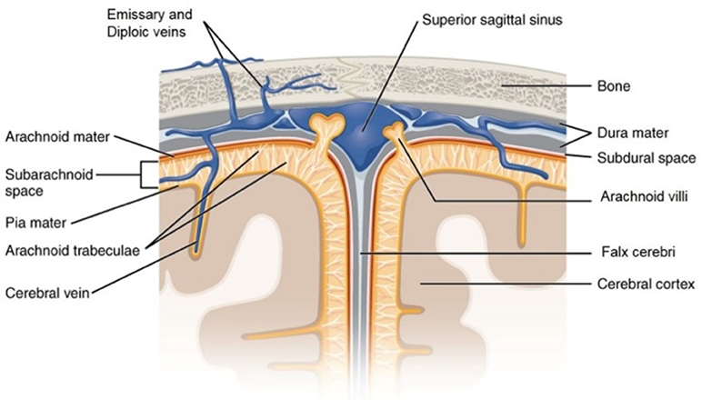
The meningeal spaces in the cranial cavity are only present between the periosteum and dura (epidural space) and dura and arachnoid (subdural space) in pathological conditions, like hematomas or abscesses. The only “real” space is the subarachnoid space that contains CSF.
Thought question
Soft tissue structures may herniate inferior to the falx cerebri or through the tentorial notch. A mass that is supratentorial can force the brain into the tentorial notch, trans-tentorial herniation. What are signs and symptoms of a trans-tentorial herniation?
- Uncal herniation
Uncal herniation can occur when a mass in the temporal lobe pushes the uncus through the tenorial notch and into the brainstem.
-
- Identify the region of the brainstem that could be affected.
- Predict signs and symptoms associated with cranial nerve damage.
Tearing of the bridging veins that connect into the superior sagittal sinus, often times accompanying a fall, can result in a subdural hemorrhage/hematoma. See Figure 4.4.
Thought question
Why does the spread of a subdural hematoma (indicated by the blue arrow) appear more diffuse on a CT or MRI than an epidural hematoma?
Meningeal spaces
See Figure 4.2.
- Epidural
- Subdural
- Subarachnoid
- Cisterns are the naturally enlarged subarachnoid spaces where CSF collects. (Figure 4.5)
- Chiasmatic (suprasellar) (anterior to the optic chiasm)
- Interpeduncular cistern (anterior to the midbrain)
- (Pre)pontine and (pre)medullary (anterior to the pons and medulla)
- Cisterna magna (large space located around the spinal cord–medullary junction surrounding the foramen magnum)
- Lumbar cistern (the subarachnoid space located from L3 to S2 after the termination of the spinal cord)
Thought question
Identify the ventricles on sagittal sections through the cerebrum and on MRI images.
Ventricles
See Figure 4.5–Figure 4.8, Interactive 4.1, and Review 4.2.
- Lateral (LV)
- Third (3V)
- Fourth (4V)
The shape of lateral ventricles changes from anterior to posterior
-
- Portions of the lateral ventricle include: the anterior horn, body, atrium, posterior horn, and inferior horn
- Septum pellucidum: The midline membrane separating the two lateral ventricles
- Interventricular foramen (foramen of Monro) opening into the 3V from the LV
- Cerebral aqueduct (Sylvian aqueduct); the inferior exit point of the 3V
- Lamina terminalis (anterior border of 3V): red arrow in Figure 4.6.
- Formed by a depression in the rhomboid fossa (Figure 3.2)
- One median foramen (Magendie) and two lateral foramina (Luschka) exit from the 4V into the subarachnoid space. These openings are often difficult to visualize.
Note
Within all the ventricles are the choroid plexuses, which are producing cerebrospinal fluid.
The ventricular system, as it relates to development, are discussed in the first large-group session prior to the these labs. Review the clinical significance of the flow and blockage of CSF.
For further reference, see The Nervous System, Volume 2: The Meninges and Ventricles.
Flow of cerebrospinal fluid
CSF is produced in all of the ventricles by choroid plexuses (w/ a total volume of ~25 ml in all the ventricles); the MRI in Figure 4.8 indicates the direction of flow of cerebrospinal fluid between the ventricles.
- Flow of cerebro-spinal fluid
CSF flows freely between the two LVs and 3V, via the interventricular foramen; from the 3V through the cerebral aqueduct into the 4V; finally out into the subarachnoid space (the direction CSF flow is indicated by the yellow arrows).
Superior sagittal sinus w/ arachnoid villi (granulations)
The arachnoid villi are the locations for the re-entry of the majority of the cerebrospinal fluid into the venous circulation (Figure 4.9).
