The basal subdivision of the brainstem is the most anterior and contains mostly the descending fiber tracts.
The tegmentum is the location of the cranial nerve nuclei, ascending fiber tracts, and the reticular formation. In the medulla, it also contains important neurons that are involved in cardiovascular and breathing reflexes.
The tectum, which is posterior to the tegmentum, is really only prominent in the midbrain and is grossly represented by the superior and inferior colliculi. It contains the nuclei that are involved in vision, hearing, and reflexive control of pupillary constriction. Tectospinal tracts originating in the tectum supply the postural muscles that directly initiate reflexive movement to auditory and visual stimuli.
Brain stem vascular lesions lead to multiple signs that may seem unrelated but can often be localized to a specific brain stem region. Knowledge of the brain stem anatomy will help with the localization. Examples of this will be presented in the functional neuroanatomy and circulation lecture.
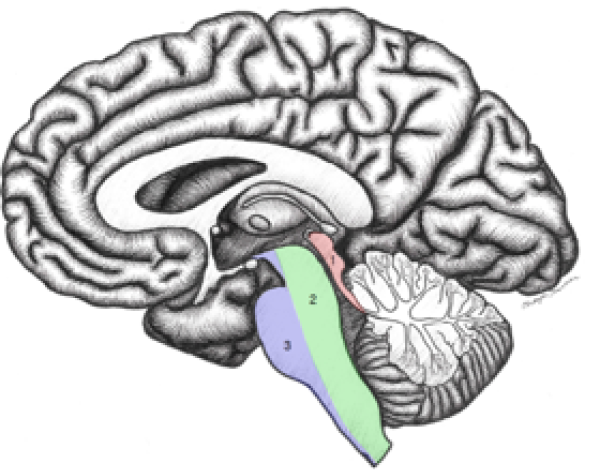
Figure 3.1. Subdivisions of the brainstem; mid-sagittal plane. Three subdivisions of the brainstem:
Tectum
Tegmentum
Basal region
Figure from Neuroanatomy: A Laboratory Guide (2e); Jansen and Lampa (2018).
Review 3.2.
Subdivisions of the brainstem; mid-sagittal plane. (Tap the right arrow for labels)
Specimen from Neuroanatomy Collection, Washington State University College of Veterinary Medicine.
Check yourself
On a sagittal image of a brainstem, locate the tegmentum and tectum (Figure 3.1 and Review 3.2).
The general regions of the brain stem will be a review from FMS 501. The portions of the brain stem and their specific important nuclear groups and tracts contained within will be discussed in the Functional Neuroanatomy and Circulation LGA sessions.
Utilize the external topography of the brain stem to locate internal structures as you a beginning to learn the cross sectional and and the related significance to lesion localization.
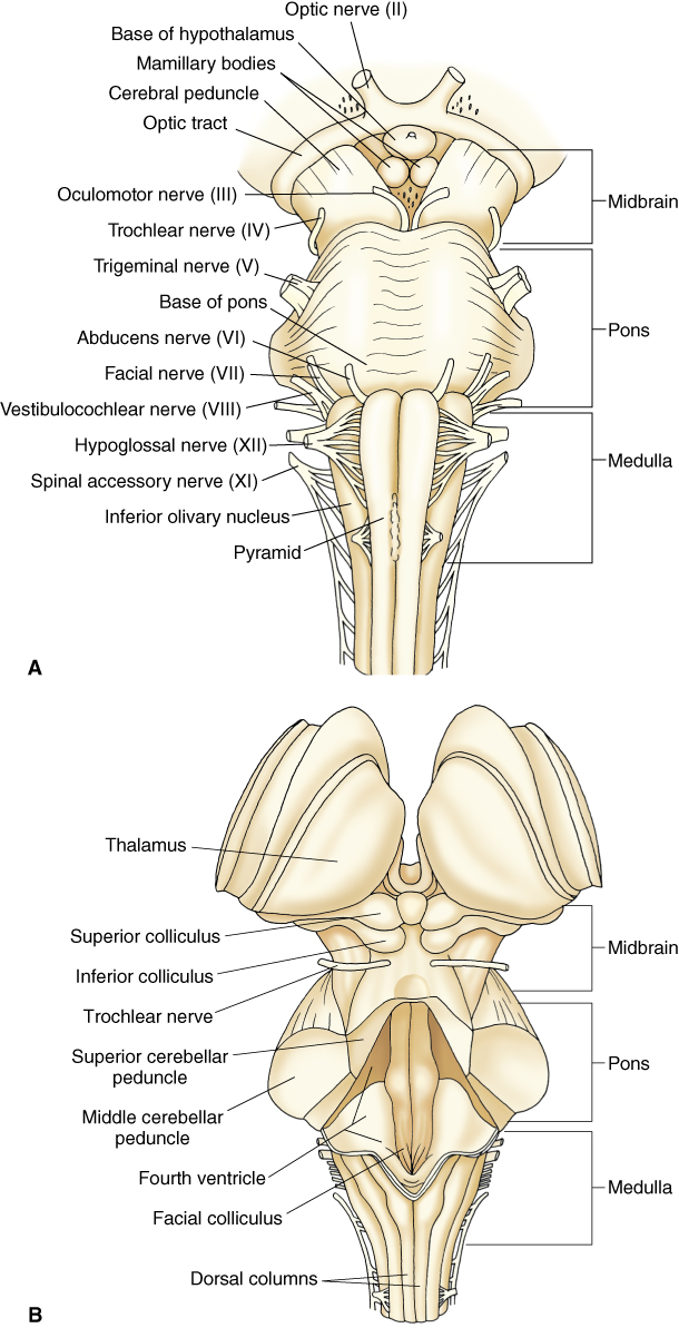
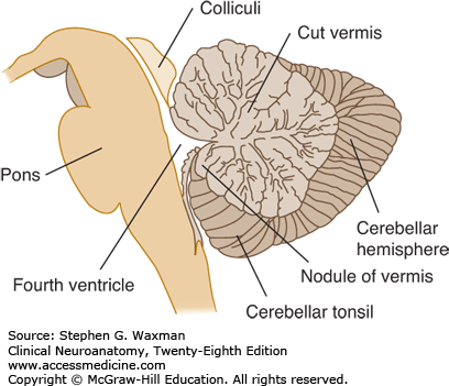
Examine the external topography of the brainstem and cerebellum and identify the following structures (Figure 3.2 and Figure 3.3):
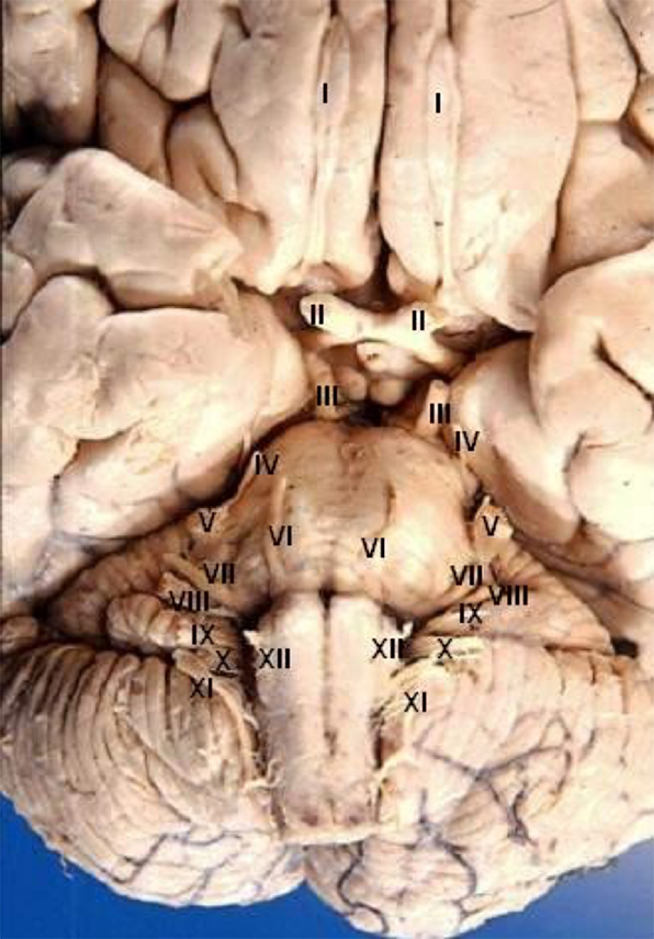
Note the exit points of the cranial nerve axons from medial to lateral on the brainstem (Figure 3.2 and Figure 3.4).
Cerebellopontine angle tumors
The cerebellopontine angle is located laterally at the junction of the caudal pons and cerebellum. A patient with a tumor in this area would have what signs or symptoms related to cranial nerve dysfunction?
Thought question Describe a common benign neoplasm that could affect structures in this region.
Tonsillar herniation
The cerebellar tonsil may herniate into the brainstem, leading to significant mortality with increases in intracranial pressure.
Figure 3.4. Inferior surface of the cerebrum and brainstem with cranial nerves. (Note the attachment points of Cranial Nerves I–XII to the cerebrum, diencephalon, and brainstem.) Wikimedia.org.
Table 3.1. Review of cranial nerves: Numbers, names, and basic functions
Number
Name
Basic function (afferent or efferent)
I
Olfactory
Transmits smell to CNS
II
Optic
Transmits visual information to CNS
III
Oculomotor
Innervates inferior rectus, superior rectus, levator palpebrae superioris, medial rectus and inferior oblique; visceral motor (parasympathetic [PS] to sphincter pupillae and ciliary muscle)
IV
Trochlear
Innervates the superior oblique muscle
V
Trigeminal
Ophthalmic
Maxillary
Mandibular
Transmits somatic afferent sensory information from the face, cornea, and anterior portion of the tongue; innervates muscles of mastication, palate, and middle ear (Mandibular n.)
VI
Abducens
Innervates lateral rectus muscle
VII
Facial
Transmits taste from anterior portion of the tongue; provides motor innervation to muscles of the face; supplies visceral motor (PS) information to mucous glands in nasal and oral cavities, salivary glands, and lacrimal gland
VIII
Vestibulocochlear
Transmits hearing and balance information from the inner ear
IX
Glossopharyngeal
Transmits taste and general sensations from posterior portion of the tongue and pharynx, innervates pharyngeal muscles, supplies visceral motor information to the parotid salivary gland
x
Vagus
Transmits sensations from larynx and pharynx, supplies visceral motor information to thoracic and abdominal viscera to descending colon; supplies innervation to laryngeal and pharyngeal muscles
XI
Accessory
Innervates trapezius and sternocleidomastoid; innervation to laryngeal muscles via vagus
XII
Hypoglossal
Innervates muscles of the tongue
Image credits
Unless otherwise noted, images are from Adobe Stock.