New—Lab 1. Cerebrum, sulci, and gyri

Steve: The scalpel icon below is an example of how we indicate exactly what the students should be doing in their dissection. I know this will be more like a prosection, but if there are specific actions that students should take, this would be a good way to indicate it. It also feeds a supplemental table of contents (it comes up on the left side of the screen), so the instructions are easy to find.

Dissection instructions: Clean off and identify branches of the internal iliac artery

Introduction to neuroanatomy

Figure 1.1. Anatomic directional terms. From Neuroanatomy: A Laboratory Guide (2e); Jansen and Lampa.

Watch this excellent Introduction to the Central Nervous System from University of British Columbia Neuroanatomy.

Cerebrum, sulci, and gyri

Lobes of the cerebrum

The limbic (limbus=margin or border L.) lobe is sometimes included as a functional lobe, but is in fact comprised of the medial portions of the frontal, parietal, and temporal lobes.

The surfaces of the cerebral hemispheres, and therefore the lobes, have a distinct topography that is formed by the sulci and gyri.

Sulcus (pl. sulci)
A groove or furrow (L.)
Gyrus (pl. gyri )
A ridge or convolution (L.)
Figure 1.2 Left cerebral hemisphere dissected to expose insula. Specimen from Neuroanatomy Collection, Washington State University College of Veterinary Medicine. The insula is outlined in yellow on the above image. (Note: inferior portions of the frontal and parietal lobes have been removed.) The insular cortex has many functions, such as emotions, motor control, and homeostasis.

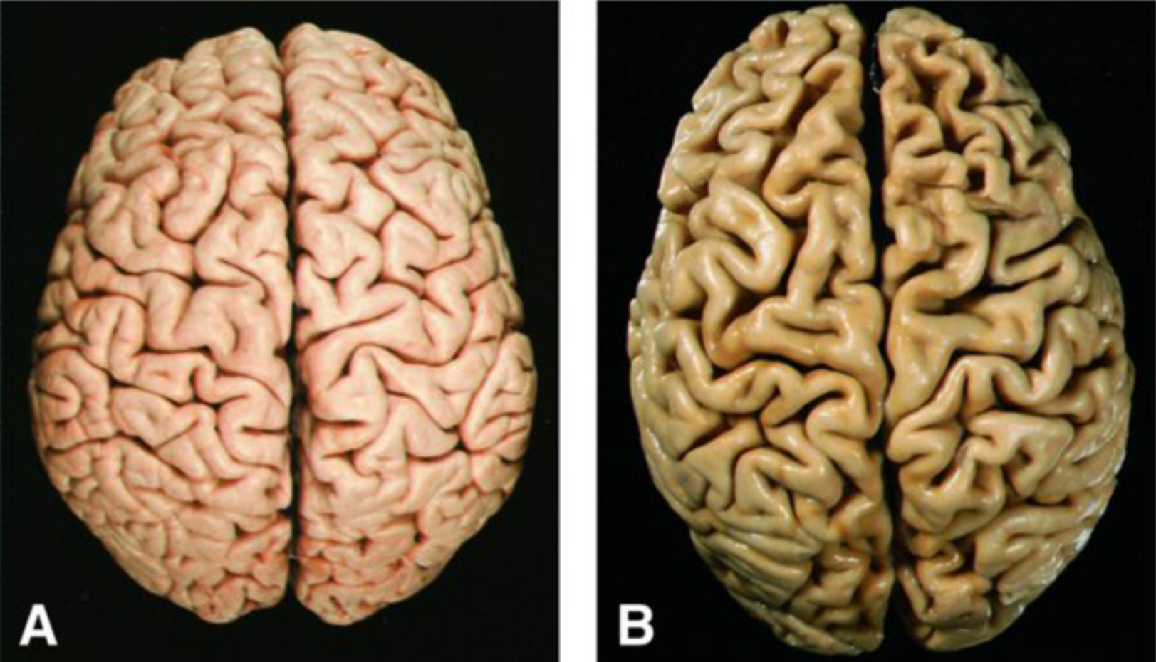
There is a loss of cortical tissue leading to narrowing of the gyri, widening of the sulci, or both in old age and disease processes. See Figure 1.3. The brain in panel A is from a normal patient, and the brain in panel B is from an older patient experiencing a loss of cortical tissue.

Thought question

List some common neurologic disorders that could lead to a loss of cortical tissue, as seen in Figure 1.3B.

Figure 1.3. From Chapter 32: The Central Nervous System, Rubin’s Pathology: Clinicopathologic Foundations of Medicine, 7e; Strayer et al. 2014; Wolters and Kluwer.

The cerebral cortex controls or receives information from the contralateral side of the body (Valsalva Doctrine). Therefore, unilateral cerebral lesions produce signs and symptoms on the opposite side of the body.

Clinical correlation

All the regions of the cortex perform different and diverse functions. Understanding the localization and nature of focal injuries to the cortex as it relates to specific clinical symptoms is an extremely important role of any clinician.

Sulci/fissures of the cerebrum

  • Longitudinal, a.k.a. sagittal fissure/sulcus
    • Separating the two cerebral hemispheres
  • Lateral sulcus (Sylvian fissure)
    • Separating temporal lobe from parietal and frontal lobe
  • Parieto-occipital sulcus
    • Separating the parietal and occipital lobes (seen medially)
  • Central sulcus
    • Separating the frontal and parietal lobes
  • Precentral sulcus
    • Anterior to the precentral gyrus
  • Postcentral sulcus
    • Posterior to postcentral gyrus
  • Cingulate sulcus
    • Dorsal (superior) to the cingulate gyrus
  • Calcarine sulcus—on medial surface of occipital lobe
    • Superior and inferior temporal sulci

Major gyri of cerebral lobes

  • Frontal lobe
    • Precentral gyrus
    • Superior, middle and inferior frontal gyri
    • Cingulate gyrus (superior to the corpus callosum)
  • Parietal lobe
    • Postcentral gyrus
    • Superior parietal lobule
    • Inferior parietal lobule (supramarginal and angular gryi are parts of inferior parietal lobule)
  • Occipital lobe
    • Lingual gyrus (inferior to the calcarine sulcus)
    • Cuneus (posterior to the parieto-occipital sulcus and superior to the calcarine sulcus)
  • Temporal lobe
    • Superior, middle and inferior temporal gyri
    • Occipito-temporal (fusiform) gyrus
    • Parahippocampal gyrus
    • Uncus
  • Insula (Figure 1.2 and Interactive 1.5)—(deep in the lateral fissure); composed of multiple gyri

Clinical correlation

From the perspective of a clinican, the anatomy of brain and spinal cord are thought of in terms of how they appear in sections or slices that are obtained by either CT or MRI imaging techniques. 

Coronal sections provide an excellent way of visualizing the anatomy of cerebrum. Topographic features that can be visualized include the depth of the sulci, differences in the gray and white matter, and the parts of the ventricular system (Interactive 1.5). Pathologic changes to neuronal tissue and the extent of masses can be noted using these planes of section.

Axonal fiber bundles of the cerebrum

Classified as commissural, association, projection fibers.

Locate the following axonal fiber bundles in the cerebrum, which can serve as good landmarks.

    • Commissural fibers connect the two hemispheres
    • Association connect within the same hemisphere
    • Projection from cortex to brainstem or spinal cord
      • Corpus callosum: A large bundle of commissural fibers located inferior to the cingulate gyrus
        • Consists of a rostrum, genu, body, and splenium
      • Anterior commissure: A small bundle of commissural fibers that connects inferior portions of the temporal lobes
      • Fornix: Association fibers which provide the major connection between the hippocampus and hypothalamus/thalamus. (This is a very important fiber tract in learning and memory and will discussed during the Limbic System Lecture.)

Check yourself 1.6.

Fiber bundles of cerebrum.
 Specimen from Neuroanatomy Collection, Washington State University College of Veterinary Medicine. (Tap the right arrow for labels)

Table of Contents
Headshot of Steve Lampa, PhD · Clinical Associate Professor
Steve Lampa
PhD · Clinical Associate Professor
envelope icon