Basilar (unpaired)—multiple pontine branches also come directly off the basilar artery
Anterior inferior cerebellar
Superior cerebellar
Posterior cerebral posterior communicating
Internal carotid
Supplying the midbrain, thalamus, hypothalamus, and cerebrum
Middle cerebral
Anterior communicating
Anterior cerebral
An episode of vascular insufficiency to neurons leads to a stroke.
Ischemic strokes are sudden blockages of blood flow to parts of the CNS are most commonly caused by a thrombus or an embolus. Clinical presentations of vascular insufficiency often present as specific functional losses.
Interactive 5.1
Color and label the different vessels forming the circle of Willis. (Tap to open; use your Apple Pencil to draw.)
From Neuroanatomy: A Laboratory Guide (2e); Jansen and Lampa.
Review 5.2.
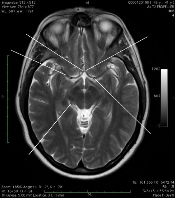
Axial MRI of cerebral circulation.
ACA: Anterior cerebral artery
ACoA: Anterior communicating artery
ICA: Internal carotid artery
MCA: Middle cerebral artery
PCA: Posterior cerebral artery.
(Tap the + for labels)
ACoA
AcoA
MCA
MCA
PCA
PCA
ACA
ACA
ICA
ICA
Image from Neuroanatomy Image Archive, Washington State University College of Veterinary Medicine.
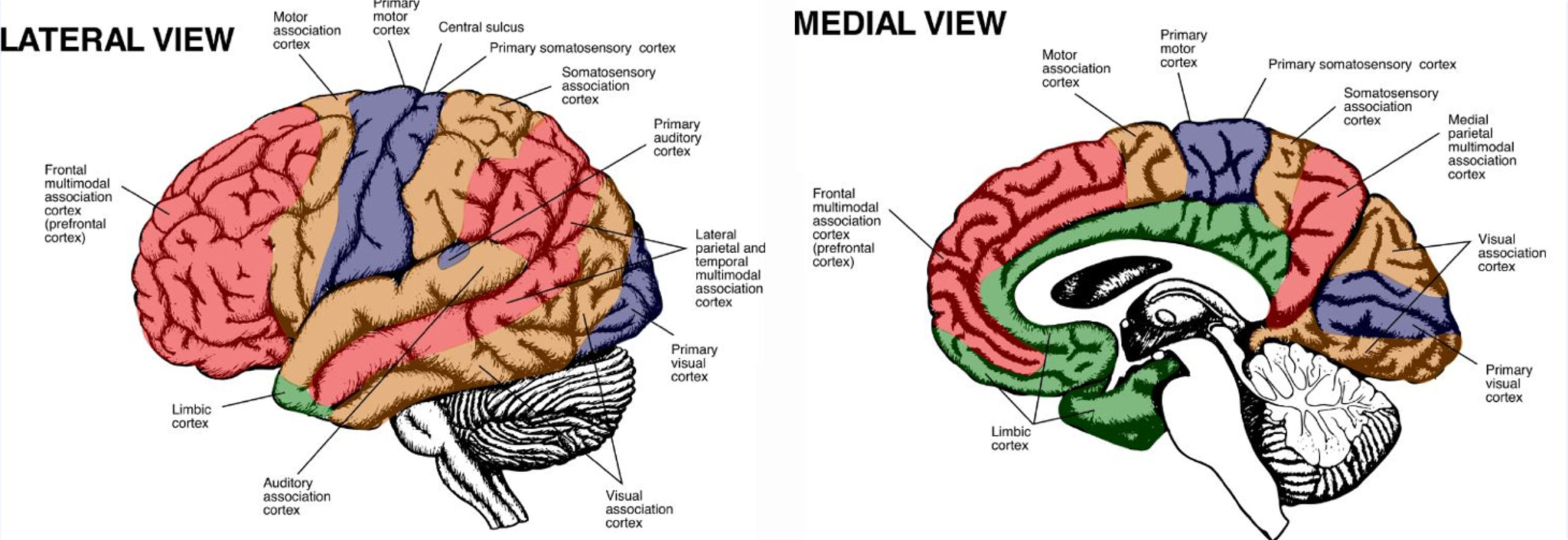
Figure 4. Lateral and medial views of functional areas of the cortex. From Neuroanatomy: A Laboratory Guide (2e); Jansen and Lampa.
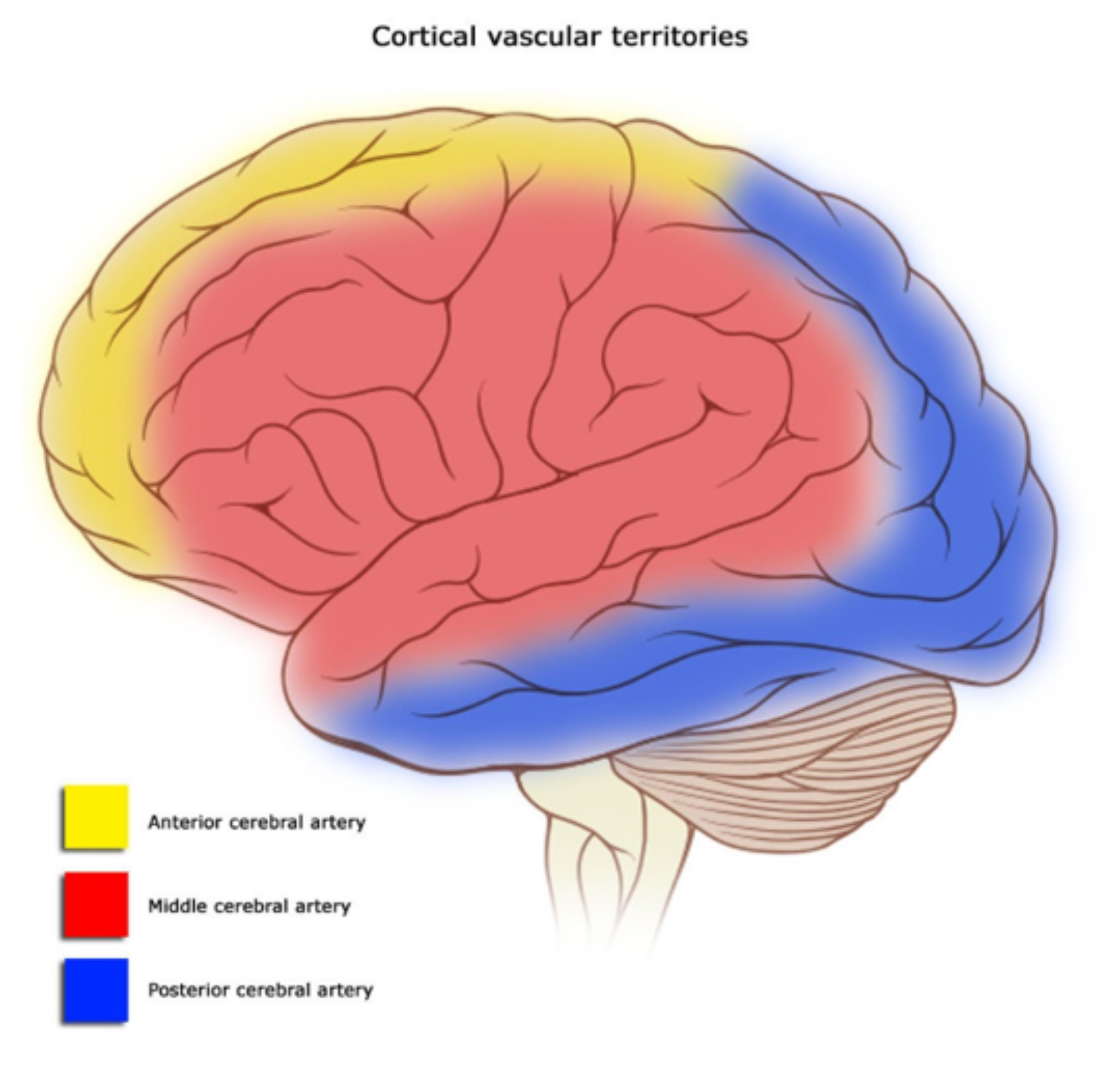
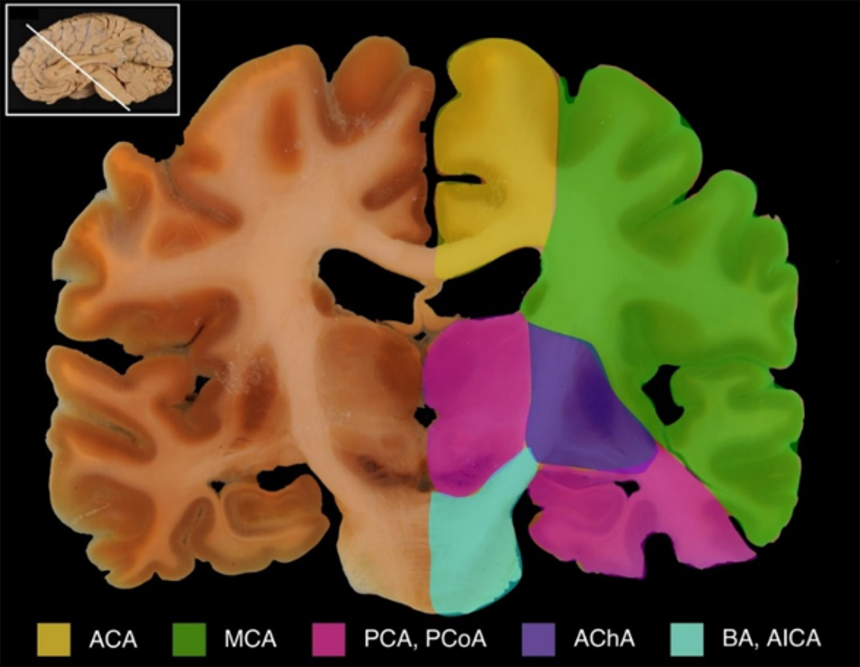
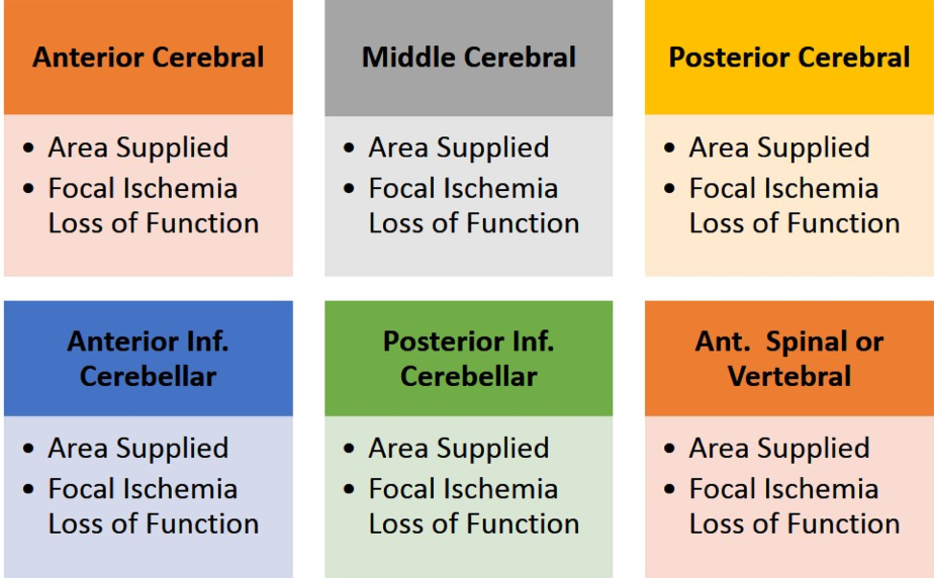
Figure 5. For each of the arteries listed here, demonstrate on a brain specimen or diagram on a whiteboard to describe the area supplied by each artery.
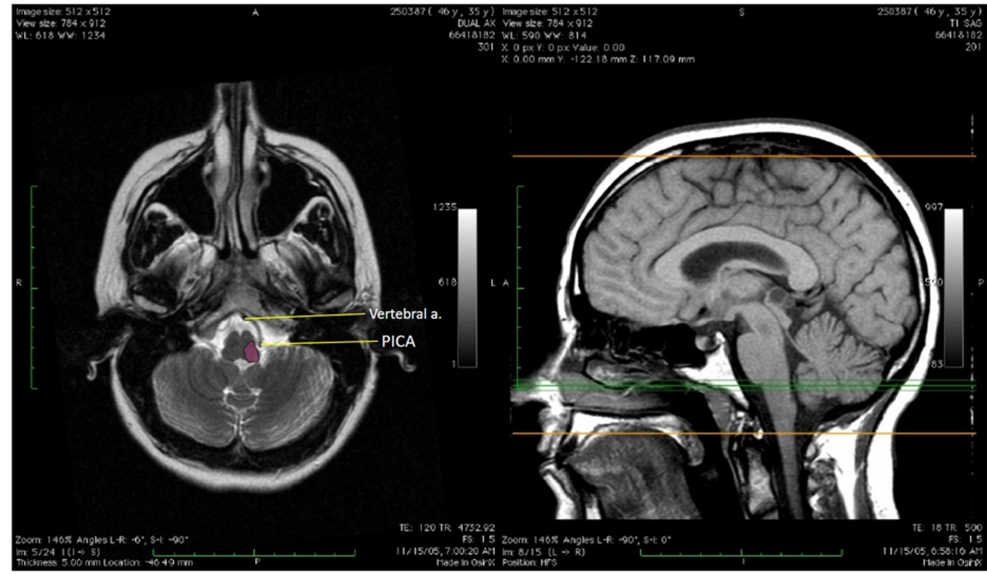
Vertigo, loss of pain and temperature from the limbs and trunk on contralateral side, loss of pain and temperature over the face on the ipsilateral side, truncal ataxia, dysphagia/dysphonia/palatal paralysis, Horner Syndrome
Inferior cerebellar peduncle
Ataxia (limb and truncal)
Vertebral artery*
Medial medulla, anterior spinal cord
Contralateral hemiparesis; touch, vibration and positional sense loss; CN XII sign
Note: Many cerebellar signs are very similar if superior cerebellar, anterior inferior cerebellar, and posterior inferior cerebellar arteries are occluded.
*Paramedian branches of the vertebral-basilar (both short and long)
**Circumferential branches of the vertebral-basilar (long)
Venous circulation
The veins of the cerebrum drain into dural venous sinuses, which mostly drain into the internal jugular vein.
Cerebral veins
Great cerebral vein (of Galen): Deep venous drainage
Formed by internal cerebral veins which drain the diencephalon and basal ganglia
Superior cerebral veins (numerous): Superficial venous drainage drains into the superior sagittal sinus
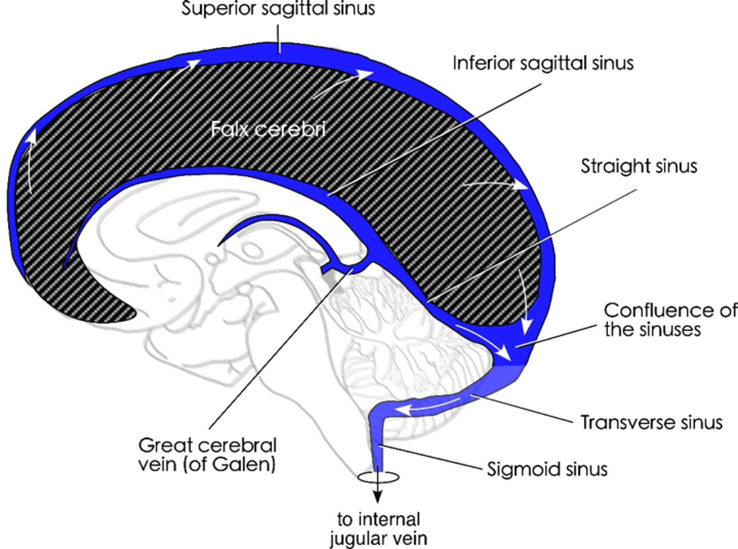
Figure 5.6. Venous sinus schematic. Figure from Neuroanatomy: A Laboratory Guide (2e); Jansen and Lampa (2018).
Review of dural venous sinuses (see Figure 5.6 and Figure 5.7); review FMS 501 notes
Superior sagittal sinus (located superior marginal of falx cerebri)
Superior cerebral veins drain into the superior sagittal sinus
Contains arachnoid villi where the CSF is re-enters the venous system
Straight sinus: Ends posteriorly in confluence of sinuses; receives blood from the great cerebral vein (of Galen) and inferior sagittal sinus
Confluence of sinuses
Transverse sinuses
Sigmoid sinuses
Jugular bulb (of internal jugular vein)
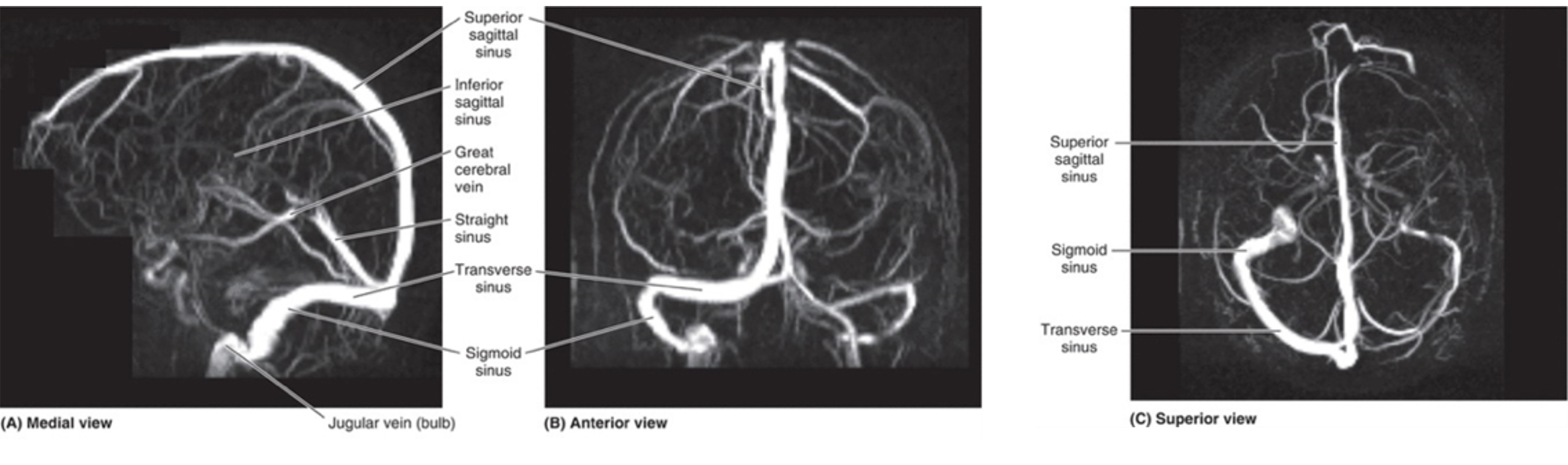
Figure 7. MR venogram of major venous sinuses. Figure from Clinically Oriented Anatomy, 8e, 2018. Accessed August 6, 2018.
Clinical significance of the dural venous sinuses
Besides returning blood from the nervous tissue of the cranial central nervous system, they are also potential routes for infections from more superficial vessels, as well as possible locations of venous thrombi. Signs from increased ICP result from occlusion of the dural sinuses.
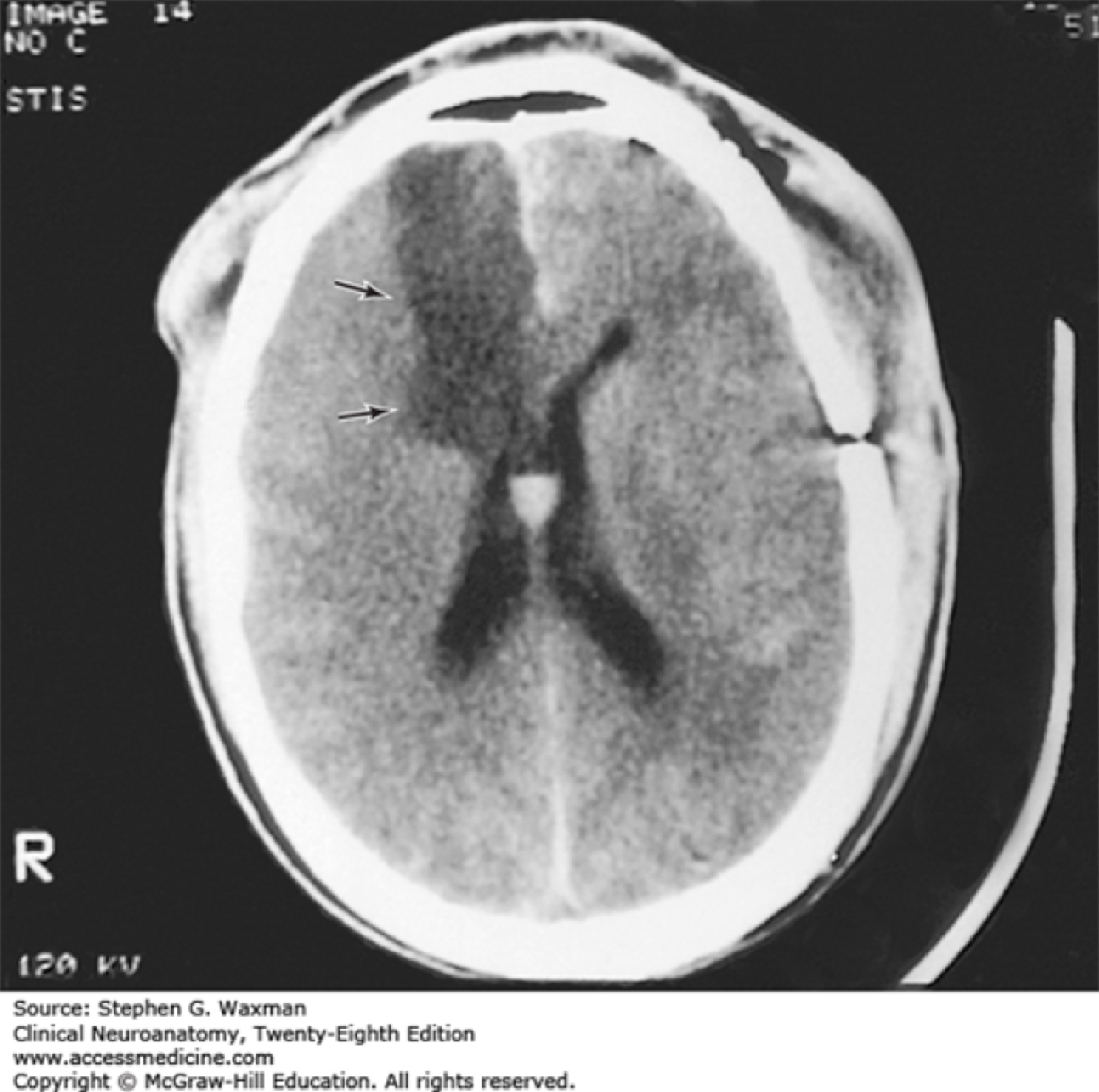
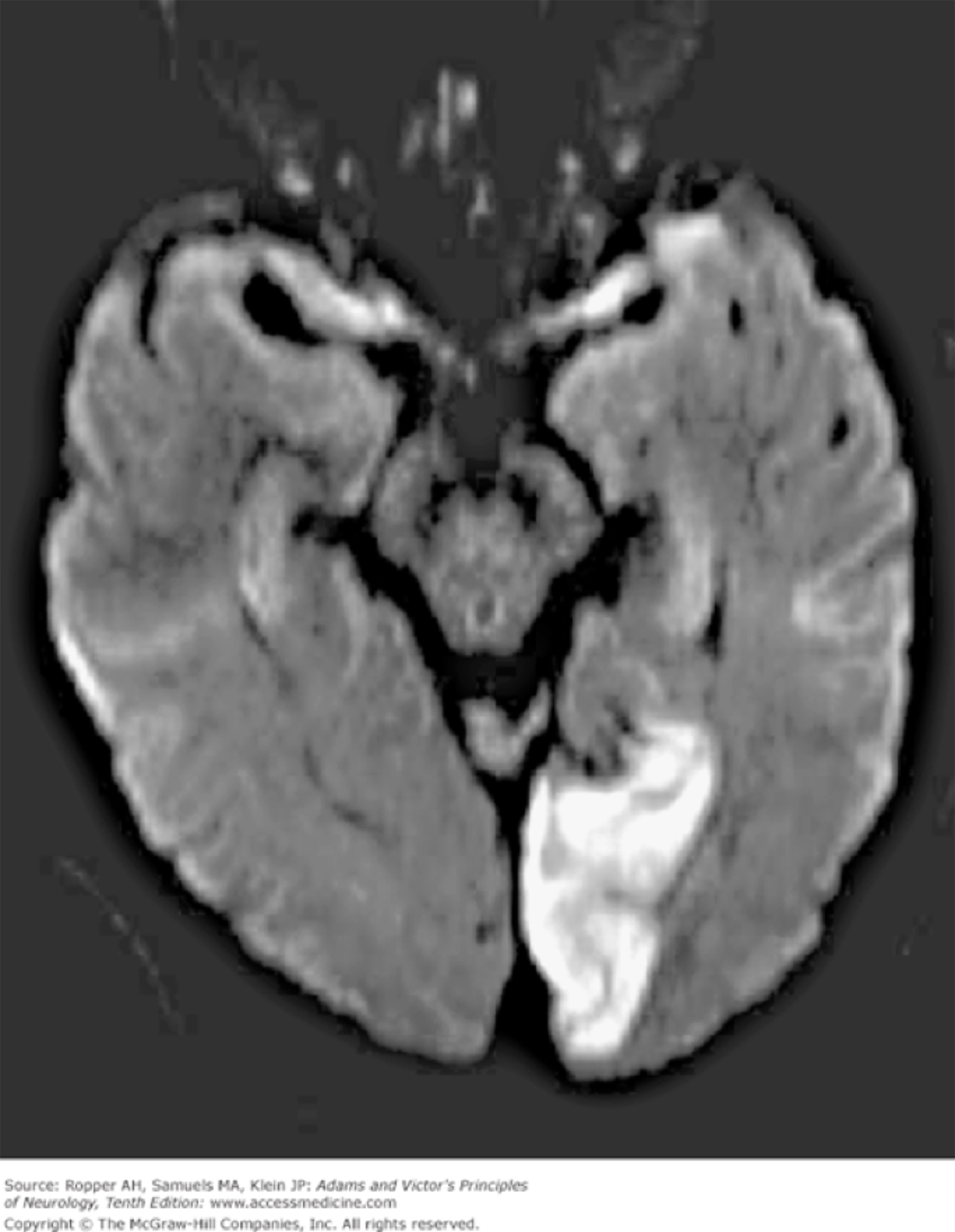
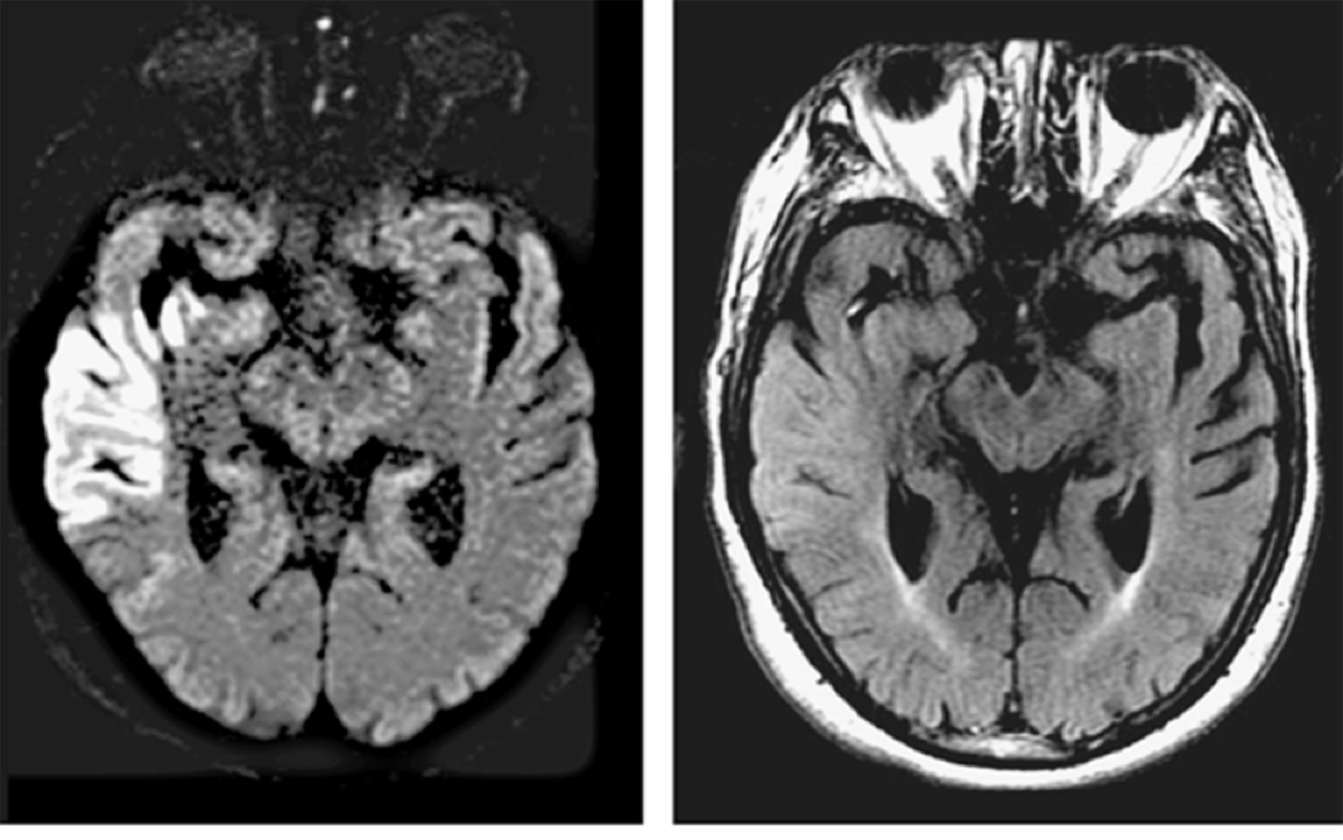
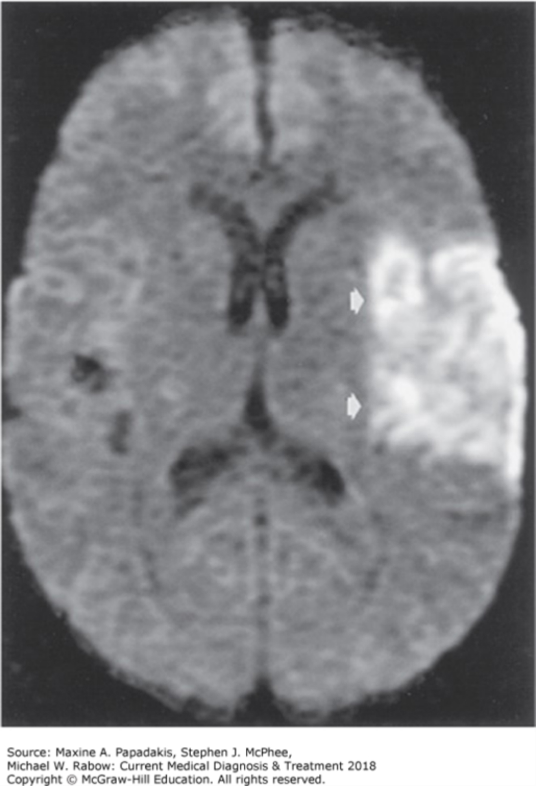
Appendix of Additional Stroke Images (MRI/CT)
For these images, try to come up with the signs/symptoms that a patient would present with.