

Neuroanatomy provides the essential framework for understanding the principles of neurology.
This lab is designed to help you build that foundation through hands-on exploration and clinical application.
Key focus areas
-
Topographic anatomy
Ventricular system, cerebrum, diencephalon, brain stem, and cerebellum
-
Three-dimensional relationships
Ventricular system and its connections to cerebral and brain stem structures
-
Clinical imaging
Using sectional anatomy and MRI to visualize and interpret these structures in context
-
Why it matters
A working knowledge of neuroanatomy is critical for accurate lesion localization, diagnostic reasoning, and applying neurological principles in patient care. By the end of this lab, you won’t just know where structures are—you’ll begin to appreciate the functional context of the content.
Instructions for this lab
What you are expected to do
Actively engage with all brain specimens, models, and images at each station.
Work collaboratively within your group to identify anatomic structures.
Use anatomic terminology when discussing structures.
Refer to the pages on the Medicine Digital Learning site to guide your learning.
Focus on structure and spatial relationships rather than the detailed clinical correlations.
Lab structure
- You will work in a group of approximately eight students.
- Your group will rotate through five stations during the lab period (~1 hour and 40 minutes).
- Remain at your assigned station until instructed to rotate.
- Manage your time efficiently so that all group members participate.
- Use the checklists provided to make sure you have identified all the relevant structures.
Faculty interaction
- Some stations are student-guided; use provided resources to guide your learning.
- Some stations are faculty-guided; listen carefully as key anatomical features will be emphasized or demonstrated.
- Goals
By the end of this lab, you should be able to identify major neuroanatomical structures, describe their three-dimensional relationships, and recognize these structures on sectional anatomy and MRI.
Station goals
Following the stations, you should be able to:
-
-
Meninges and ventricles
-
-
-
-
Identify the lateral, third, and fourth ventricles
-
Trace ventricular connections
-
Identify major meningeal layers
-
-
-
-
Cerebrum, sulci, and gyri
-
-
-
-
Identify major lobes
-
Locate key sulci and gyri
-
-
-
-
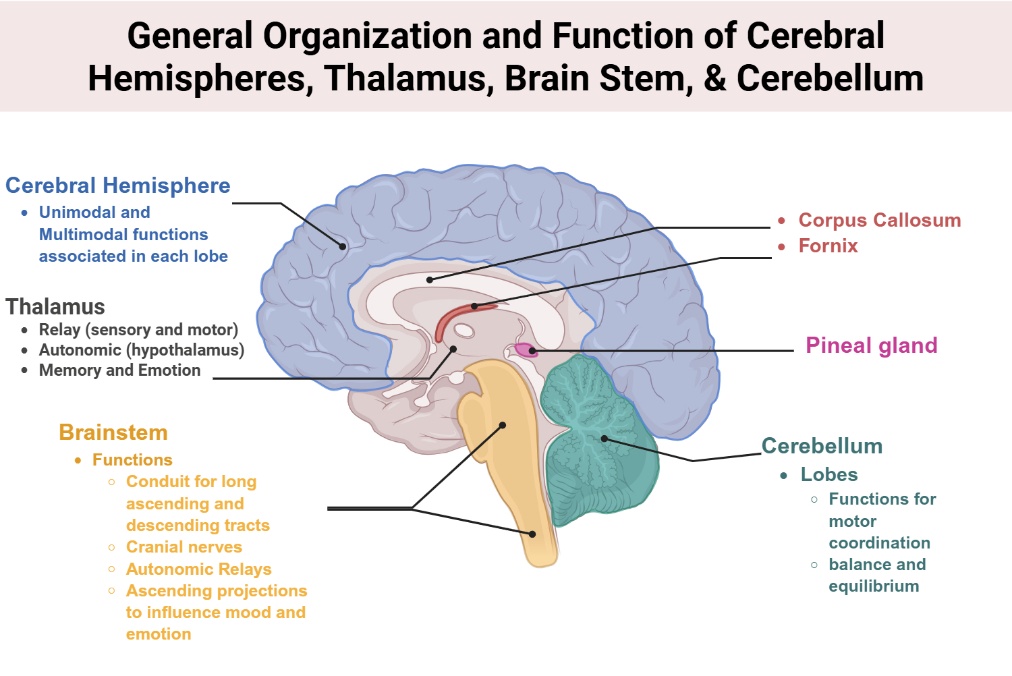
Diencephalon and brainstem
-
-
-
-
Identify midbrain, pons, and medulla structures
-
Locate the attachments of the cranial nerves and describe their general functions
-
Identify thalamus and hypothalamus and associated structures
-
Describe spatial relationships between these regions
-
-
-
-
Cross-sectional anatomy
-
-
-
-
Identify major structures on coronal sections
-
Correlate sectional anatomy with whole-brain views and MRI
-
-
-
-
Blood supply and circulation
-
-
-
-
Identify major arteries of the anterior and posterior circulations
-
Describe the territories supplied by Circle of Willis vessels
-
-
Key reminder
The functional and clinical significance of these structures will be addressed in large-group sessions. Your primary goal in this lab is for anatomical recognition and spatial understanding of the brain.
