Blood supply to the Central Nervous System
An understanding of the brain’s blood supply is crucial to understanding normal CNS function. Conversely, the consequences of cerebrovascular disease affecting the brainstem and cerebrum leading to a loss of function is also vital.
Two major arteries supplying branches to the brain, brain stem, cerebellum, and upper spinal cord are the internal carotid (ICA) and vertebral/basilar.
Interactive 3.1
Color and label the different vessels forming the circle of Willis. (Tap to open; use your Apple Pencil to draw.)
The cerebral arterial circle (of Willis) is located at the base of the brain and serves as an important anastomosis between the vertebral/basilar and internal carotid arteries supplying the brain.
-
- The arterial circle is formed by the following paired arteries that anastomose around the base of the brain:
- posterior cerebral (PCA)
- posterior communicating (PCoA)
- internal carotid (ICA)
- anterior cerebral (ACA)
- anterior communicating arteries (ACoA)
- The various components of the arterial circle also give rise to numerous smaller branches to the brain.
- The arterial circle is formed by the following paired arteries that anastomose around the base of the brain:
Blood supply to the cerebrum and diencephalon
Portions of both the precentral (motor) and postcentral (sensory) gyri lie on the medial surface of the frontal and parietal lobes, so therefore a restriction of blood flow through the ACA can cause deficits in contralateral motor and somatosensory modalities usually only affecting the lower extremity. (Look at the sensory and motor homunculus to understand these deficits. See Figure 3.1.)
Most of the precentral and postcentral gyri are located on the lateral surface of the cerebral hemisphere and are supplied by the MCA. Therefore, an occlusion of this vessel may cause major motor and somatosensory deficits. Another possible functional deficit seen if the MCA were occluded could include language or motor speech. (See Figure 3.1.)
Anterior choroidal arteries, either direct branches off the internal carotid artery or sometimes the MCA, supply blood to the deep regions of the thalamus and cerebrum, and in particular the internal capsule. The importance of these small arteries will become apparent when considering the functional connections to and from the thalamus.
Smaller branches supplying the basal ganglia and internal capsule are the medial striate (off ACA); the lenticulostriate and medial striate (off MCA). Blockage of these arterial branches leads to serious motor dysfunction.
The posterior cerebral artery supplies the occipital lobe and medial portions of the temporal lobe. Occlusion of the posterior cerebral artery at its origin leads to visual field losses, other deficits are seen affecting midbrain and diencephalon structures.
Review 3.2
Identify each artery on this axial MRI of cerebral circulation. (tap for labels)
-
- ACA: Anterior cerebral artery
- ACoA: Anterior communicating artery
- ICA: Internal carotid artery
- MCA: Middle cerebral artery
- PCA: Posterior cerebral artery.

Anterior cerebral artery
Anterior communicating artery
Internal carotid artery
Middle cerebral artery
Posterior cerebral artery.
Image from Neuroanatomy Image Archive, Washington State University College of Veterinary Medicine.
Review 3.3.
Identify the gyrus of cerebrum receiving its main arterial supply from the posterior cerebral artery.
Review 3.3.
Which artery supplies branches to the posterior limb of the internal capsule?
Blood supply to the brain stem
The brain stem, cerebellum, and cervical spinal cord receives its blood supply from the vertebral-basilar system.

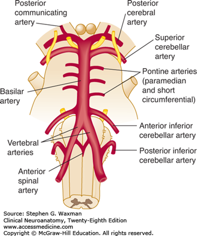
Paired vertebral arteries and the basilar artery pass along the anterior surface of the brain stem giving rise to the arteries supplying the brain stem. (See Figure 3.2 and Figure 3.3.)
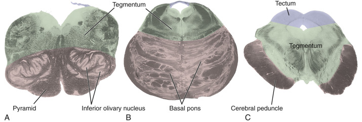
Each region of the brain stem is supplied in a series of wedge-shaped territories. The concept of these territories is very important to the understanding of brain stem infarcts; where medial or lateral areas may be affected selectively.
- The antero-lateral caudal medulla and cervical spinal cord are supplied by the anterior spinal artery and small branches of the vertebral artery.
- Postero-lateral portions of the caudal medulla are supplied by the posterior spinal artery and small branches of the posterior inferior cerebellar artery (PICA), both of which branch off the vertebral.
- The anterior and medial structures in the rostral medulla may receive direct branches from either the vertebral artery or the anterior spinal artery, a branch of vertebral.
- Lateral and posterior structures in the rostral medulla, such as the spinothalamic tract and the inferior cerebellar peduncle, are supplied by branches of the vertebral artery and the PICA.
Examples of the anterior and medial structures are:
-
- Pyramids
- Medial lemniscus
- Anterior pontine blood supply is from branches directly off the basilar artery.
- The anterior inferior cerebellar artery (AICA) and the superior cerebellar artery contribute branches to the middle and superior cerebellar peduncles and to parts of the posterior and lateral portions of the pontine tegmentum.
- The midbrain blood supply is chiefly from the posterior cerebral artery, with some contribution from the basilar and superior cerebellar arteries more caudally.
- In addition, the posterior communicating arteries will send branches that supply the cerebral peduncle and rostral-most portion of the midbrain.
Review 3.4.
The anterior pons receives its primary blood supply from the:
Gross organization of the brain stem
Three general regions can be identified when looking at cross section and may be described in relation to portions of the ventricular system. A brief description of each of these regions is found in Figure 3.4.
First, there is a small region dorsal/posterior to ventricular system.
The midbrain is the only region of the brain stem where there is a substantial amount of neural tissue posterior to the ventricular system. The region of the midbrain it is referred to as the tectum (Latin: roof), which consists of the superior and inferior colliculi. Nuclei which are responsible for relaying information about vision and hearing are located deep to the colliculi.
In both the pons and rostral medulla, the fourth ventricle is covered posteriorly by the superior and inferior medullary vela (plural of vellum) and the cerebellum, but neither of these regions have a distinct tectum in comparison to the midbrain.
Immediately ventral/anterior to the ventricular system is a region called the tegmentum.
The tegmentum in every portion of the brain stem contains axons and cells bodies associated with the reticular formation, cranial nerve nuclei and tracts, and ascending and descending pathways.
The final region is ventral to tegmentum.
Structures ventral to the tegmentum are responsible for forming prominent external features of the brain stem.
For example: the cerebral peduncles (crus cerebri), the basal pons (basis pontis), the medullary pyramids.
These ventral regions contain descending axonal tracts from the cerebral cortex to the spinal cord (corticospinal tracts), to certain cranial nerve nuclei (corticobulbar), or to pontine nuclei (corticopontine).
Description of internal anatomy of the brain stem
- The medulla
- The pons
- The midbrain
The caudal (closed) medulla extends from the caudal edge of the pyramidal decussation to the caudal end of the fourth ventricle.
-
- In the caudal medulla there are prominent structures that have a similar appearance to the posterior horn of the spinal cord these are the spinal trigeminal tract and nucleus. These two nervous structures are the head’s equivalent of Lissauer’s tract and the substantia gelatinosa of the spinal cord.
- The internal arcuate fibers traverse the reticular formation in the caudal medulla.
The rostral (open) medulla extends from the caudal extent of the 4th ventricle, called the obex, to the inferior cerebellar peduncle.
-
- In transverse section, the rostral medulla no longer looks like the spinal cord because of a widening of the floor of the 4th ventricle, the rhomboid fossa.
- The inferior olivary nucleus is a very prominent nucleus forming the olive externally. The inferior olivary nucleus is the source of climbing fibers to cerebellar cortex.
- The inferior cerebellar peduncle is located dorso-laterally in the rostral medulla and grows progressively larger rostrally. In a transverse section, axons can be seen leaving the medial portion of the inferior olivary nucleus, arching across the midline, and joining the contralateral inferior cerebellar peduncle. The inferior cerebellar peduncles carry information associated with equilibrium and balance from the vestibular nuclei.
- Medial to the inferior olivary nucleus is the medial lemniscus, which appears as a flattened band with a ventral-dorsal axis. Ventral to the medial lemniscus is the medullary pyramid. Hypoglossal (CN XII) axons pass lateral to the pyramid and medial to the inferior olivary nucleus.
- Dorsal to the medial lemniscus, near the floor of the fourth ventricle, is a distinctive bundle of fibers, the medial longitudinal fasciculus that can be traced as they ascend to the level of the midbrain. The medial longitudinal fasciculus (MLF) is involved in the coordination of the head with extraocular eye movements.
In the rostral medulla, there are also prominent cranial nerve nuclei; from medial to lateral, these include:
-
- the hypoglossal nucleus
- the vagal dorsal motor nucleus
- the solitary nucleus and tract
- the vestibular nuclei
The caudal pons
-
- Extending from the widest portion of the 4th ventricle to the rostral edge of the middle cerebellar peduncle is the caudal pons. The inferior olivary nucleus ends and inferior cerebellar peduncle fibers bend dorsally to enter the cerebellum at the level of the caudal pons. As the inferior olivary nucleus ends, the medial lemniscus becomes more oval.
- The MLF occupies the same relative position, adjacent to the midline and the floor of the 4th ventricle.
- Ventrally, the corticospinal tracts are still longitudinally oriented. Most of the other longitudinal fibers are corticopontine fibers which originate in the cerebral cortex and terminate in ipsilateral pontine nuclei. The corticopontine fibers arising from the pontine nuclei cross the midline and form the massive middle cerebellar peduncle connecting into the cerebellum.
- A large facial motor nucleus is in the ventro-lateral tegmentum of the caudal pons. In the caudal pons, facial nerve axons wrap around the prominent abducens nucleus and form the genu (L. knee) of the facial nerve before turning back ventrally to exit from the pons.
- The abducens nucleus together with the genu of the facial nerve, are responsible for formation of the facial colliculus an elevation in the floor of the 4th ventricle.
Descending tracts of the pons
- The corticospinal tracts are dispersed within the basis pontis (basal pons).
- The corticopontine fibers of basal pons give rise to the prominent bundles of longitudinally-oriented fibers, the transverse pontine fibers, with interspersed pontine nuclei among the bundles.
- There are also corticobulbar fibers, supplying descending information to the cranial nerve motor nuclei in the basal pons as well.
The rostral pons
-
- The rostral pons extends from the rostral edge of the middle cerebellar peduncle to the beginning of the cerebral aqueduct. It includes parts of the basal pons and 4th ventricle. The trigeminal nerve (CN V) axons emerge at the mid-pontine level, and the trochlear nerve axons (CN IV) emerges at the dorsal portion of the pons-midbrain junction.
- The trigeminal nuclei, both motor and sensory, are located medial to the middle cerebral peduncle and anterior to 4th ventricle.
- The MLF is still visible in the rostral pons, as is the caudal pons.
- The 4th ventricle narrows near the cerebral aqueduct and the superior cerebellar peduncle becomes apparent in the wall of the 4th ventricle. The superior cerebellar peduncle is the major efferent nerve bundle from the cerebellum, which projects to the red nucleus, thalamus, as well as other brain stem structures.
- The corticospinal axons are still seen descending through the rostral pons as multiple longitudinally oriented bundles of fibers accompanied by numerous corticopontine bundles.
The caudal midbrain
-
- The caudal midbrain contains the inferior colliculi posteriorly and extends from where the trochlear nerve emerges to the inter-collicular groove separating the inferior and superior colliculi. The nuclei in the inferior colliculus, a major component of the ascending auditory pathway.
- The superior cerebellar peduncles begin to decussate and the MLF continues in its usual position in the tegmentum of the caudal midbrain.
- Medial and ventral to the inferior colliculus and encircling the cerebral aqueduct is a pale-staining region of gray matter called the periaqueductal gray (PAG). The PAG is an essential part of the descending pain modulation system.
- The medial lemniscus appears as a flattened band of fibers curving dorsally, and the spinothalamic tract lies dorsal to the medial lemniscus at the surface of the midbrain. The two tracts eventually terminate in the overlapping regions of the somatosensory relay nucleus of the thalamus, the ventral posterolateral nucleus (VPL).
- In the caudal midbrain, forms a cerebral peduncle on each side, through which the corticospinal and corticopontine fibers travel.
The rostral midbrain
-
- The rostral midbrain contains the superior colliculi and extends to the posterior commissure.
- At this level of the brain stem the both MLF and the decussation of the superior cerebellar peduncles end.
- In the tegmentum, the red nucleus is visible bilaterally. Some fibers from the contralateral superior cerebellar peduncle synapse here, but most fibers continue to the thalamus. The red nucleus is the origin of the rubrospinal tract. The rubrospinal tract in humans and non-human primates is not as important as in quadrupeds.
- The nuclear complex of oculomotor nerve is present anterior to the cerebral aqueduct and axons of the oculomotor (CN III) nerve can be seen passing through the red nucleus.
- Ventral to the red nucleus is the substantia nigra. The darkly pigmented cells found in the dorsal part of the substantia nigra use dopamine for their neurotransmitter.
Key concept
Even though some of the axons of the cranial nerves exit/enter the brain stem at one level, the nuclei associated with that information may span multiple regions of the brain stem (i.e., the spinal trigeminal nucleus/tract).
Summary of major functional structures in the brain stem
Descending pathways
-
- Corticospinal and corticobulbar (corticonuclear)
- Corticopontine
- Rubrospinal
- Reticulospinal
- Vestibulospinal
Ascending pathways
-
- Posterior column: Medial lemniscus
- Anterolateral system (spinothalamic tracts)
- Spinocerebellar (posterior, anterior, and cuneocerebellar)
- Pontine nuclei: Source of fibers from the cerebral cortex into the cerebellum via the middle cerebellar peduncle
- Red nucleus: Efferent targets of the superior cerebellar peduncle
- Vestibular nuclei: Input into cerebellar vermis (inferior cerebellar peduncle)
- Inferior olivary nucleus (source of input to cerebellum
- Dispersed cholinergic, noradrenergic, serotonergic, and dopaminergic nuclei; periaqueductal gray, rostral ventral lateral medulla (RVLM); raphe nuclei
- Sleep and consciousness
- Brain stem control systems
- Postural and locomotion
For example: Vestibular nuclei, reticular nuclei, red nucleus, substantia nigra. - Visceral reflex functions
For example: Heart rate, respiration, cough nausea, vomiting, sneezing, blood pressure, sphincter control
(See Figure 3.5.)
- Motor and sensory nuclei in the medulla
- Motor and sensory nuclei in the pons
Motor and sensory nuclei in the midbrain
Auditory and vestibular function

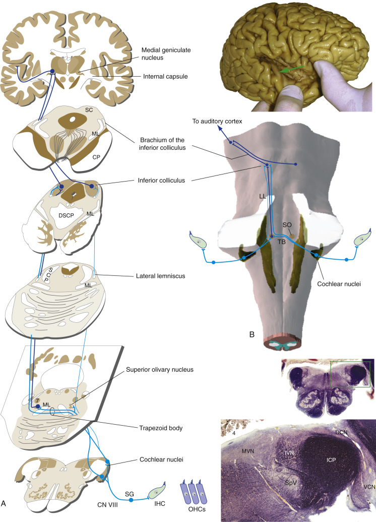
Hearing and balance are very different sensations functionally, but they both originate peripherally from the inner ear. (See Figure 3.6.) The eighth cranial nerve carries both sensory components, one in the cochlear division and one in a vestibular division.
Both divisions of CN VIII have special sensory receptors, hair cells, but the accessory structures in the semi-circular canals and cochlea respond to different types of mechanical stimuli. The cochlear division carries information about sound, whereas the vestibular division signals position and movement of the head.
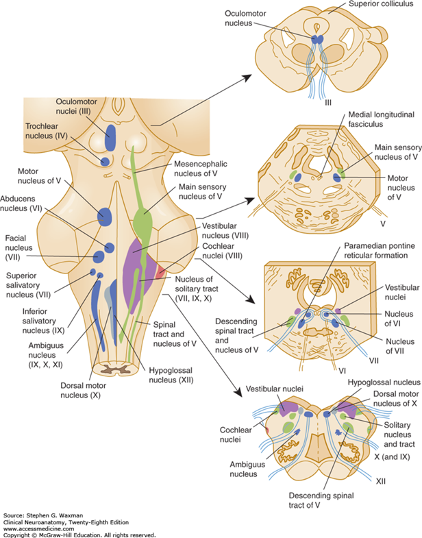
Both the vestibular and cochlear nuclei where axons in the vestibulocochlear nerve axons terminate are located in the posterior-lateral part of the rostral medulla and caudal pons.
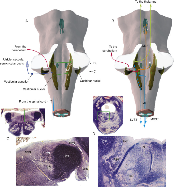
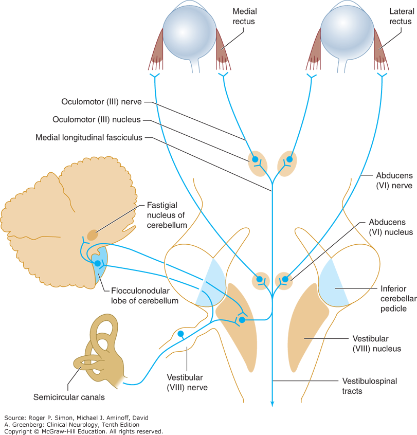
review 3.5.
Fill in the text to help with understanding the primary auditory pathway. (Tap the right arrow for answers)
Gross anatomy of the cerebellum
Each hemisphere of the cerebellum can be divided into the anterior and posterior lobes, separated by the primary fissure, the vermis, and the flocculonodular lobe (made up of the flocculus and part of the vermis) (see Figure 3.9).
-
- The inferior portion of the cerebellar vermis and flocculonodular lobes help regulate balance and eye movements via interactions with the vestibular system (e.g. vestibular nuclei). It is generally assumed that these regions, along with other parts of the vermis, are involved in control of the proximal trunk muscles, whereas the more lateral portions of the cerebellar hemispheres help control the distal muscles in the extremities.
- The lateral cerebellar hemispheres are also involved in motor planning.
- The cerebellar tonsils are located infero-laterally and herniate through the foramen magnum in patients with Chiari malformations, they are also potentially involved in herniating during increases in intracranial pressure that affects neuroanatomical structures found in the infratentorial compartment.
Key concept
The white matter tracts connecting the cerebellum to the brain stem include:
-
- the superior cerebellar peduncle (cerebellum to midbrain)
- middle cerebellar peduncle (pons to cerebellum)
- inferior cerebellar peduncle (medulla to cerebellum)
Cerebellar peduncles connections
- Vestibular receptors from the inner ear (Figure 3.10)
- Body surface and muscle receptors (spinocerebellar pathways)
- The inferior olivary nucleus
It also contains axons that carry output from the cerebellum into brain stem upper motor neurons that regulate proximal muscles (vestibular nuclei and reticular nuclei) is via the vestibulospinal and reticulospinal tracts.
- Widespread areas of cerebral cortex (corticopontine fibers that synapse in pontine nuclei)
- Cerebral cortex
- Thalamus
- Red nucleus
Key concept
The vascular supply to the cerebellum is by branches of the posterior circulation:
-
- posterior inferior cerebellar
- anterior inferior cerebellar
- superior cerebellar
- posterior cerebral arteries
Equilibrium and balance
Equilibrium is the state of physical balance of the body and its relation to the external environment. To maintain this physical balance there needs to be visual, vestibular input from the semicircular canals, and proprioceptive input.
The input associated with equilibrium is integrated at the level of the brain stem and cerebellum. Disorders of equilibrium result from diseases that can affect central or peripheral vestibular pathways, the cerebellum, or integrated sensory pathways involved in proprioception.
Disorders of equilibrium and balance usually present with one of two clinical problems: vertigo or ataxia.
- Peripheral vertigo
- Central vertigo
- Ataxia
Lesions affecting the semicircular canals and vestibule of the inner ear or the vestibular division of the vestibulocochlear (VIII) nerve can cause peripheral vertigo.
Vertigo from peripheral lesions tends to be intermittent, lasts for briefer periods, and produces more distress than vertigo of central origin (see next section).
Nystagmus (rhythmic oscillation of the eyes) is always present with peripheral vertigo. Peripheral lesions can also produce additional symptoms of inner ear or cranial nerve VIII nerve dysfunction, such as hearing loss and tinnitus.
Central nervous system origin of vertigo is the result of lesions that affect the vestibular nuclei in the brain stem or their connections into cerebellum. Another rare cause of vertigo is produced by a lesion of cortical structures, such as when it occurs as a symptom of complex partial seizures (see chapter 12, Seizures and Syncope).
Central vertigo may occur with or without nystagmus; if nystagmus is present, it can be vertical, unidirectional, or multidirectional and may differ in character in the two eyes.
Vertigo from central lesions may be accompanied by intrinsic brain stem or cerebellar signs, such as:
-
- motor or sensory deficits
- hyperreflexia
- extensor plantar responses
- dysarthria (slurred, slow speech)
- limb ataxia
Ataxia is incoordination or clumsiness of movement that is not the result of muscle weakness. It can be caused by vestibular input, cerebellar, or sensory (proprioceptive) disorders. Ataxias may either be inherited (for example: spinocerebellar ataxias, Friedreich’s Ataxia) or acquired (for example: a tumor, stroke, alcohol-induced damage, or vestibular function).
Vestibular: Originating associated with inner ear, vestibular nuclei, or inferior
cerebellar peduncle
Cerebellar: Gait or truncal ataxia (vermis (truncal) or lateral (limb ataxia)
Sensory: Proprioceptive information is interrupted, affecting both the
spinocerebellar pathways and posterior columns
Cerebellopontine Angle Syndrome

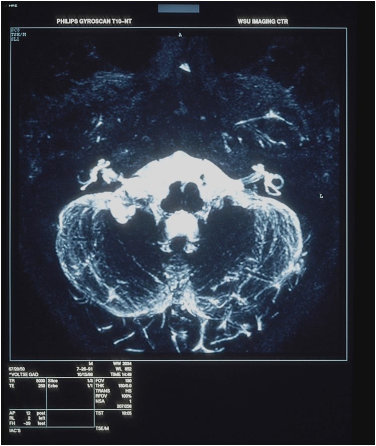
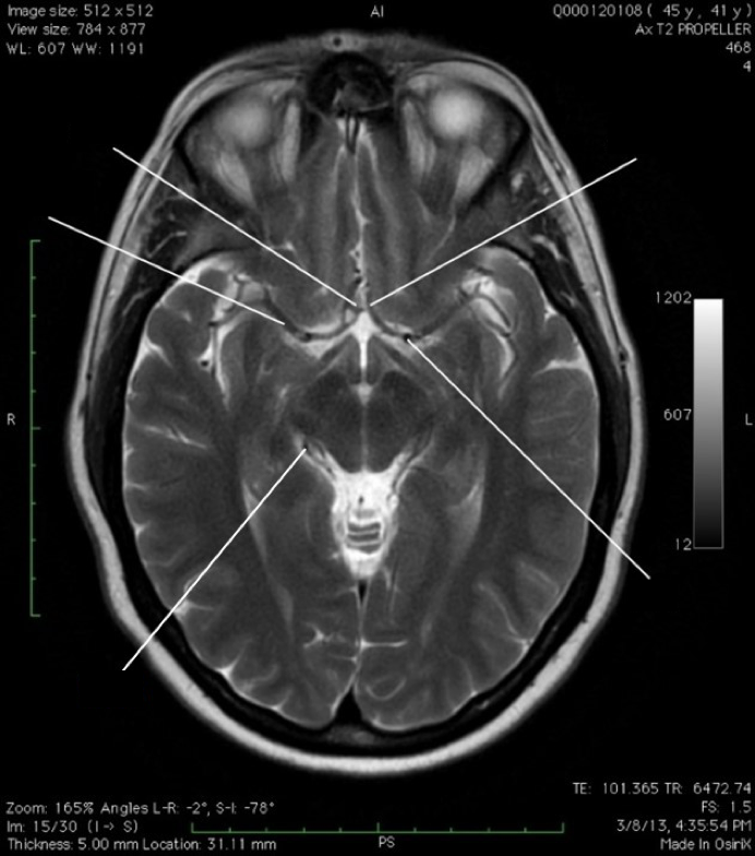
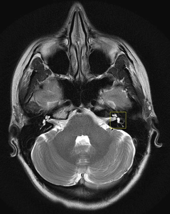
The most common tumor in the region of cerebellopontine angle, bordered by the cerebellum, lateral pons, and the temporal bone is a benign tumor, an acoustic neuroma. (See Figure 3.11 as an example.)
This tumor typically arises from the nerve sheath of the vestibular portion of the vestibulocochlear (VIII) nerve in the internal auditory canal. Less common tumors at this site include meningiomas, epidermoid tumors, and lipomas.
Cerebellopontine angle tumors produce symptoms associated with damage to nearby cranial nerves, brain stem, ventricular, and cerebellar structures.
Thought question
List some common clinical signs of an acoustic neuroma.