


- Suggested reading
- Need to Know
The reading attached here is to supplement this session. Everyone comes into medical school with different background. The lessons listed below help you understand the videos and short passages in the module. They are not required and do not feel as though you must read them. However, do not have the impression that all you need to do is go through the module, and you will be fine. Read the objective carefully, and ask yourself if you truly understand the material. If not, look at the readings for further clarification. You should have all the resources you need for the objectives listed between these short videos, the in-class session, and the suggested reading.
This pdf is a short chapter on inheritance patterns. It does an excellent job of covering objective 2.
This pdf is a bit longer, and it covers the remaining objectives. However, I believe the Osmosis videos do a great job on these objectives as well. Do not get caught up in the details; think “big picture” regarding genetics and genetic techniques.
This is my “need to know” list If you have a good understanding of everything here, you will be fine for the exam, Step 1, and your clerkship years. I use these guides for you to organize your thoughts when learning objectives.
Diagram and distinguish between the five different types of modes of inheritance: autosomal dominant, autosomal recessive, X-linked recessive, X-linked dominant, and mitochondrial inheritance
-
Autosomal dominant
-
Autosomal recessive
-
Mitochondrial inheritance
-
X-linked dominant
-
X-linked recessive
Describe genomic imprinting, trinucleotide repeat disorders, chromosomal abnormalities, and how they can precipitate disease states
-
Prader-Willi Syndrome
-
Angelman Syndrome
-
Trinucleotide repeat diseases
-
Genomic Imprinting (using PW and Angelman as examples)
-
Aneuploidy and diseases/disorders/syndromes associated with aneuploidy
Describe key molecular techniques used to analyze DNA and chromosomes and explain how they aid in diagnosing genetic and chromosomal disorders
-
Microarray Analysis technique
-
PCR technique
-
Karyotyping
-
In Situ Hybridization, Fluorescence (FISH)
-
Comparative genome hybridization
-
Whole genome sequencing
Compare and contrast meiosis and mitosis and describe what can occur when there are defects in these processes.
-
Mitosis
-
Meiosis
-
Nondisjunction in meiosis
Compare and contrast meiosis and mitosis and describe what can occur when there are defects in these processes
This Osmosis video does a very nice job of discussing mitosis and meiosis.
Mitosis
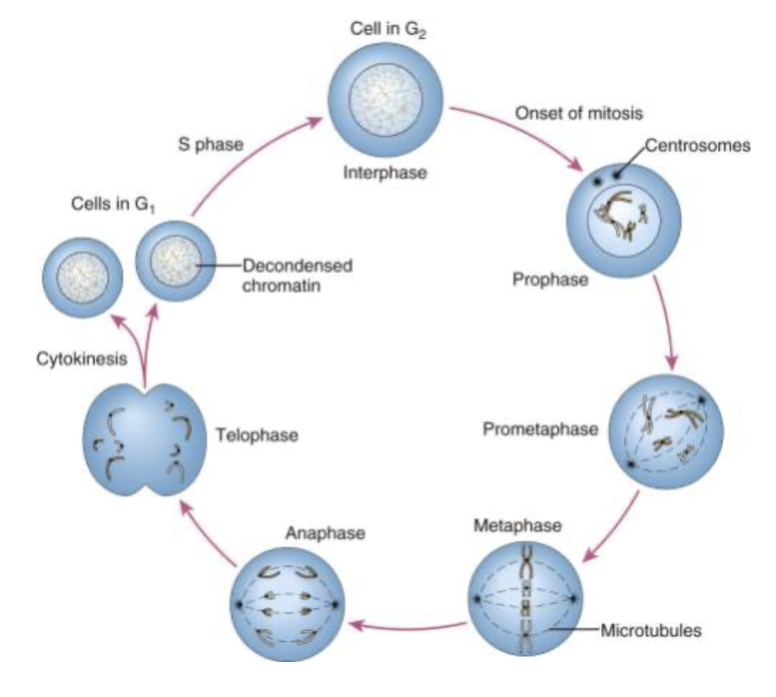
Taken from: Nussbaum RL, McInnes RR, Willard HF. Thompson & Thompson Genetics in Medicine. Eighth ed. Philadelphia: Elsevier; 2016.
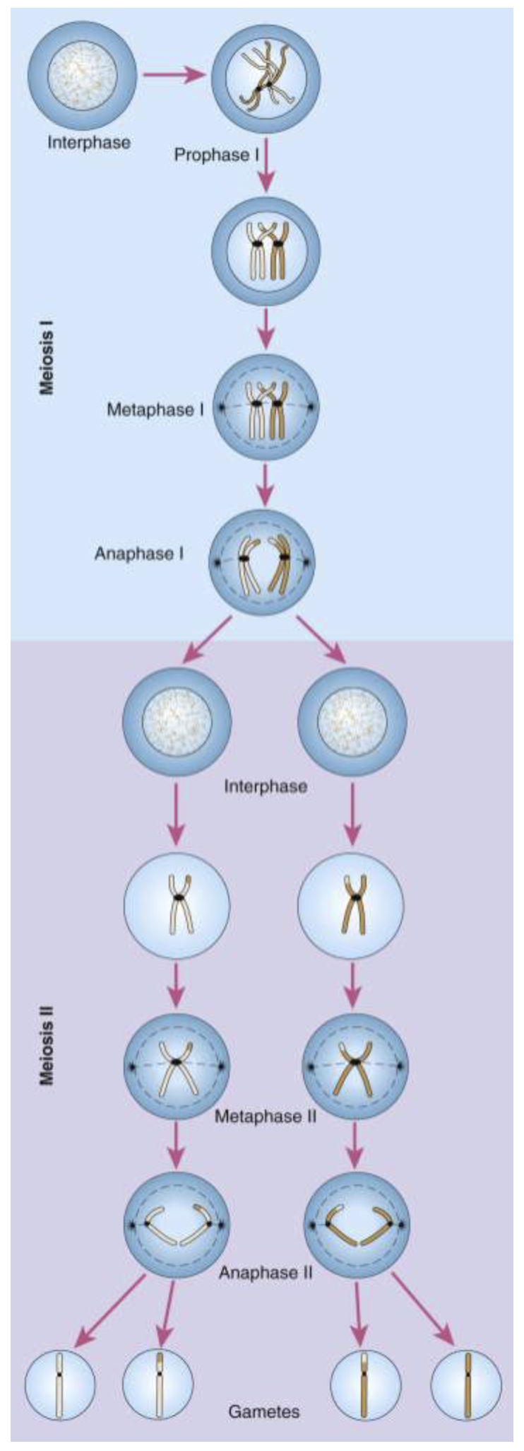
Meiosis
A single chromosome pair and a single crossover are shown, leading to formation of four distinct gametes. The chromosomes replicate during interphase and begin to condense as the cell enters prophase of meiosis I. In meiosis I, the chromosomes synapse and recombine. A crossover is visible as the homologues align at metaphase I, with the centromeres oriented toward opposite poles. In anaphase I, the exchange of DNA between the homologues is apparent as the chromosomes are pulled to opposite poles. After completion of meiosis I and cytokinesis, meiosis II proceeds with a mitosis-like division. The sister kinetochores separate and move to opposite poles in anaphase II, yielding four haploid products.
Figure and text taken from: Nussbaum RL, McInnes RR, Willard HF. Thompson & Thompson Genetics in Medicine. Eighth ed. Philadelphia: Elsevier; 2016
Nondisjunction in meiosis
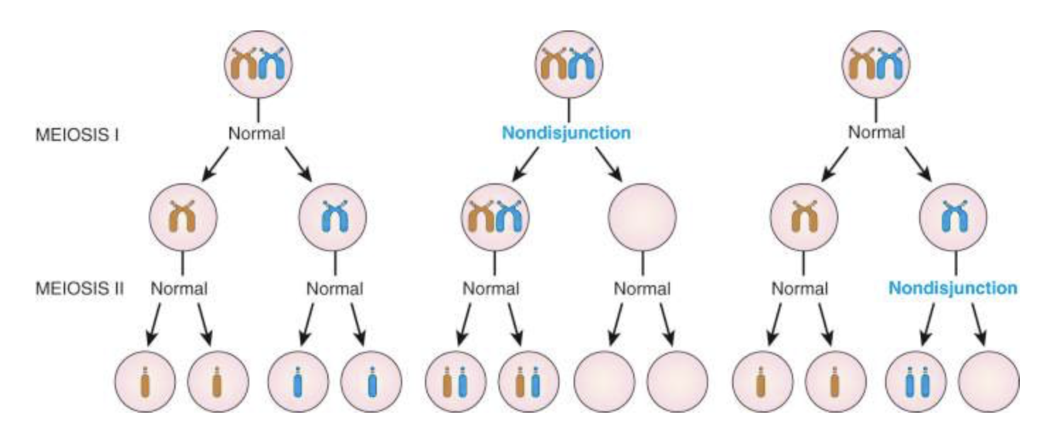
Diagram and distinguish between the five different types of modes of inheritance: autosomal dominant, autosomal recessive, X-linked recessive, X-linked dominant, and mitochondrial inheritance
This Osmosis video does an excellent job of describing the different inheritance patterns.
There are five different modes of inheritance you should be concerned with:
-
- autosomal dominant
- autosomal recessive
- X-linked recessive
- X-linked dominant
- mitochondrial inheritance
Quiz Yourself
Can you match these statements with the proper mode of inheritance?
Often structural genes, both male and female affected, affects many generations
Autosomal Dominant
Fatherly transmission only occurs to daughters
X-linked dominant
Often enzyme deficiencies, both male and female affected
Autosomal Recessive
Transmitted only through the mother
Mitochondrial inheritance
Skips generations and no male-to-male transmission
X-linked recessive
Quiz Yourself
The one feature that is most helpful in determining in a pedigree if a particular condition, which was occurring in multiple generations of a family is likely to be X-linked dominant rather than autosomal dominant is:
Describe genomic imprinting, trinucleotide repeat disorders, chromosomal abnormalities, and how they can precipitate disease states
Prader-Willi Syndrome Osmosis video.
Angelman syndrome Osmosis video. Angelman Syndrome and Prader-Willi syndrome are high-yield examples of genomic imprinting.
There are many different nucleotide repeat syndromes. A high-yield one to remember, which will come up again during neuro, is Huntington disease.
There are many different chromosomal abnormalities that lead to pathologies. The Osmosis video above is about Edwards Syndrome.
There are other Osmosis videos that explain these abnormalities. Some high-yield ones include (but are not limited to):
Describe key molecular techniques used to analyze DNA and chromosomes and explain how they aid in diagnosing genetic and chromosomal disorders
This video does a great job of explaining PCR. PCR is something that will come up all the time, as a diagnostic tool, during your career.
FISH is another technique you should know how it works.