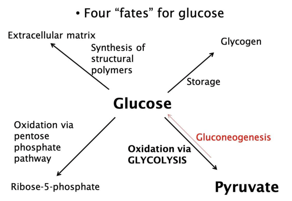
- Suggested reading
- Reading guide
- Need to Know
If you read Chapter 8: Glycolysis from Lippincott, you should have no issue with the objectives. However, use your time wisely. If you have a strong biochemistry background and glycolysis, this chapter may not be needed. A “reading guide” is also posted (you’ll see the pattern). This reading guide is for people who have a difficult time focusing on these types of chapters. Most students DO NOT need to use the guide.
Describe the four metabolic fates of glucose
-
Pyruvate Formation
-
Glycogen synthesis
-
Extracellular matrix synthesis
-
Formation of Ribose-5-phosphate
Diagram and describe the steps of glycolysis, including key regulatory enzymes and how they are controlled by energy status and hormonal signals
-
Arsenic poisoning (glycolysis mechanism)
-
Glycolysis
-
Regulation of glycolysis
-
Phosphofructokinase-1
-
Glyceraldehyde-3-phosphate dehydrogenase
Differentiate between hexokinase and glucokinase in terms of their biochemical properties, tissue distribution, regulatory mechanisms, and physiological roles in glycolysis, particularly in the context of glucose homeostasis
-
Hexokinase
-
Glucokinase
Describe the formation of pyruvate from glucose via glycolysis and its conversion to lactate under anaerobic conditions, including key enzymes and clinical relevance
-
Pyruvate kinase
-
Pyruvate kinase deficiency
Describe the four metabolic fates of glucose
It’s important to realize that glucose does a lot of different things. The simple figure below demonstrates this. All of these will be discussed over the next few sessions.
Diagram and describe the steps of glycolysis, including key regulatory enzymes and how they are controlled by energy status and hormonal signals
This Osmosis video does a great job of explaining the pertinent points and steps of glycolysis.
Differentiate between hexokinase and glucokinase in terms of their biochemical properties, tissue distribution, regulatory mechanisms, and physiological roles in glycolysis, particularly in the context of glucose homeostasis
The Osmosis video touches on these two enzymes a bit. However, there is much more than that video tells you. Both enzymes are kinases that take glucose and make glucose-6-phosphate. This is an irreversible reaction and “traps” the glucose molecule in the cell. Glucokinase is a “glucose sensor” and is active in the liver and the pancreas.
The take-home point here is that glucokinase only is active after a high carbohydrate meal when blood glucose levels are elevated.
Describe the formation of pyruvate from glucose via glycolysis and its conversion to lactate under anaerobic conditions, including key enzymes and clinical relevance
In the last step of glycolysis, phosphoenolpyruvate is converted to pyruvate. This reaction is catalyzed by pyruvate kinase (PK) and is the third irreversible reaction of glycolysis. ATP is synthesized at this step using substrate-level phosphorylation.
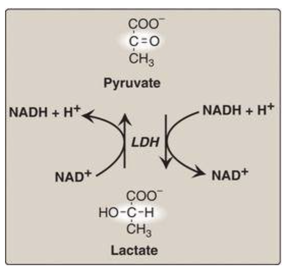
Lactate is formed from pyruvate by lactate dehydrogenase (LDH). Lactate is the final product of anaerobic glycolysis in eukaryotic cells (see the figure here). Reduction to lactate is the major fate for pyruvate in poorly vascularized tissues or in erythrocytes, which lack mitochondria.
The direction of the LDH reaction depends on the relative intracellular concentrations of pyruvate and lactate and on the ratio of NADH/NAD+. For example, in the liver and heart, this ratio is lower than in exercising muscle. Consequently, the liver and heart oxidize lactate (obtained from the blood) to pyruvate. In the liver, pyruvate is either converted to glucose by gluconeogenesis or converted to acetyl CoA that is oxidized in the TCA cycle. Heart muscle exclusively oxidizes lactate to carbon dioxide and water via the TCA cycle.

