Nematodes

Headshot of Joanna Breems, MD, FACP · Clinical Assistant Professor
Joanna Breems
MD, FACP · Clinical Assistant Professor
envelope icon phone icon
Table of Contents

check yourself

Fill in these summary tables to check your learning.

Round worms

GI

Enterobius (Pin worm)

Transmitted by ingestion of eggs, which develop into adults within the gut.

Females migrate to rectum/anus to lay eggs on skin. Symptoms are associated with this migration and lead to intense anal pruritis (itchy butt). Re-infection is common when hands are used to scratch the pruritic area, carrying eggs that are then incidentally ingested.

See also in the Micro-ID session guide

Trichuris (Whip worm)

Intestinal nematode transmitted to humans by ingestion of eggs which develop into adults in gut.

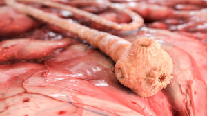
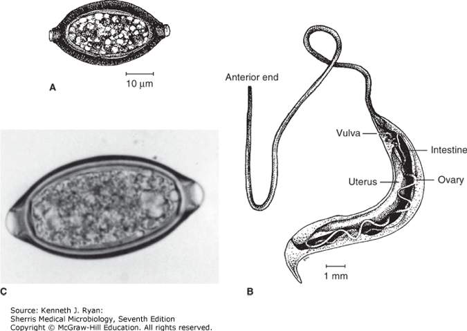
The thin ‘whip’ end embeds in the GI mucosa. Eggs are shed into soil where they embryonate and become infectious. With heavy egg burden, anemia from occult blood loss and rectal prolapse can occur. Adult worms can often be seen embedded in the prolapsed mucosa.

Ascaris lumbricoides (Round worm)

Intestinal nematode transmitted to humans by ingestion of eggs which develop into larvae in gut.

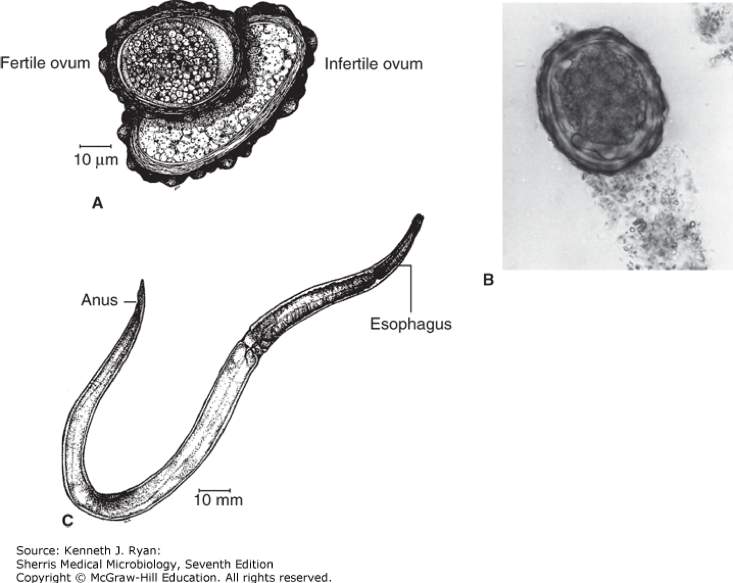
Larvae penetrate the gut to the blood where they migrate to the lungs, are coughed up and swallowed, then develop into adult worms in the gut. Adults are big round worm (30 cm) and very common worldwide. Causes intestinal obstruction with worm balls. Also invade appendix or biliary tree. Pulmonary migration stage can cause transient asthma like symptoms, or eosinophilic pneumonia (Loeffler’s syndrome).

See also in the Micro-ID session guide

Hookworm (Ancylostoma and Necator)

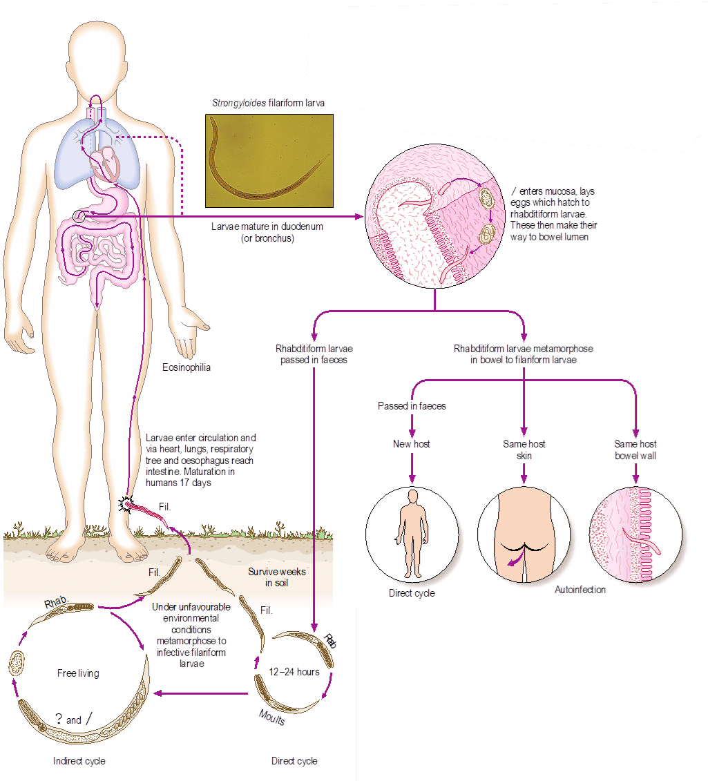
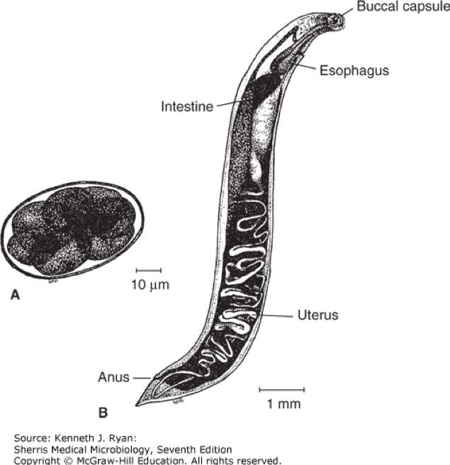
Filarial form penetrates intact skin, then enters the blood and eventually migrates to the lung. After entering alveoli, larvae pass up trachea and are swallowed. They mature into adult worms in the GI tract. Adult worms attach to the walls of the small intestine via teeth and cutting plates.

Clinical disease includes cutaneous larvae migrans, a pruritic serpentine rash that occurs during the cutaneous phase, known as “ground itch. ” Pulmonary migration can lead to eosinophilic pneumonia and Loeffler’s syndrome. And adult worms lead to chronic iron deficiency anemia due to microscopic blood loss. The iron-deficiency anemia has been linked with poor cognitive development in children and perpetuation of chronic impoverished states.

See also in the Micro-ID session guide

Strongyloides

Filarial form penetrates intact skin then enters the blood and eventually migrates to the lung. After entering alveoli, larvae pass up trachea and are swallowed. They mature into adult worms in the GI tract. Adults produce eggs that hatch in colon to rhabditiform larvae (non-infectious). Rhabditiform larvae can molt in the gut forming infectious filariform larvae that can directly reenter the blood stream—autoinfection. Autoinfection can allow persistence of infection for decades. Rhabditiform larvae that are passed into the soil can mature into adults and have several free-living cycles.

Clinical manifestations include asthma like pulmonary disease and eosinophilia during migration. Autoinfection allows for high worm burden leading to hyperinfection, which is more common with immunosuppression. Hyperinfection can cause polymicrobial bacteremia when bacteria translocate across bowel with larvae. Hyperinfection can be fatal.

Blood and tissue

Cause disease through their presence in tissues and lymphohematogenous system. Some migrate through the human GI tract, but that is temporary part of the life cycle.

Trichinosis (Trichinella spiralis)

Transmission to humans when ingesting undercooked meat that contains encysted larvae which mature into adult worm in small intestine. The adult worms release larvae that enter blood and migrate to tissues; they are able to encyst in skeletal muscle and brain. Reservoir in pigs. Clinical manifestations include severe myalgia/myositis. Late stages result in calcified cysts in muscle.

Filaria

Transmitted to humans through the bite of mosquito which deposits larvae into skin where they develop into microfilariae that migrate to lymphatic where they develop into adult worm. Adult worm lies coiled in lymphatic for the entirety of decade-long lives. Clinical disease is filariasis which results from long-term lymphatic obstruction. Tropical pulmonary eosinophilia can occur.

Dx by finding microfilaria in night-time blood smear. Serology also helpful.

Tx: Diethylcarbamazine for microfilariae. There is no treatment for adult worms.

Transmitted by Simulium black flies that live near rivers (“River blindness”). Larvae are deposited into skin where they mature to adult worms within skin nodules. Females produce microfilariae that migrate in interstitial fluid. Microfilariae in the eye ultimately causes blindness. Affected skin is described as “lizard-like,” itchy, and hyperpigmented.

Dx: Skin snip reveals microfilaria.

Tx: Ivermectin for microfilariae. Suramin for adult worms.

Toxocara (Dog and cat ascarids)

Visceral larva migrans.

Transmitted by ingestion of eggs in soil or food contaminated with dog feces. Eggs hatch into larvae in small intestine then enter blood and migrate to organs —brain, liver, eyes—where they are trapped and die. Clinical manifestations include fever, eosinophilia, hepatitis, and chorioretinitis. Exudative endophthalmitis mimics retinoblastoma.