Recall that the somatic division of the peripheral nervous system (PNS) supplies the body wall and limbs = things we have conscious control over (skeletal muscles) and conscious perception of (pain, touch, and temperature of skin, for example).
Now we examine another part of our peripheral nervous system—the Autonomic Nervous System (ANS)—which supplies visceral structures, like organs in the body cavities. These are things that are operated upon at a subconscious level and produce sensations that are poorly perceived or do not come to consciousness at all.
Although somatic and visceral structures function quite differently, there are some similarities in the architecture of their innervations:
Nuclei in the brain are involved in innervating both somatic and visceral structures.
Both somatic and visceral structures are supplied by efferent (motor) and afferent (sensory) nervous pathways.
Sensory neurons serving both somatic and visceral structures have their cell bodies in sensory ganglia.
Bydefinition,theautonomicnervoussystemisamotorsystem. Visceral structures are supplied with sensory neurons too, but visceral sensory pathways are technically not part of the ANS. However, we will also discuss these pathways in this chapter.
Whereas the somatic nervous system innervates skeletal muscle, the targets of the ANS are smooth muscle, cardiacmuscle, and glands, regardless of embryonic origin.
Recite the mantra
Smooth muscle, Cardiac muscle, and Glands!
The ANS has two divisions
Parasympathetic Division"resting and digesting"
Functions in energy conservation activities and increasing the motility and secretions in the gut. Also called the craniosacral outflow, describing the regions of the central nervous system where parasympathetic axons exit (brainstem and sacral spinal cord segments).
Sympathetic Division"fight, flight, fright"
Functions in emergency or stressful situations. Also called the thoracolumbar outflow, describing the regions of the central nervous system where sympathetic axons exit (thoracic and lumbar spinal cord segments).
Normally, visceral organs receive both sympathetic and parasympathetic innervations and in most cases the two divisions work in a competitive balance. The heart and pupil of the eye are examples (Heart: S = speeds it up; PS = slows it down) (Eye: S = dilates the pupils; PS = constricts pupils). Some structures receive only sympathetic innervation (blood vessels, sweat glands). And in a few cases, there is cooperation between the two divisions, such as genital functions, where parasympathetic stimulation produces erection and sympathetic causes ejaculation.
Table 4.1 Functions of autonomic nervous system (ANS)
Organ, tract, or system
Effect of sympathetic stimulation†
Effect of parasympathetic stimulation††
Eyes
Pupil
Ciliary body
Dilates pupil (admits more light for increased acuity at a distance)
Constricts pupil (protects pupil from excessively bright light)
Contracts ciliary muscle, allowing lens to thicken for near vision (accommodation)
Skin
Arrector muscles of hair
Peripheral blood vessels Sweat glands
Causes hairs to stand on end (“gooseflesh” or “goose bumps”) Vasoconstricts (blanching of skin, lips, and turning fingertips blue)
Promotes sweating*
No effect (does not reach)‡
No effect (does not reach)‡
No effect (does not reach)‡
Other glands
Lacrimal glands Salivary glands
Slightly decreases secretion**
Secretion decreases, becomes thicker, more viscous**
Promotes secretion
Promotes abundant, watery secretion
Heart
Increases the rate and strength of contraction; dilates coronary vessels**
Decreases the rate and strength of contraction (conserving energy); promotes constriction of coronary vessels in relation to reduced demand
Lungs
Inhibits effect of parasympathetic system, resulting in bronchodilation and reduced secretion, allowing for maximum air exchange
Constricts bronchi (conserving energy) and promotes bronchial secretion
Digestive tract
Inhibits peristalsis, and constricts blood vessels to digestive tract so that blood is available to skeletal muscle; closes sphincters
Promotes peristalsis and secretion of digestive juices Contracts the rectum, relaxes sphincters
Liver and gallbladder
Promotes breakdown of glycogen to glucose (for increased energy)
Promotes building/conservation of glycogen; increases secretion of bile, contracts gallbladder
Urinary tract
Vasoconstriction of renal vessels slows urine formation; internal sphincter of bladder contracted to maintain urinary continence
Inhibits contraction of the internal sphincter of the bladder, contracts detrusor muscle of the bladder wall causing urination
Genital system
Causes ejaculation and vasoconstriction resulting in remission of erection
Produces engorgement (erection) of erectile tissues of the external genitals
Suprarenal medulla
Release of adrenaline into blood
No effect (does not innervate)
*With the exception of the sweat glands, glanular secretion is parasympathetically stimulated.
**With the exception of the coronary arteries, vasoconstriction is sympathetically stimulated; the effects of sympathetic stimulation on glands (other than sweat glands) are the indirect effects of vasoconstriction.
†In general, the effects of sympathetic stimulation are catabolic, preparing the body for the fight-or-flight response.
††In general, the effects of parasympathetic stimulation are anabolic, promoting normal function and conserving energy.
‡The parasympathetic system is restricted in its distribution to the head, neck, and body cavities (except for erectile tissues of genitalia); otherwise, parasympathetic fibers are never found in the body wall and limbs. Sympathetic fibers, by comparison, are distributed to all vascularized portions of the body.
FROM MOORE ET AL., CLINICALLY ORIENTED ANATOMY, TABLE 1.2.
The ANS has a two-neuron pathway from CNS to target
Figure 4.1 Comparison of ANS and somatic motor pathways.
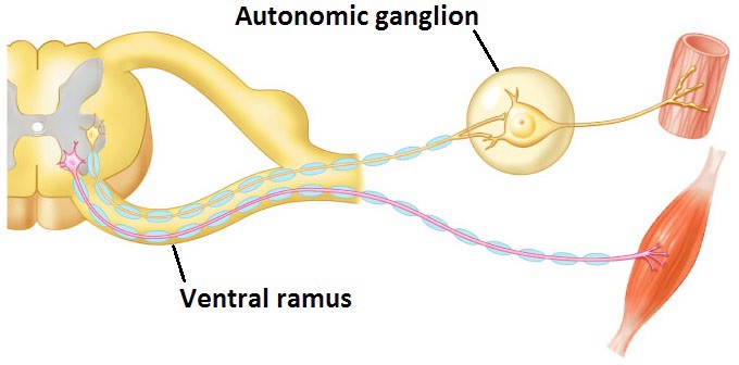
The primary feature that distinguishes the autonomic NS from the somatic system is the presence of ganglia in the pathway. These autonomic ganglia are motor ganglia—something we have not encountered before in our discussions on the peripheral nervous system.
Compare this to the somatic motor pathway. Only one neuron in the pathway from CNS to skeletal muscle. No ganglia. See Figure 4.1.
Regardless of where they are located, the autonomic ganglion cells are always the second cells in the ANS pathway. The first neuron is called the pre-ganglionic (or pre-synaptic) neuron. The second neuron is the post-ganglionic (or post- synaptic) neuron.
Preganglionic neuron
Cell body in a nucleus in the CNS axon leaves the CNS synapses on a postganglionic neuron cell body in an autonomic ganglion.
Postganglionic neuron
Cell body in an autonomic ganglion axon terminates on the target organ (cardiac muscle, smooth muscle, or gland)
Figure 4.2 CORNELIUS ROSSE, M. D., D. SC., STUDY GUIDE FOR GROSS ANATOMY AND EMBRYOLOGY, UNIVERSIT Y OF WASHINGTON. USED WITH PERMISSION.
Where are the cell bodies of the preganglionic ANS neurons located?
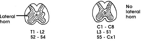
These are in nuclei within the central nervous system. A nucleus is a cluster of functionally related neuron cell bodies in the CNS. Autonomic nuclei associated with ANS preganglionic neurons are in the spinal cord and brainstem.
Spinal cord segments T-1 to L-2 and S-2 to S-4.
Located in the intermediolateral cell column of the spinal cord.
This column appears in cross section as the lateral horn of gray. See Figure 4.2.
ANS preganglionic axons leave the spinal cord through ventral roots, pass through spinal nerves, and enter the ventral rami of spinal nerves.
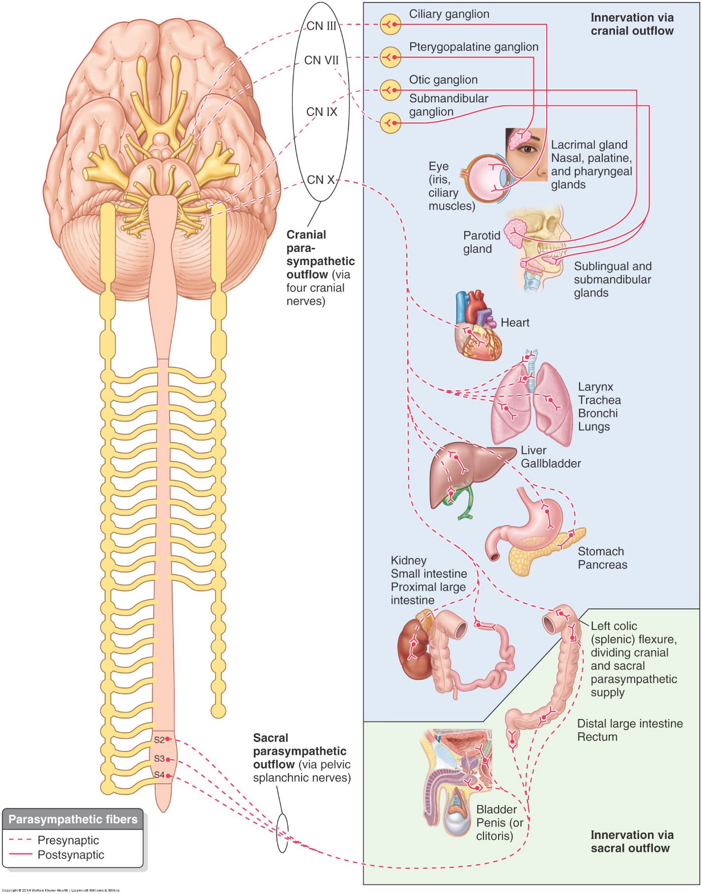
Brainstem
Located in the visceral motor nuclei of cranial nerves III (oculomotor), VII (facial), IX (glossopharyngeal), and X (vagus).
ANS preganglionic axons leave the brainstem in cranial nerves III, VII, IX, and X.
Where are the cell bodies of the postganglionic ANS neurons located?
These are in autonomic ganglia. All autonomic ganglia are derived from neural crest.
In the parasympathetic division, these ganglia are:
in the head (associated with cranial nerves III, VII, IX, and X) and
in or near the wall of target organs in the abdominopelvic cavity. The later ganglia are called intramural ganglia.
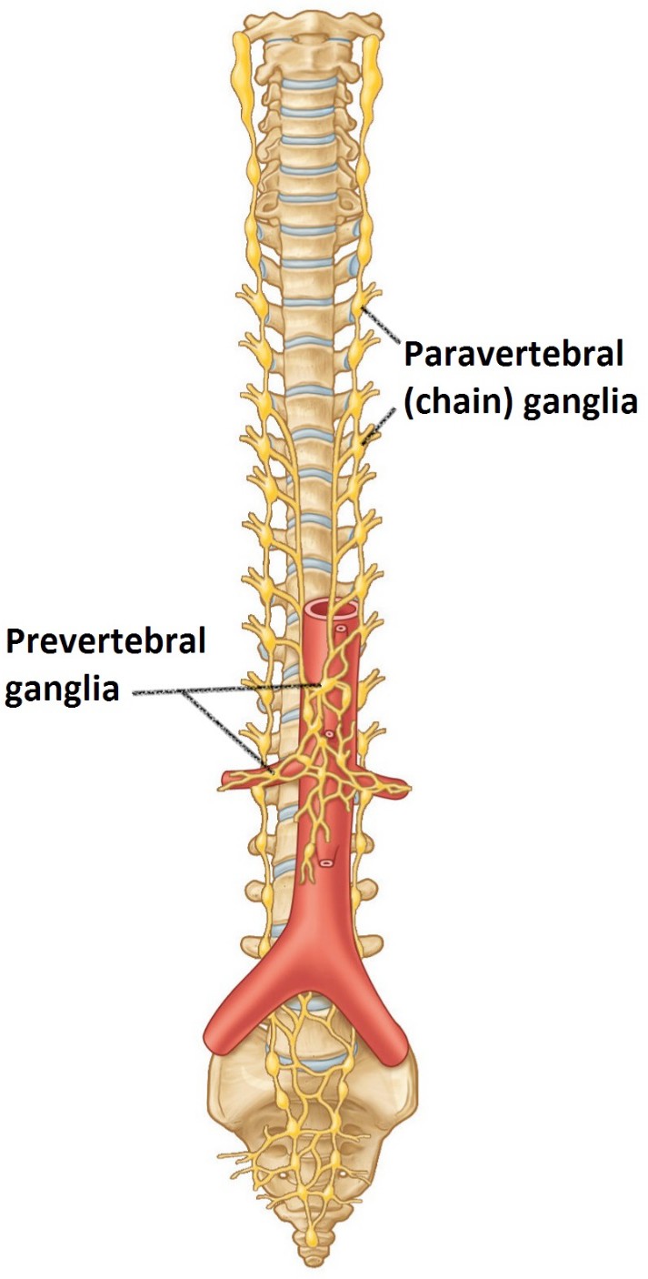
In the sympathetic division, these ganglia are:
arranged in chains along the left and right sides of the vertebral column (paravertebral ganglia) and
in the abdominopelvic cavity, near the aorta (prevertebral ganglia).
Autonomic ganglia: Sympathetic vs. parasympathetic pathways
Parasympathetic preganglionic fibers
Synapse on ganglia in the head and in intramural ganglia in the trunk. All these ganglia are near the target organ. Therefore, parasympathetic preganglionic fibers are generally long; postganglionic fibers are short.
Sympathetic preganglionic fibers
Synapse in paravertebral or in prevertebral ganglia. All these ganglia are relatively close to the vertebral column. Therefore, sympathetic preganglionic fibers are generally shorter; postganglionic fibers are generally longer.
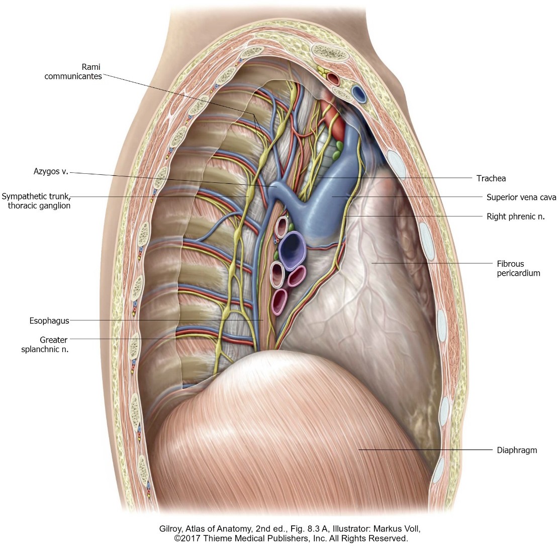
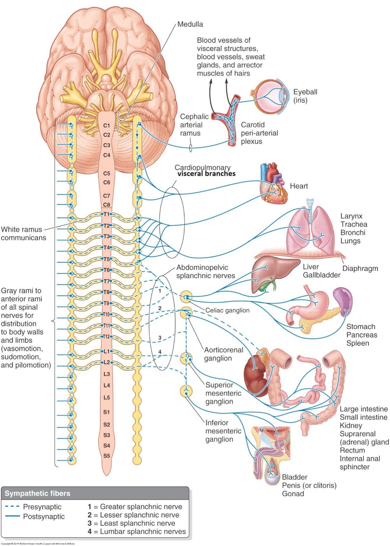
The sympathetic trunks
Figure 4.3.
Similar to dorsal root ganglia, paravertebral ganglia (left and right) originally developed for each somite of the body. Thus, there were 31 pairs of paravertebral ganglia. Fusion of ganglia occurs in the cervical and lumbar regions, so in the adult there are fewer than 31. Paravertebral ganglia are connected vertically by interganglionic branches, forming a chain of ganglia on each side of the vertebral column. This is called the sympathetic trunk. Paravertebral ganglia are also known as sympathetic chain ganglia. Thesympathetic trunks extend from the base ofthe skull all the way to the coccyx.
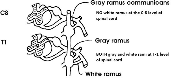
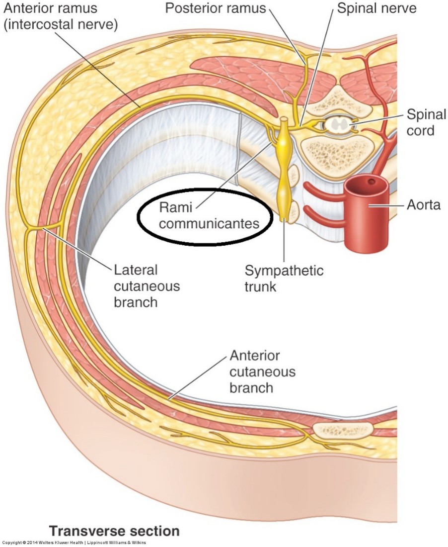
Sympathetic trunk: Gray and white rami communicantes
Figure 4.4 CORNELIUS ROSSE, M. D., D. SC., STUDY GUIDE FOR GROSS ANATOMY AND EMBRYOLOGY, UNIVERSITY OF WASHINGTON. USED WITH PERMISSION.
Each sympathetic chain ganglion has at least one branch that connects the ganglion to a corresponding ventral ramus of a spinal nerve. This branch transmits postganglionic sympathetic fibers from the chain ganglion to the spinal nerve. Since postganglionic fibers are non-myelinated (do not look white), this branch is called the gray ramus communicans.
Note
ALL sympathetic chain ganglia have one or more gray rami communicantes
ONLY chain ganglia T-1 to L-2 have a second branch connecting the ganglion to a corresponding ventral ramus of a spinal nerve. This branch transmits preganglionic fibers from the spinal nerve to the chain ganglion. Preganglionic visceral fibers are myelinated—therefore, this branch is called the white ramus communicans.
All chain ganglia have gray rami.
Only chain ganglia T-1 to L-2 have white rami.
Therefore, chain ganglia T-1 to L-2 have both gray and white rami. See Figure 4.4.
The gray ramus is medial to the white ramus and generally smaller.
Figure 4.5 MOORE ET AL., CLINICALLY ORIENTED ANATOMY, FIGURE 1.15.
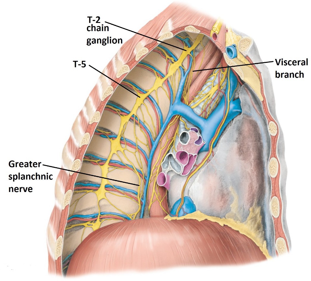
Sympathetic chain ganglia: Visceral branches and splanchnic nerves
Figure 4.6 MOORE ET AL., CLINICALLY ORIENTED ANATOMY, FIGURE 1.70.
All sympathetic chain ganglia have a branch directed medially that supplies visceral organs in the body cavities. Medial branches of cervical and upper thoracic (T-1 to T-5) chain ganglia contain postganglionic fibers supplying thoracic organs. These are simply called visceral branches. Medial branches from lower thoracic, lumbar, and sacral chain ganglia contain preganglionic fibers that supply abdominopelvic organs. By definition, medial branches of chain ganglia containing preganglionic fibers are called splanchnic nerves.
Visceral branches
Medial branches of chain ganglia that contain postganglionic fibers. They supply thoracic organs. Those supplying the heart are called cardiac branches.
Splanchnic nerves
Medial branches of chain ganglia that contain preganglionic fibers. They supply abdominopelvic organs.
Splanchnic nerves in the sympathetic division are named as follows:
Thoracic splanchnic nerves
Lumbar splanchnic nerves
Sacral splanchnic nerves
Medial branches of T-5 to T-12 chain ganglia. Thoracic splanchnic nerves are three in number:
Greater splanchnic nerve (T-5 to T-9)
Lesser splanchnic nerve (T-10 to T-11)
Least splanchnic nerve (T-12)
Medial branches of lumbar chain ganglia.
Medial branches of sacral chain ganglia.
Sympathetic pathways are different for thoracic and abdominal organs
Figure 4.7 MOORE ET AL., CLINICALLY ORIENTED ANATOMY, FIGURE I.44.
Preganglionic sympathetic fibers that supply thoracic organs synapse in paravertebral ganglia. Postganglionic fibers reach thoracic organs via visceral branches.
Preganglionic sympathetic fibers that supply abdominal and pelvic organs synapse in prevertebral ganglia located near the abdominal aorta. Preganglionic fibers pass through paravertebral (chain) ganglia without synapsing and reach the prevertebral ganglia via splanchnic nerves.
Visceral branches and splanchnic nerves are medial branches of chain ganglia aimed at the organs in the body cavities. Visceral branches contain postganglionic fibers; splanchnic nerves contain postganglionic fibers.
Practice drawing these pathways with Interactive 4.1.
Interactive 4.1
Draw sympathetic pathways innervating a thoracic organ and an abdominal organ. Compare. CORNELIUS ROSSE, M.D., D.SC., STUDY GUIDE FOR GROSS ANATOMY AND EMBRYOLOGY, UNIVERSITY OF WASHINGTON. USED WITH PERMISSION. (Tap to open; use your Apple Pencil to draw.)
Pelvic splanchnic nerves
Pay close attention
This topic is always a source of confusion for students.
Splanchnic nerves exist in the parasympathetic division too. Preganglionic parasympathetic fibers reach the intramural ganglia of pelvic organs (and the distal colon) as direct medial branches of the ventral rami of spinal nerves S-2, S-3, and S-4 (NOT through medial branches of paravertebral ganglia = remember that paravertebral ganglia are sympathetic!). These are pelvic splanchnic nerves. The cell bodies of the preganglionic neurons are in the intermediolateral cell column of spinal cord segments S-2 to S-4 (the sacral part of the craniosacral outflow). We will discuss these more in the pelvis. A template for drawing this pathway is in Interactive 4.2.
Figure 4.8 Summary diagram of parasympathetic division of ANS. MOORE ET AL., CLINICALLY ORIENTED ANATOMY, FIGURE I.48.
Interactive 4.2
Draw a parasympathetic pathway innervating an organ in the pelvis. CORNELIUS ROSSE, M.D., D.SC., STUDY GUIDE FOR GROSS ANATOMY AND EMBRYOLOGY, UNIVERSITY OF WASHINGTON. USED WITH PERMISSION. (Tap to open; use your Apple Pencil to draw.)
Autonomic innervation of structures in somatic regions (skin, for example)
Ah-ha!
That’s what gray rami are for!
Only the sympathetic division of the ANS supplies smooth muscle and glands in the somatic regions of the body (body wall and limbs). Functionally, these serve to:
Constrict blood vessels in the skin.
Dilate blood vessels in skeletal muscles.
Stimulate contraction of arrector pili muscles in the skin. These raise the hairs in your skin and cause “goose bumps.”
Stimulate secretion of sweat glands.
Preganglionic cell bodies of sympathetic neurons that supply somatic regions are located in the intermediolateral cell column of spinal cord segments T-1 to L-2. Axons leave T-1 to L-2 spinal cord segments through ventral roots, pass through spinal nerves and ventral rami, and enter the corresponding T-1 to L-2 paravertebral ganglia through white rami communicantes. Since these first neurons in the pathway supply all parts of the body, from head to toe, some fibers ascend or descend in the sympathetic trunk to reach their target levels in the body – kind of like an elevator ride.
The cell bodies of the postganglionic neurons that supply somatic regions are located in paravertebral (chain) ganglia in the cervical, thoracic, lumbar, and sacral regions.
Postganglionic sympathetic fibers leave the chain ganglia through gray rami communicantes (that’s what gray rami are for. And that’s why ALL sympathetic chain ganglia have gray rami!). After passing through the gray rami, postganglionic fibers are distributed to the trunk, neck, and limbs via dorsal and ventral rami of spinal nerves. In the head, postganglionic sympathetic fibers follow arteries to reach their target structures. Interactive 4.3 is a template for drawing this pathway.
Figure 4.9 Summary diagram of sympathetic division of the ANS. MOORE ET AL., CLINICALLY ORIENTED ANATOMY, FIGURE I.46.
Interactive 4.3
Draw 2-neuron sympathetic pathways innervating a sweat gland (A) in the back (use a dorsal ramus) and (B) on the chest or upper limb (use a ventral ramus). CORNELIUS ROSSE, M.D., D.SC., STUDY GUIDE FOR GROSS ANATOMY AND EMBRYOLOGY, UNIVERSITY OF WASHINGTON. USED WITH PERMISSION. (Tap to open; use your Apple Pencil to draw.)
Visceral afferent neurons and pathways
Sensations from visceral structures are carried in visceral sensory neurons. Structurally these are typical pseudo-unipolar neurons. Visceral sensory (afferent) fibers travel in tandem with efferent nerve fibers of both the sympathetic and parasympathetic divisions—but since the information is flowing to the CNS it is traveling in a retrograde direction to the autonomic motor fibers. Technically, theseafferent pathways are not considered part of the autonomic nervous system.However, they are very important since they carry information concerning the body’s internal conditions, including:
Organ pain
Caused by violent smooth muscle contraction or excessive distension, chemical irritation, or lack of blood (ischemia)
Stretch
In blood vessels and air sacs in the lungs, initiating visceral reflexes that adjust blood pressure and respiratory cycles.
Other sensations
Nausea, hunger, satiation (fullness).
Visceral sensations may not come to consciousness (as in the case of visceral reflexes), and those that do are perceived as poorly localized and diffuse.
Let’s generalize a bit to help us get a handle on concepts concerning visceral afferent pathways.
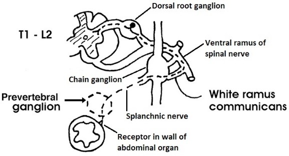
Pain from most organs in the thoracic and abdominopelvic cavities flows to the spinal cord alongsympathetic pathways, but in a reverse direction. The peripheral processes (axons) of the visceral sensory neurons pass to sympathetic chain ganglia through splanchnic nervesand visceral branches, then through white rami communicantes to spinal nerves. The cell bodies of these sensory neurons are in the dorsal root ganglia associated with T-1 to L-2 spinal nerves. Note that since this is a sensory pathway (not an autonomic motor pathway),there is only one neuron involved, not two. There is no synapse within prevertebral or chain ganglia—the sensory fibers are only passin’ through.
Figure 4.10 CORNELIUS ROSSE, M.D., D.SC., STUDY GUIDE FOR GROSS ANATOMY AND EMBRYOLOGY, UNIVERSITY OF WASHINGTON. USED WITH PERMISSION.
Figure 4.10 is an example of a visceral afferent pathway relaying information to the CNS from a generic abdominal organ. Notice the pathway flows in a retrograde direction using the same nervous structures as the sympathetic motor pathway (splanchnic nerve, ganglia, white ramus, etc.).
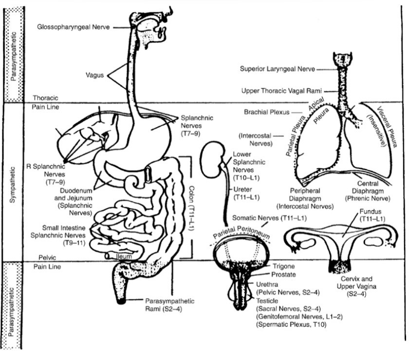
Figure 4.11 “Pain lines” demarcating the ANS divisions that organ pain pathways accompany.
Some organ pain does make its way centrally to the CNS accompanying parasympathetic fibers. These organs, most of which are in the neck and pelvis, are exceptions to the rule. The visceral sensory fibers in these cases flow retrograde through the vagus (Cranial nerve X) and pelvic splanchnic nerves. A useful tool for understanding visceral organ pain pathways is the concept of “pain lines.” A diagram depicting these is shown here. See Figure 4.11. The pelvic pain line roughly follows the course of the peritoneum that dips down into the pelvic cavity, where it drapes over pelvic organs, like a tarp thrown over furniture when the living room walls are being painted. Pelvic organs or their parts that are at or above the pain line (above the peritoneum or touching the peritoneum) send their pain impulses to the CNS via sympathetic pathways in reverse. Pelvic organs or their parts that are below (and not touching) the peritoneum send pain to the CNS along parasympathetic pathways (via pelvic splanchnic nerves). For example, the upper part of the bladder is covered by peritoneum as it drapes from the anterior abdominal wall and dips down into the pelvis. Pain follows sympathetic routes here. The lower bladder is subperitoneal and devoid of peritoneum, so pain here follows pelvic splanchnic nerves.
Non-pain sensations, both conscious (nausea, for example) and subconscious (visceral reflex information, like blood pressure) travel to the CNS accompanying parasympathetic fibers. The glossopharyngeal and vagus nerves (cranial nerves IX and X) for example carry stretch information from the aortic and carotid artery baroreceptors (stretch receptors), respectively.