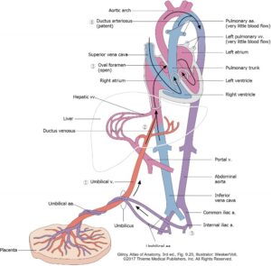
Figure 32.29. Prenatal circulation.
Oxygenated blood reaches the fetus from the placenta via the umbilical vein (within the umbilical cord).
Once in the fetus, the umbilical vein reaches the liver by passing through the falciform ligament.
Much of this blood bypasses the liver through the ductus venosus.
Oxygenated blood enters the right atrium through the inferior vena cava, then passes to the left atrium through the one-way valve in the interatrial septum (foramen ovale).
Blood from the fetus that does enter the right ventricle and pulmonary trunk is shunted to the aorta via the ductus arteriosus.
After passing through the fetal systemic circulation, blood is returned to the placenta through left and right umbilical arteries, branches of the internal iliac arteries in the pelvis.
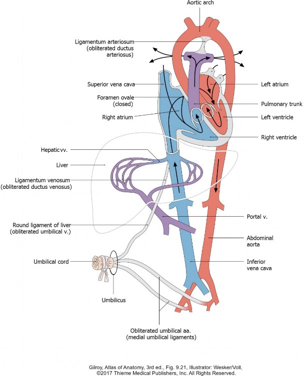
Postnatal circulation
Figure 32.30. Postnatal circulation.
The umbilical vein closes and becomes fibrotic between umbilicus and liver, becoming the round ligament of the liver. This vestigial ligament is located within the falciform ligament.
The ductus venosus and ductus arteriosus close, forming the vestigial ligamentum venosum and ligamentum arteriosum, respectively.
Increased pressure in the left atrium seals the flap-valve in the interatrial septum and the overlapping septa primum and secundum fuse. The fossa ovalis on the right side of the interatrial septum is a landmark indicating the site of the foramen ovale.
The segments of the umbilical arteries between urinary bladder and umbilicus obliterate, forming the medial umbilical ligaments of the anterior abdominal wall.