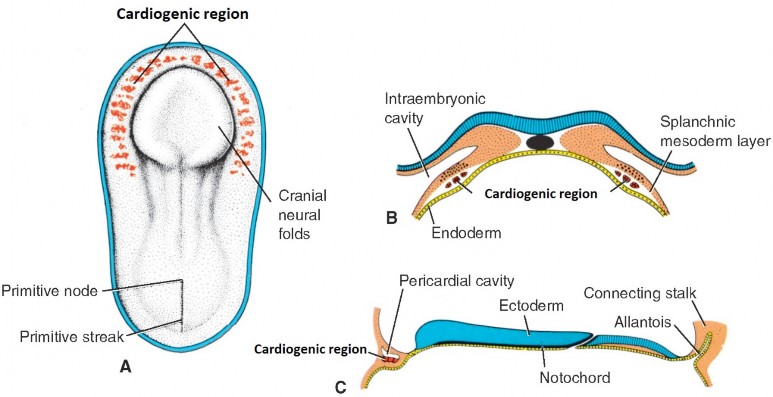
During the 3rd week, clusters of cells derived from splanchnic mesoderm assemble in a horseshoe-shaped cord of tissue located cranial and lateral to the developing neural plate. This is the cardiogenic region. The solid ribbon of cells hollows out to form a horseshoe-shaped primitive endocardial tube.
Figure 32.1. LANGMAN’S MEDICAL EMBRYOLOGY, FIGURE 31.1.
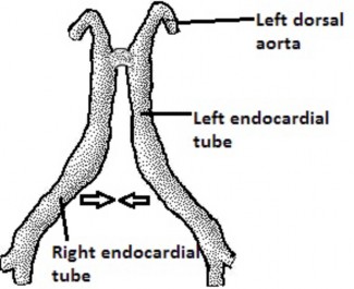
The head fold positions the endocardial tube in the thoracic region cranial to the septum transversum (future diaphragm). Lateral folding brings the two sides of the horseshoe to the midlinewhere they line up side-by-side ventral to the foregut and fuse, producing a single primitive heart tube.
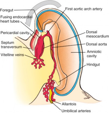
The primitive heart tube (and the adult heart!) has venous and arterial ends. These are the parts of the heart where blood enters and leaves (venous blood in; arterial blood out). The primitive vessels (veins and arteries) that connect to the venous and arterial ends attach to the heart tube at about the same time as the endocardial tubes fuse—so the blood-in and blood-out “plumbing” is established early on.
Figure 32.2.
Figure 32.3. LARSEN’S HUMAN EMBRYOLOGY, FIGURE 12.4.
Blow-your-mind facts
The primitive heart begins beating about Day 22.
Remodeling and septation (division of the heart into right and left halves) occurs while the embryonic heart is pumping blood!
To make our lives easier, let’s develop the heart in two steps: First, we will develop its external form, then we will work on its internal anatomy.