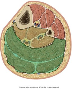
Figure 27.1
7 muscles arranged in a superficial layer (darker green in Figure 27.1) and deep layer (yellow in Figure 27.1)
Superficial muscles: powerful plantar flexors because they support and move body weight
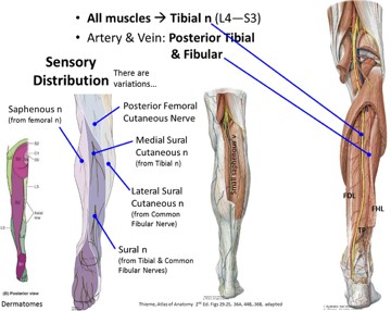
All muscles in the posterior leg are innervated by the tibial nerve and supplied with blood by the posterior tibial vessels
Superficial muscle group
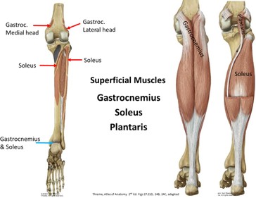
Figure 27.2
The gastrocnemius, soleus, and plantaris are the superficial muscles. The gastrocnemius and soleus form a three part muscle, the triceps surae, and form the prominence of the calf. These muscles act together to plantarflex the foot at the ankle joint, and can easily raise the heel against the weight of the body. It is the gastrocnemius that produces the rapid movements during running and jumping.
Most superficial muscle which is fusiform, two-headed, and two-jointed (crosses two joints)
Origin: Condyles of femur
Medial head: Is slightly larger and extends more distally than lateral head
Lateral head: Sometimes (3–30%) contains a sesamoid bone, the fabella (L. bean), close to its origin and is often mistaken for a loose body or osteophyte
Insertion: Calcaneus via calcaneal (Achilles) tendon, shared with soleus muscle
Action: Vertical fibers cause rapid movements during running and jumping; assist in steadying the legs; active during standing, even at ease; very active when standing on the toes; acts on both knee and ankle joints, but can’t exert full force on both at the same time
Nerve: Tibial nerve (S1,2); test S1,2 by walking on toes
Broad, flat, multipennate fatigue-resistant, slow-twitch muscle very powerful, but its contractions are slower than those of the gastrocnemius
Can be palpated on each side of gastrocnemius, inferior to the mid-calf, when a person is standing on tiptoes (or squatting)
Horseshoe shaped origin on tibia and fibula: Popliteal artery dives deep to the soleus and divides into anterior and posterior tibial arteries
I: Calcaneus with gastrocnemius via Achilles tendon
A: With gastrocnemius, plantarflexion
Maintenance of posture: Steadies the leg on the foot (prevents your body from falling anteriorly when standing, antagonistic to dorsiflexors, by pulling on the tibia and fibula)
Small fusiform belly, might be absent (10%) or even double
Long and slender tendon runs between soleus and gastrocnemius lateral to medial
Can be used as an allograft for the hand
A: Foot proprioception (high density of muscle spindles)
N: Tibial nerve
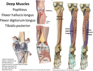
Deep muscles in the posterior leg
Four muscles comprise this group: the popliteus, flexor digitorum longus, flexor hallucis longus, and tibialis posterior. The popliteus acts on the knee joint, whereas the other muscles act on the ankle and foot joints (but are poor plantar flexors).
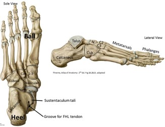
Figure 27.3 Use this figure to reacquaint yourself with the foot bones.
Flat triangular muscle that forms the floor of the inferior part of the popliteal fossa
O: Arises inside the knee joint capsule, from the lateral condyle of femur in joint capsule (deep to lateral collateral ligament), and lateral meniscus
I: Posterior tibia
Action: Probably the most important function of the popliteus muscle is to “unlock” the knee from the close-pack (maximally congruent) position when the knee is fully extended. This has to happen before flexion of the knee can begin. The popliteus can laterally rotate the femur or medially rotate the tibia, depending on which bone is stabilized. The rotation produced by the popliteus is not dramatic, only a few degrees, but enough to allow for knee joint flexion to begin.
Tendon passes posterior to medial malleolus, deep to flexor retinaculum (within the tarsal tunnel)
As it travels to the great toe, it runs between 2 sesamoid bones (protection from pressure of the head of the first metatarsal) in the tendons of the flexor hallucis brevis (intrinsic foot muscle)
A: Powerful “push-off” muscle during walking, running, and jumping. It provides much of the spring to the step and is important in holding the leg in the normal position on the foot.
Flexes the great toe (at all joints), plantarflexes the foot, and supports the medial longitudinal arch of the foot.
Largest, deepest, fusiform muscle between flexor digitorum and hallucis longus muscles
I: Navicular tuberosity, cuneiform, cuboid, base of metatarsals 2–4
Tendon seen and felt just posterior to the medial malleolus,especially when the foot is everted against resistance
A: Inverts and plantarflexes the foot; in gait, it fixes the medial longitudinal arch in the sole of the foot during weight bearing
N: Tibial nerve (T4,5)
Nerves and vessels of the posterior leg (and sole of the foot )
Figure 27.5All posterior compartment muscles are supplied by the tibial nerve. (Yay!)
Cutaneous supply: Posterior cutaneous nerve of the thigh, lateral sural cutaneous nerve (from the common fibular nerve), medial sural cutaneous nerve (from the tibial nerve), and saphenous nerve (from the femoral nerve)
Arteries
Veins of posterior leg
Posterior tibial artery, the continuation of the popliteal artery, and the fibular artery, a branch of the posterior tibial artery in the deep posterior compartment
The posterior tibial vessels, tibial nerve, and fibular vessels run in the connective tissue plane that separates the superficial and deep layers of the posterior compartment
The small (short) saphenous vein begins on the lateral side of the dorsum of the foot, passes behind the lateral malleolus, then ascends almost straight up the center of the posterior leg in the superficial fascia. It penetrates the deep fascia over the popliteal fossa and drains in to the popliteal vein. It is accompanied in the calf by the sural nerve. A pair of deep veins, the posterior tibial veins, accompany the artery of the same name in the posterior compartment. The posterior tibial veins are tributaries of the popliteal vein.
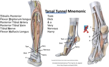
Flexor retinaculum of the ankle
Figure 27.6
Y-shaped thickening of the deep fascia of the leg, and passes from the medial side of the calcaneus to themedial malleolus.
Tendons of the long deep muscles pass deep to theflexor retinaculum, within the tarsal tunnel
At medial malleolus: tendons, nerve, andvessels are regularly arranged from superficial to deep and are important in surgery of this area (e.g., provide a posterior tibial nerve block at the ankle)