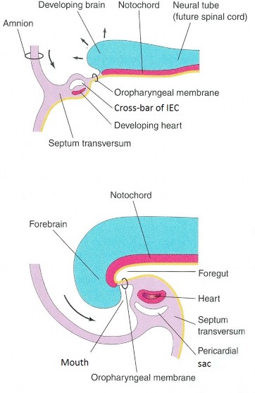
The rapid growth of the cranial part of the neural tube (brain) induces the cranial rim of the embryonic disc to fold and “tuck under.” This produces the ventral surface of the future head, neck, and chest.
Within the “head” mesoderm are subsets of tissues that will form the heart (cardiogenic mesoderm) and the diaphragm (septum transversum). These primordial tissues along with the oropharyngeal membrane (future mouth) are located cranial to the developing neural tube before folding. After head folding,the definitive locations of the diaphragm and heart in the thorax, and the opening of the mouth on the face, are established, caudal to the brain. See the before and after in Figure 2.22.
Summary: Results of the head fold
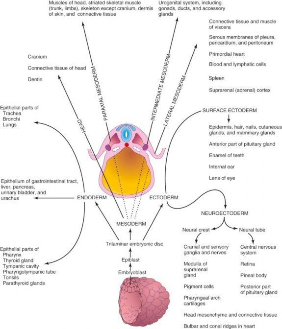
- The ventral parts of the head, neck, and thorax are created from ectoderm and somatic mesoderm. The ventral wall of the thorax encloses the upper part of the body cavity = the thoracic cavity. Three serous sacs will develop within the thoracic cavity.
- The endoderm and associated splanchnic mesoderm in the cranial region of the embryonic disc are rolled into a tube and located within the embryo dorsal to the mouth and heart, producing the cranial part of the digestive tube (= the foregut). Endoderm gives rise to the inner epithelium and splanchnic mesoderm surrounds the endoderm, giving rise to smooth muscle, connective tissues, and blood vessels of the foregut.
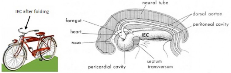
- The “cross-bar” portion of the intra-embryonic coelom and the developing heart are placed ventral to the foregut, the developing heart being dorsal to the IEC, between it and the gut. The part of the IEC adjacent to the heart will become a serous sac known as the serous pericardium. The cross-bar part of the intra- embryonic coelom is bent forward and now takes on the shape of (old-fashioned) “bicycle handlebars,” rather than a horseshoe. The cross bar portion of the handlebars will be partitioned into three serous sacs = the serous pericardium and two pleural sacs.
Figure 2.22 MOORE ET AL., THE DEVELOPING HUMAN, FIGURE 8-4. - The septum transversum, the cranial-most part of the head mesoderm, is moved caudal to the future heart and pericardium, between these structures and the neck of the yolk sac. The septum transversum will give rise to a major part of the future diaphragm.
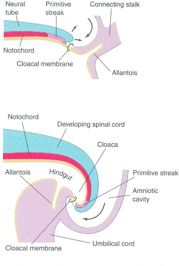
Similar to the head fold, the growing neural tube, amniotic sac, and somites overgrow the caudal rim of the embryonic disc. The caudal portion of the embryonic disc containing the cloacal membrane folds under the dorsal embryonic surface, becoming part of the embryo’s ventral surface. This explains the location of the anus anterior to the tip of the coccyx (and you thought the anus was on the “backside” of the body!). The tail fold also carries the connecting stalk (connecting the caudal end of the embryonic disc to the developing placenta) cranially until it abuts the yolk sac. See the before and after figures in Figure 2.23.
Summary: Results of the tail fold
-
The lower part of the ventral body wall is formed from ectoderm and somatic mesoderm.
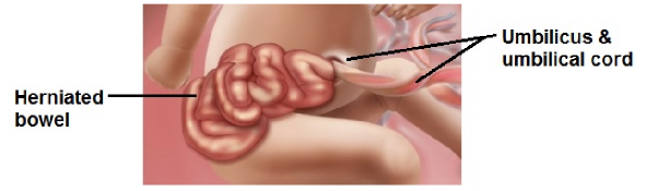
- The connecting stalk attaches to the ventral body wall. The connecting stalk (and other tissues) gives rise to the umbilical cord. The tail fold translocates the developing umbilical cord to the ventral body wall, thus producing the region of the region of the abdominal wall that contains the future umbilicus (“belly button”).
- Endoderm in the caudal region of the embryonic disc is rolled into a tube and placed within the folded embryo, giving rise to the epithelial lining of the tail-end of the embryonic digestive tube (= the hindgut). Splanchnic mesoderm surrounds the endoderm, forming smooth muscle, blood vessels, connective tissues, and serous membranes of organs of the hindgut (e.g., descending colon, rectum).
Note: The allantois is a diverticulum of the hindgut that figures later in the development of the urinary bladder.
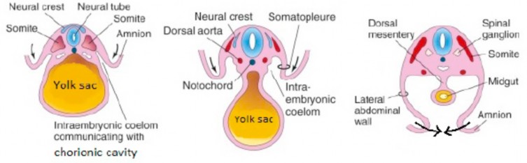
Concurrent with caudal and cranial folding, the embryonic disc is also folding ventrally along its left and right edges. Lateral folding of the disc will give the embryo its oval shape as viewed in transverse section. The two lateral folds, the head fold, and the tail fold all tuck under, ventrally; converging like the edges of a purse being closed by a purse- string around the yolk sac and umbilical cord. Folding of the embryonic disc causes the yolk sac to become elongated and constricted where the caudal, cranial, and lateral rims of the embryonic disc meet and impinge upon it. This connection between yolk sac and folded embryo is called the vitelline duct. It later regresses and should disappear.
Summary: Results of lateral folding
-
Ectoderm and somatic mesoderm from the left and right sides of the embryonic disc fuse in the midline completing the ventral body wall and enclosing the body cavity. Later, the development of the diaphragm separates the single body cavity into thoracic and abdominopelvic cavities.
- The right and left coelomic ducts (of the IEC) come together and fuse to form a serous sac in the abdominopelvic cavity (= the peritoneal sac). Somatic mesoderm gives rise to the parietal peritoneum; splanchnic mesoderm forms the visceral peritoneum.
- Endoderm in the central region of the embryonic disc is rolled into a tube forming the epithelial lining of the mid-portion of the primitive digestive tube (= the midgut). Splanchnic mesoderm surrounds the endoderm, forming the smooth muscle, blood vessels, connective tissues, and serosa of midgut organs.
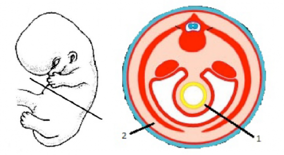
Note that the tube-within-a-tube design of the vertebrate body is now complete. The gut tube (tube 1) is located within the body cavity surrounded by the body wall (tube 2). This distinction between the “inner” and “outer” tubes is important because they have different origins and innervations: