Trophoblast goes wild
- Early in the second week, some trophoblast cells lose their cell membranes and coalesce into a mass of cytoplasm with many scattered nuclei (= a syncytium). Called the syncytiotrophoblast, this invasive mass burrows into the spaces between endometrial cells, pulling the blastocyst into the endometrium.
- The trophoblast cells internal to the syncytiotrophoblast and adjacent to the blastocyst cavity maintain their cell membranes. They are collectively known as the cytotrophoblast. As these cells divide, some lose their cell membranes and “seed” the syncytiotrophoblast.
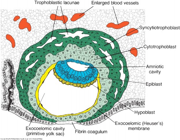
Figure 2.6 LANGMAN’ S MEDICAL EMBRYOLOGY, 12TH ED., FIGURE 4.3.
The bilaminar disc
Cells of the embryoblast differentiate into two layers:
-
- The cells facing the embryonic pole become columnar in shape—this layer is the epiblast. The epiblast defines the dorsal side of the disc.
- The cells facing the blastocyst cavity are cuboidal—this layer is the hypoblast. The hypoblast defines the ventral side of the disc.
- We now have a two-layered embryoblast = the bilaminar embryonic disc.
Amniotic cavity and yolk sac
- Epiblast cells proliferate and a fluid-filled space develops within the cell mass = the amniotic cavity. The dome-shaped roof of epiblast cells around the cavity is the amnion. The amniotic cavity and amnion together produce the amniotic sac.
- On the other side, cells derived from the hypoblast migrate away from the bilaminar disc, in a direction opposite from the amniotic sac. These cells form a membrane that lines the inner aspect of the cytotrophoblast, adjacent to the blastocyst cavity. This new cell layer of hypoblast origin and the cavity it surrounds (former blastocyst cavity) are referred to as the primitive yolk sac. Later it will be modified to form a definitive yolk sac. The yolk sac has several important functions:
- The yolk sac is the site of early hematopoiesis (= formation of blood cells). Blood vessels that form around the yolk sac are called vitelline vessels. These are the precursors of the blood vessels that supply the adult gastro- intestinal tract.
-
- The yolk sac gives rise to the primordial sex cells (= cells that give rise to sperm and oocytes) that later migrate and populate the developing gonads.
The double bubble
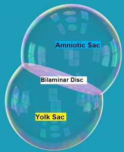
Figure 2.7
The bilaminar embryonic disk is situated between two fluid-filled sacs. Facing toward the endometrium is the amniotic sac. On the other side, facing toward the uterine cavity, is the yolk sac. This stage of development can be thought of as a “double bubble”, with the bilaminar disc sandwiched between the two sacs.
Extra-embryonic mesoderm (EEM)
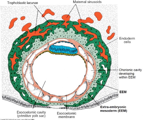
Figure 2.8 LANGMAN’ S MEDICAL EMBRYOLOGY, 12TH ED., FIGURE 4.4.
- A new, loosely organized tissue layer develops between the cytotrophoblast and double bubble = the extra-embryonic mesoderm (EEM). Its origin is uncertain—it may originate from hypoblast. Fluid-filled cavities appear in the EEM and coalesce into a single space called the chorionic cavity. The EEM has now been split into two layers—one lines the outside of the double bubble and the other layer lines the inside of the cytotrophoblast.
- A three-layered membrane is formed that surrounds and defines the chorionic cavity = the chorion. It is composed of EEM, cytotrophoblast, and syncytiotrophoblast. The chorion is the membrane that ruptures when a mother’s “water breaks.”
- Formation of the chorionic cavity is an important step = tissues that will form the embryo (double-bubble) are now physically separated from tissues that will form the placenta (chorion). The developing embryo now has room to grow within the chorionic cavity.
- The double bubble is not free floating in the chorionic cavity—it is attached to the chorion via a connecting stalk made of extra-embryonic mesoderm. The connecting stalk is the forerunner of the umbilical cord. The double bubble resembles an astronaut taking a “space walk,” tethered to the spaceship via the connecting stalk.
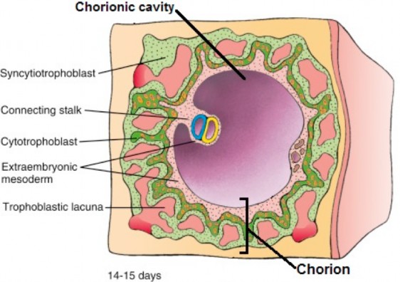
Figure 2.9 LARSEN’ S HUMAN EMBRYOLOGY, 5TH ED., FIGURE 2-6.
Question
The trophoblast differentiates into which two parts?
Cytotrophoblast and syncytiotro-phoblast.
Question
The embryoblast arranges into which two layers?
Epiblast and hypoblast.
interactive
Embryonic and extraembryonic structures of Week 2. Can you identify these? (tap to open; use your Apple Pencil to draw and make notes)
HAND-DRAWN CONLEY- GRAM.
Meanwhile, back at the trophoblast
- The syncytiotrophoblast is an invasive and digestive mass—it secretes proteolytic enzymes that break down proteins in the endometrial stroma (the connective tissue framework that supports the endometrial cells), allowing the syncytiotrophoblast to invade deeper into the endometrium.
- Spaces (lacunae) appear within the syncytiotrophoblast that fill with blood, glandular secretions, lipids, and glycogen —from maternal capillaries, uterine glands, and the decidual cells, respectively. These spaces form a lacunar network within the syncytiotrophoblast, giving it a sponge-like appearance. Maternal blood containing oxygen and nutrients enters the lacunar network from uterine capillaries eroded by the syncytiotrophoblast, and these “goodies” reach the embryonic disc by diffusion through the chorion and double bubble. Waste products from the embryo, as well as hCG from the syncytiotrophoblast, enter the lacunar network and are drained away by maternal endometrial veins.
- This primitive circulation is sufficient for the needs of the tiny embryonic disc. However, as the embryo grows it will no longer suffice. Toward the end of the second week a more complex circulation begins to develop within the connecting stalk that transfers blood between capillaries in the chorion (these are adjacent to the lacunar network, but separated from maternal blood by a thin membrane) and blood vessels in the embryo. These vessels that link placenta and embryo are the umbilical arteries and veins.
It is important to recognize, however,
that fetal and maternal blood never actually mix.