- Optional Reading
- Embryology resources
- Langman’s Medical Embryology, 15th ed., chapters 3, 4, 5, and 6.
There are a number of good Internet sites dealing with human embryology, some with animations. We have included a few here:
-
- Medical Animations: YouTube channel devoted to embryology animations.
- Embryology: G’day! Dr. Hill’s site in Australia has a staggering amount of material ranging from serial sections to movies.
- Human Embryology Animations: Nice cartoons and animations of development (especially the heart!).
- The Visible Embryo: This site also has a nice summary timeline of human development.
Why study embryology?
 You will discover that the study of the developing human illuminates gross anatomy. That is, you will understand why body structures have their anatomic relationships if you know something about their development. For example, why is the origin of the recurrent laryngeal nerve from the vagus nerve so much lower on the left side of the body than on the right?
Hmmm . . .
This will become clear in a few short weeks.
Knowledge of human development also explains anatomic variations (which you will see plenty of in the gross lab) and the causes of certain congenital anomalies (birth defects). Variation in human anatomy is obviously important clinically. You will discover that variation among bodies is actually the norm, rather than the exception.
Terminology
The period of intra-uterine development of the human.
A period of three calendar months. Clinicians divide the gestation period into three trimesters. The most dramatic and critical stages of development occur during the 1st trimester.
Using the last normal menstrual period as the reference point. Gestation lasts 40 weeks using this calendar.
Using the estimated day of fertilization as the reference point. Gestation lasts 38 weeks. In our discussions, we will use fertilization age when describing development.
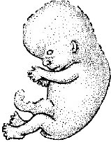
The first eight weeks of development. This is a busy time with the most striking advances in development. All of the body’s organ systems have formed by the end of the embryonic period. This period is of most interest to us as we study gross anatomy.
Fetus is the term used to describe the developing human after the completion of the embryonic period. The fetal period therefore is between the ninth week and birth.
Let’s be clear that development doesn’t cease at birth—birth is simply a dramatic change in environment—you’d cry, too, if you had to leave the warmth and security of the uterus to enter the cold cruel world! Development of the human isn’t completed until the mid-twenties. We will use the term “adult” when describing the anatomy of the fully developed human.
Descriptive terms
Many terms of direction and relationship used for adult anatomy are also used in embryos. However, since it is hard to place an embryo in the anatomical position, there are a couple sets of terms that are of particular importance when describing embryonic anatomy:
Measuring the size of embryos and fetuses
Crown-rump length is most commonly used. Ultrasound crown-rump length measurements are useful for estimating the age of fetuses. (See Figure 2.1.)
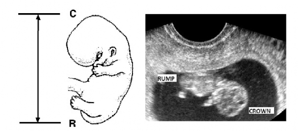
Figure 2.1 Examples:
- A 4-week-old embryo has a crown-rump length of 4mm;
- at 8 weeks the crown-rump is 30mm;
- at 17 weeks the crown-rump is 15 cm!!
Rapid growth in the fetus!
Embryogenesis begins at fertilization
That’s where our story begins. Fertilization normally occurs in the ampulla of the uterine (Fallopian) tube.
The results of fertilization:
- Stimulates the oocyte (“egg”) to complete the second meiotic division.
- Through mingling of maternal and paternal chromosomes, it produces a genetically unique unicellular embryo called a zygote.
- Restores the zygote to a diploid number of chromosomes (46).
- Determines the sex of the embryo.
- Initiates cleavage (cell division) in the zygote.
Fertilization is the zero time point of embryonic development. The fertilization age of the embryo is calculated from this point.
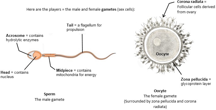
Figure 2.2
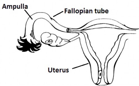
Figure 2.3