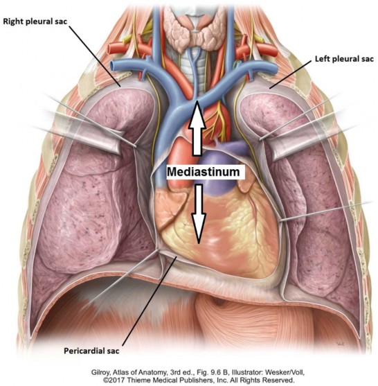
The mediastinum is the mass of tissue between the two pleural sacs. It essentially contains all the internal organs of the thorax, except for the lungs and pleural sacs. The overall boundaries of the mediastinum are:
Superior thoracic aperture above: The mediastinum communicates with the neck through the aperture.
Diaphragm below: Openings in the diaphragm allow limited communication between mediastinum and abdominal cavity.
Sternum and costal cartilages anteriorly
Thoracic vertebral bodies posteriorly
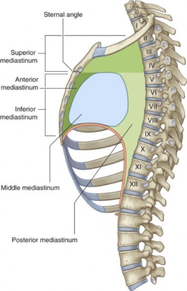
Figure 14.2 Subdivisions of mediastinum. GRAY’S ANATOMY FOR STUDENTS, 3RD ED., FIGURE 3.52.
The mediastinum is divided into superior and inferior portions. The inferior mediastinum is further divided into: anterior, middle, and posterior parts.
Students will need to know the contents of the mediastinum and their relationships. This is especially important for interpreting radiologic images of the thorax. Here are a few pointers to help you get the lay of the land:
The trachea and esophagus are in the midline—these are structures derived from the primitive gut tube.
Major veins predominate on the right side of the mediastinum. If you recall the two left-to-right venous shunts discussed previously in the chapter on heart development, and the fact that deoxygenated blood needs to reach the right side of the heart, this will make sense.
Major arteries predominate on the left. This is due to asymmetrical development of the aorta and the aortic arch arteries, discussed in the chapter on heart development.
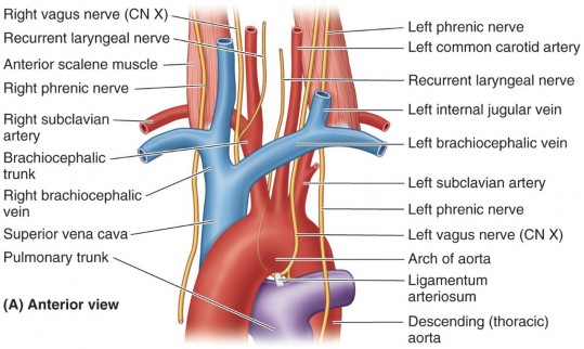
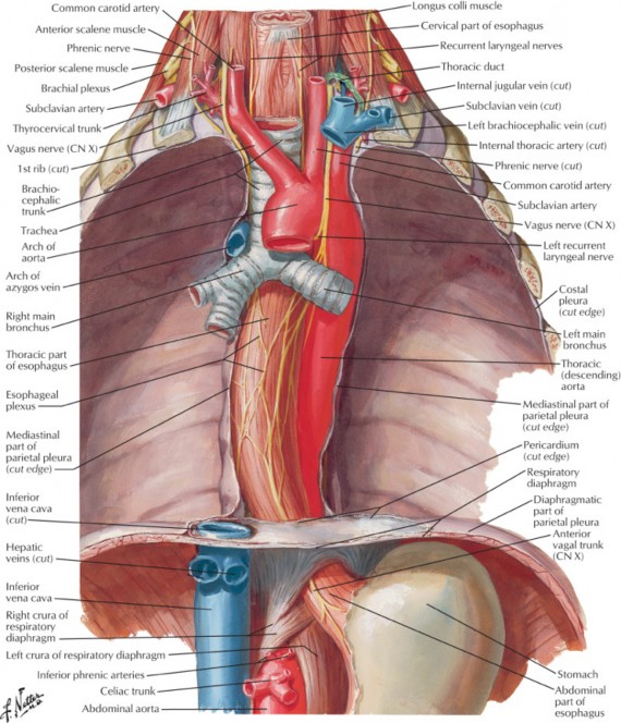
The two major nerves of the mediastinum are the phrenic and vagus nerves. Their courses through the mediastinum relate to the roots of the lungs.
The phrenic nerves target the diaphragm—they course anterior to the lung roots, curving around the pericardium.
The vagus nerves target the esophagus (they are motor to the gut tube). They pass posterior to the lung roots to reach the esophagus.
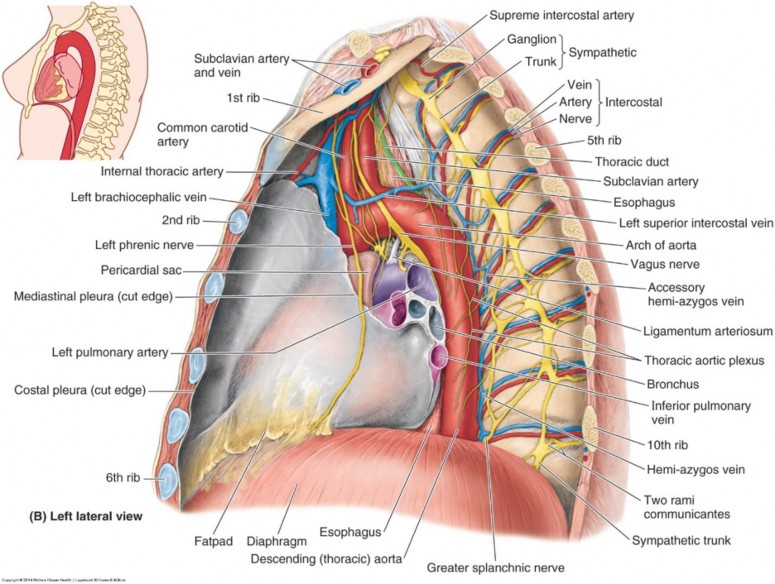
Figure 14.3 Left lateral view of mediastinum. CLINICALLY ORIENTED ANATOMY, 7TH ED., FIG. 1.70.
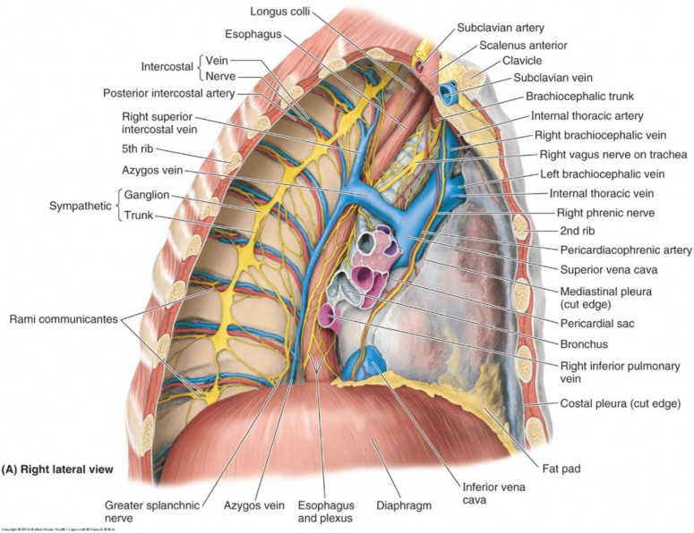
Figure 14.4 Right lateral view of mediastinum. CLINICALLY ORIENTED ANATOMY, 7TH ED., FIG. 1.70.
Superior mediastinum
Boundaries:
Above: Superior thoracic aperture
Below: Horizontal plane at T4/T5 (through the sternal angle)
Continuous with the neck superiorly. This is important clinically as infections from the neck can reach the mediastinum via this continuity.
Main contents:
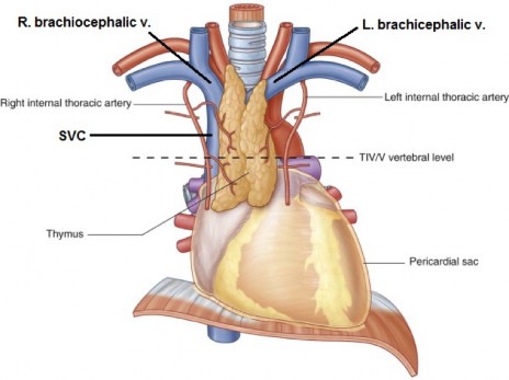
Thymus
Superior vena cava (SVC)
Right and left brachiocephalic veins (RBCV and LBCV)
Arch of aorta and branches = brachiocephalic artery, left common carotid artery, and left subclavian artery
Vagus nerves, phrenic nerves, cardiac plexus, and left recurrent laryngeal nerve (most unlabeled)
Trachea, esophagus, and thoracic duct
Figure 14.5 Contents of superior mediastinum. GRAY’ S ANATOMY FOR STUDENTS, 3RD ED., FIG. 3.81.
Figure 14.6 Nerves of superior mediastinum. CLINICALLY ORIENTED ANATOMY, 7TH ED., FIGURE 1.68.
The inferior mediastinum is below the superior mediastinum: its boundary above is a plane that passes through the sternal angle and the intervertebral disc between T-4/T-5; the boundary below is the diaphragm. The inferior mediastinum is subdivided into three regions, with reference to the pericardium.
A narrow space between sternum and pericardium. Not too important to us in gross anatomy. It contains the sternopericardial ligaments and part of the thymus. The internal thoracic vessels are nearby, traveling in the neurovascular plane of the body wall adjacent to the anterior mediastinum. It is continuous with the superior mediastinum.
Defined as the pericardium and its contents. These include the heart and the roots (proximal portions) of the great vessels: ascending aorta, pulmonary trunk, SVC, IVC, and pulmonary veins. The phrenic nerves are tethered to the fibrous pericardium, so they should probably be included in the middle mediastinum.
Narrow space between the posterior side of the pericardium and the vertebral column. It contains tubular structures oriented vertically, many of which pass between the neck and abdominal cavity. Contents include: Thoracic aorta and its branches, thoracic duct, bifurcation of trachea and main bronchi, tracheobronchial lymph nodes, azygos and hemiazygos veins, esophagus, esophageal plexus, vagus nerves, sympathetic trunk, and greater splanchnic nerves. It is continuous with the superior mediastinum.
Details of mediastinal contents
Great vessels
Aorta
Ascending: comes directly out of the left ventricle and gives off the coronary arteries. It passes anterior to the right pulmonary artery.
Arch of aorta (in the superior mediastinum)
Begins and ends at the level of the sternal angle, arches superoposteriorly to the left. It passes superior to the right pulmonary artery.
3 branches supply the head, neck, and upper extremity, and arise posterior to the manubrium in the superior mediastinum. Ordered as they branch from the arch:
Right brachiocephalic trunk/artery
Left common carotid artery
Left subclavian artery
Descending thoracic aorta
Begins on the left side of T4 and continues inferiorly on the left sides of the thoracic vertebral bodies.
Gives off the left and right posterior intercostal arteries, which supply the body wall. Which of these vessels is longest: left or right?
Bronchial arteries (to air tubes) and esophageal arteries are normally branches.
Pulmonary trunk
Carries deoxygenated blood from the right ventricle to the lungs.
Located to the left of the ascending aorta.
Bifurcates into the left and right pulmonary arteries. Left pulmonary artery is short and vertical. Right pulmonary artery is longer and horizontal. It passes to the right under the arch of the aorta. The tracheal bifurcation is directly posterior to the right pulmonary artery.
Ligamentum arteriosum: connects the left pulmonary artery to the aortic arch.
Left recurrent laryngeal nerve: courses around the ligamentum arteriosum to ascend between the esophagus and trachea toinnervate the larynx.
Superior Vena Cava (SVC)
Formed at the level of the right 1st costal cartilage, from the union of the right and left brachiocephalic veins
Brachiocephalic veins: formed by the union of the internal jugular and subclavian veins. Left brachiocephalic vein: longer,passes anterior to the three vessels that come off the aortic arch (recall the embryology of this vessel!).
Receives blood from all regions of the body above the diaphragm (except the heart)
Enters the right atrium at about the level of the 3rd costal cartilage
Located in both the superior and middle mediastina
Inferior Vena Cava (IVC): is very short in the thorax; it enters the thoracic cavity through the caval opening in the diaphragm anddrains into the right atrium at about the right 5th costal cartilage.
Drains blood from the abdominopelvic cavity and lower extremities
Located in the middle mediastinum
Inferior Vena Cava (IVC)
Very short in the thorax, it enters the thoracic cavity through the caval opening in the diaphragm anddrains into the right atrium at about the right 5th costal cartilage.
Drains blood from the abdominopelvic cavity and lower extremities
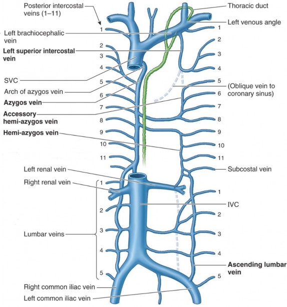
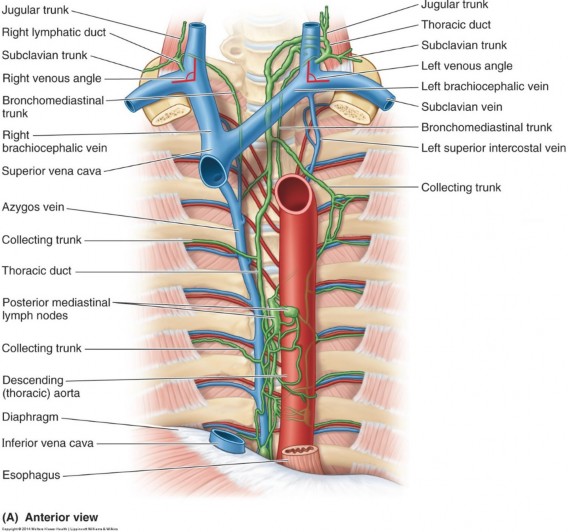
In the posterior mediastinum, the azygos system of veins consists of two vertical venous channels on either side of the vertebral column: the azygos vein proper on the right and the hemi-azygos veins on the left. These channels receive the right and left posterior intercostal veins. The azygos system drains the vertebral column, thoracic walls, and the viscera of the mediastinum. It connects to the lumbar veins in the posterior abdominal wall.
The upper three intercostal spaces on the left are usually drained by an independent left superior intercostal vein. It may communicate below with the accessory hemiazygos vein or above with the left brachiocephalic vein. Clear as mud, eh?
Nerves of the mediastinum
Figure 14.9 Left vagus and phrenic nerves. GRAY’S ANATOMY FOR STUDENTS, 3RD ED., FIG. 3.88.
Travel in both the superior and middle mediastina and have a sensory (from the parietal pleura and pericardium) anda motor (to the diaphragm) component
Right phrenic: Enters the superior mediastinum lateral to the right brachiocephalic vein, and it descends along the SVC. Then it courses between the pericardium and lung before entering the diaphragm.
Left phrenic: Descends behind the left brachiocephalic vein and crosses the arch of the aorta, anterior to the vagus. It runs between the pericardium and lung, superficial to the left atrium and left ventricle.
Both: Pass anterior to the root structures of the lung
Exit the base of the skull and pass through the neck behind the common carotid arteries. Pass through both the superior and the posterior mediastina.
The vagi supply PRE-ganglionic parasympathetic branches to the cardiac, pulmonary, and esophageal plexuses
Right vagus: Crosses the right subclavian artery; gives off a right recurrent laryngeal nerve that loops posterior to the right subclavian artery and ascends superiorly along the trachea to supply the larynx. In the superior mediastinum, the right vagus isadjacent to the trachea, then slides behind the arch of the azygos vein and root of the lung to reach the esophagus.
Left vagus: Enters the superior mediastinum between the left common carotid and left subclavian artery, crosses the aortic arch, and gives off the left recurrent laryngeal nerve that passes under the arch of the aorta and posterior to the ligamentum arteriosum as stated earlier. The left vagus then moves posterior to the root of the lung to reach the esophagus.
Both: in posterior mediastinum, they initially descend on left and right sides of the esophagus. Here they ramify, providing many branches that contribute to the esophageal plexus. On the distal esophagus, the vagal fibers reform, but change positions and names becoming the anterior and posterior vagal trunks. These pass with the esophagus through the diaphragm to supply organs in the abdomen.
This is a chain of interconnected autonomic ganglia (post-ganglionic cell bodies); located in the posterior mediastinum alongside the vertebral column, where the heads of the ribs articulate with the vertebral bodies. See the lateral views of the mediastinum in the figures earlier in this chapter.
These are branches of the sympathetic trunk, containing pre-ganglionic sympathetic fibers from the chain ganglia located at T-5 to T-9. These pass through the diaphragm to supply abdominal organs. More on these and the autonomic nervous system to come in a later chapter. See the lateral views of the mediastinum in the figures earlier in this chapter.
Thoracic duct
Figure 14.10 Course of thoracic duct. CLINICALLY ORIENTED ANATOMY, 7TH ED., FIG. 1.73.
Largest lymph vessel in the body—it returns 75% of the body’s interstitial fluid to the heart from the lower extremities, pelvis, abdomen, left side of the thorax, left upper extremity,and left side of the head and neck. Passes through both superior and posterior mediastina.
Small and beaded (many valves); originates from a saccular structure in the abdomen (the cisterna chyli) at T12. Enters the posterior mediastinum through an opening in the diaphragm.
Lies on the vertebral bodies in the posterior mediastinum. It is covered by the esophagus and sandwiched between the thoracic aorta and the azygos vein.
At the level of the T-4 vertebra, it veers to the left side of the superior mediastinum.
Enters the neck through the superior thoracic aperture.
Hooks anteriorly to drain into the left venous angle (otherwise known as the junction of the left internal jugular vein and the left subclavian vein, or the jugulosubclavian angle). During its course, it will receive lymph from many lymphatic collecting trunks.
The other 25% of the body’s lymph is drained by the right lymph duct.
Thoracic esophagus
Figure 14.11 Relationships of esophagus. NETTER, ATLAS OF HUMAN ANATOMY, 7TH ED., PLATE 236.
The esophagus has three parts: cervical, thoracic, and abdominal.
The esophagus enters the mediastinum via the superior thoracic aperture. It passes through both superior and posterior mediastina. For most of its course, it is centered over the thoracic vertebral bodies. The thoracic duct and azygos vein are behind it. The tracheal bifurcation is anterior. Distally, it inclines to the left and leaves the posterior mediastinum through the esophageal hiatus in the diaphragm.
When empty, the esophagus is collapsed (has no lumen).
It has important relationships in the mediastinum:
To its left is the descending aorta.
Below the tracheal bifurcation, the anterior wall of the esophagus is related to the pericardium, and through it, the oblique pericardial sinus and the left atrium of the heart (base of the heart).
On its anterior surface is the esophageal plexus. This contains a mixture of sympathetic, parasympathetic, and visceral afferent nerve fibers.