Pleura and pleural sacs
Pleura is a serous membrane associated with the lungs. Pleura comes in two varieties:
- Costal pleura named for the ribs it lines.
- Diaphragmatic pleura covers the superior surface of the diaphragm.
- Mediastinal pleura covers the lateral sides of the mediastinum.
- Cervical pleura (a.k.a. cupula) projects above rib 1, where it forms a dome above the apex of the lung.

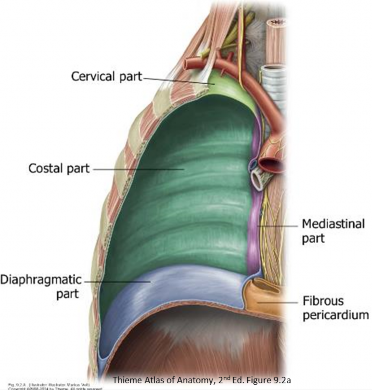
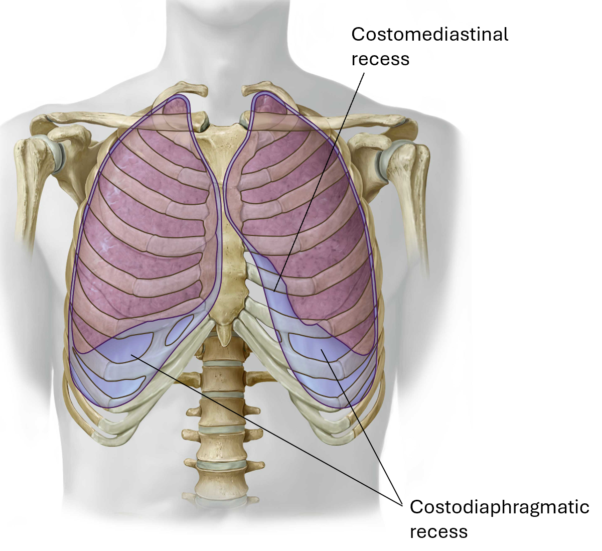
The four parts of the parietal pleura form a continuous layer. Sharp transitions occur where one part changes direction to become another, which forms recesses of the pleural cavities:
-
- Costomediastinal recess: Costal pleura leaves the anterior thoracic wall to become mediastinal pleura.
- Costodiaphragmatic recess: Costal pleura reflects onto the diaphragm
Pleural Cavity

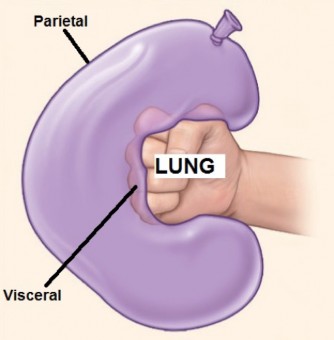
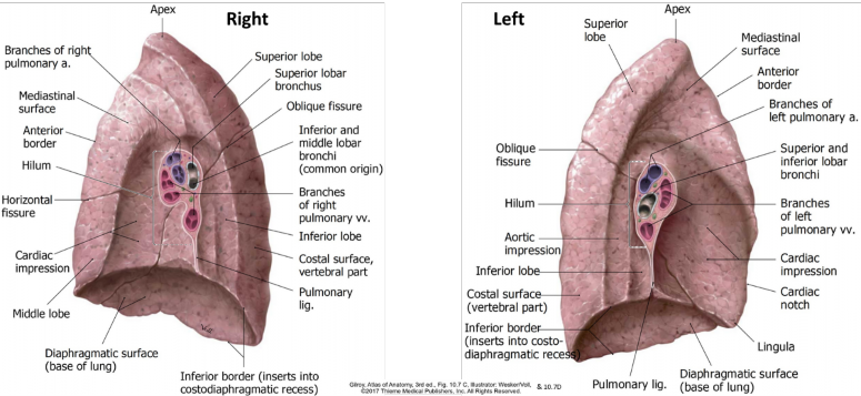
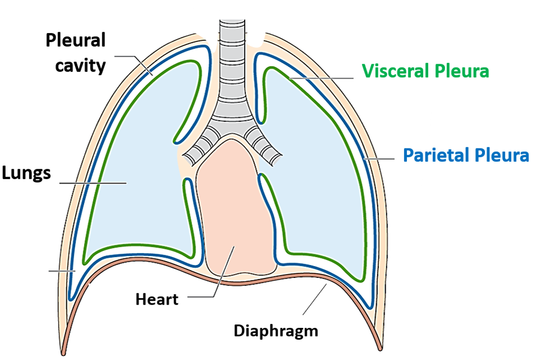
The visceral and parietal layers of pleura are continuous. This continuity occurs where the mediastinal pleura forms a cuff around the structures entering and leaving the lung (root of the lung), and from here, doubles back onto the surface of the lung to become visceral pleura. The parietal and visceral layers of pleura thus form a closed sac around the lungs = the pleural sac. Within the sac is the pleural cavity, which contains a small amount of lubricating serous fluid that allows the parietal and visceral pleurae to slide across each other without friction during respiration.
Surface projections of the pleural cavities and lungs
In quiet breathing, the inferior borders of the lungs remain about two ribs higher than that of the inferior extent of the parietal pleura in the costodiaphragmatic recesses. Use the “rule of twos” to locate the positions of lung and parietal pleura on the body wall, as illustrated in Figure 13.5.
Question
Why would knowing the two-rib gap between lung and parietal pleura be clinically useful?
Placement of needles for thoracentesis and percutaneous liver biopsy are two examples.
Lungs
- The lungs are paired, cone-shaped organs that function in exchanging gases between atmospheric air and blood.
- Air enters the lungs through a series of branching air tubes called the bronchial tree.
- The lungs are lobulated structures:
Lower (inferior)
Middle
Lower (inferior)
Oblique and horizontal on the right
Figure 13.6
External anatomy of the lungs
Figure 13.7 GILROY, ATLAS OF ANATOMY, 3RDED., FIGS. 10.7C AND 10.7D.
- The apex projects above the clavicle; the base faces the diaphragm.
- Three surfaces:
- Costal
- Mediastinal
- Diaphragmatic
- Two sharp borders:
- Anterior (where costal and mediastinal surfaces meet)
- Inferior (where diaphragmatic and costal surfaces meet)
- Root of the lung: The collection of structures that enter/exit the lung: main bronchus, pulmonary artery, superior and inferior pulmonary veins, autonomic nerves, and lymphatic vessels.
- Suspends the lung from the mediastinum. It is the “wrist” of the “fist in a balloon” analogy. The root is surrounded by a “cuff ” of mediastinal pleura.
- Pulmonary ligament: A redundant double layer of pleura hanging below the root. It resembles a baggy shirt sleeve around the wrist. It supports the lung and allows for expansion of blood vessels, avoiding constriction within the cuff of pleura around the root.
- Hilum: The depression on the medial surface of the lung where the root structures enter and leave the lung. It is the “portal” to the lung.