The heart and pericardium are located in the mediastinum, resting atop the diaphragm, between the lungs and pleural cavities. Owing to its development, most of the heart is located to the left side of the body’s midline. The size of the heart is described as that of the person’s fist.
The pericardium
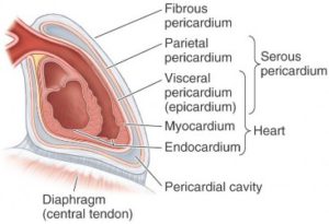
The pericardium is a sac that encloses the heart and roots of the great vessels (ascending aorta, superior vena cava, inferior vena cava,and pulmonary arteries and veins) as they leave/enter the heart. The outer layer of the sac, the fibrous pericardium, consists of dense connective tissue. The fibrous pericardium is attached to the sternum and diaphragm.
Figure 13.9 MOORE ET AL., CLINICALLY ORIENTED ANATOMY, 7TH ED., FIGURE 1.43.
Inside the fibrous pericardium and surrounding the heart is the serous pericardium, a typical serous sac having parietal and visceral layers. The parietal layer of the serous pericardium is fused to the inside of the fibrous pericardium and reflects on to the surface of the heart around the roots of the great vessels. Thus, the “sac” surrounding the heart that is visible in the cadaver when the chest wall is removed is composed of two fused layers. From embryology you learned that the fibrous pericardium was peeled away from the embryonic body wall and is formed from somatic mesoderm. Thus, if you consider the fibrous pericardium to be a body wall equivalent, the location of the parietal layer of the serous pericardium adjacent to it makes perfect sense. The outer layer of the heart itself is the visceral layer of the serous pericardium (aka epicardium). Between the visceral and parietal layers is the pericardial cavity, a potential space containing a small amount of serous fluid. The function of this fluid is to lubricate the surfaces of the serous pericardium to prevent friction and irritation to the tissues.
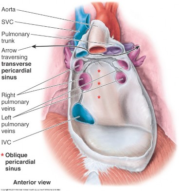
The left and right sides of the pericardial cavity are connected behind the ascending aorta and pulmonary trunk by a channel called the transverse pericardial sinus. This is useful to surgeons who can pass clamps or ligatures through the sinus in order to occlude the great arteries during cardiac surgery.
Figure 13.10 MOORE ET AL., CLINICALLY ORIENTED ANATOMY, 7TH ED., FIGURE 1.46.
Another modification of the pericardial cavity, the oblique pericardial sinus, is located posterior to the left atrium. It can be explored by elevating the apex of the heart and sliding the fingers behind the heart until they are stopped by the reflection of serous pericardium onto the major veins (SVC, IVC, and pulmonary veins) that surround the sinus. Rather than a passageway, the oblique sinus is a blind cul-de-sac.
The pericardial sac (fibrous pericardium + parietal layer of serous pericardium) is a somatic structure. Sensory fibers are conveyed via the phrenic nerves. Indeed, inflammation of the pericardium (pericarditis) is characterized by sharp substernal pain that may be referred to the shoulders. Blood supply to the pericardium is through the pericardiacophrenic arteries, small branches of the internal thoracic arteries that course with the phrenic nerves.
External anatomy of the heart
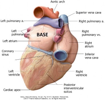
The heart resembles a short cone, having a base and apex. The base of the heart faces to the right and posteriorly. It consists primarily of the left atrium. The base is fixed to the pericardium since all the great veins enter the heart at its base. From the base, the heart projects down and to the left to terminate at the apex, which is located in the left 5th intercostal space, about 3 inches from the sternum, near the nipple.
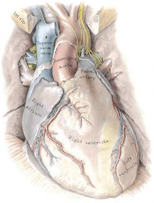
Figure 13.11 GRANT ’ S ATLAS OF ANATOMY, 14TH ED., FIGURE 3.45.
Figure 13.12
Surfaces
Borders
The diaphragmatic surface is formed by both the left and right ventricles. This surface extends from the base to the apex of the heart and faces the diaphragm.
The anterior (sternocostal) surface faces anteriorly and is formed primarily by the right ventricle with small contributions from the right atrium and left ventricle.
The right pulmonary surface is formed by the right atrium and faces the right lung.
The left pulmonary surface consists of the left ventricle and faces the left lung. The pulmonary surfaces are rounded whereas the other surfaces are somewhat flat.
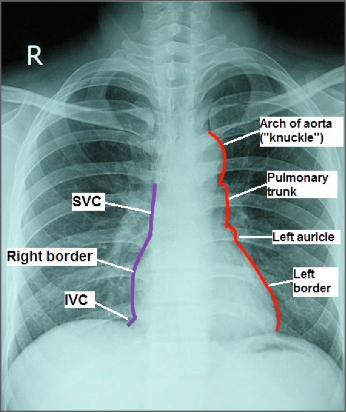
Borders (a.k.a. = margins) are not well defined on the gross heart, since it is conical in shape. However, they are important clinical entities used when examining the heart on X-rays or describing the radiological anatomy of the heart. On a chest X-ray, the silhouette cast by the heart next to the black lung fields (lungs are filled with air = black on X-rays) is defined by three borders:
The right border is formed by the right atrium abutting the black field of the right lung.
The left border is formed by the left ventricle and left auricle abutting the left lung.
The inferior border is formed mainly by the right ventricle and a small bit of left ventricle. The shadow of the diaphragm obscures the inferior border.
Figure 13.13
Note also that on a chest X-ray the shadows of great vessels are seen merging with the right and left borders of the heart. From superior to inferior:
On the right: SVC Right border of heart IVC
On the left: Arch of aorta (seen as a “knuckle”—with its convexity to the left) Pulmonary trunk Left border of heart Apex of heart
The internal division of the heart into four chambers is indicated on its external surface by the coronary sulcus (divides atria from ventricles) and the interventricular sulci on the sternocostal and diaphragmatic surfaces.