Autonomic motor and visceral afferent fibers reach and leave thoracic organs through autonomic nerve plexuses.
Concept
In autonomic plexuses, parasympathetic and sympathetic fibers and visceral afferent nerve fibers are intermixed. This is true of all autonomic plexuses in the body cavities.
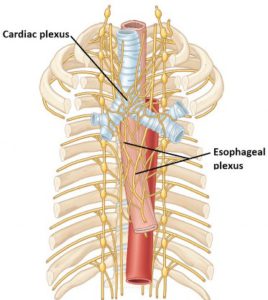
Figure 13.14 MOORE ET AL., CLINICALLY ORIENTED ANATOMY, FIGURE 1.75.
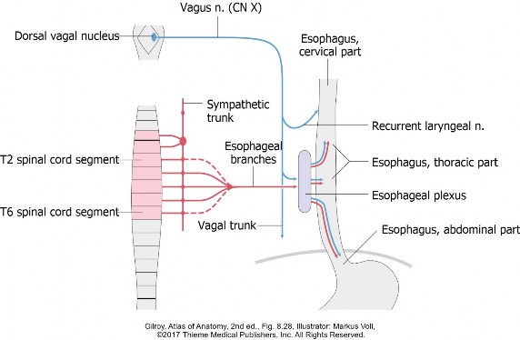
The two major thoracic autonomic plexuses are the cardiac plexus, located near the tracheal bifurcation, and the esophageal plexus, on the surface of the esophagus.
From the cardiac plexus, two smaller pairs of plexuses are derived: the pulmonary plexuses, which follow the bronchi into the lungs.
Sympathetic innervation of thoracic organs
Cell bodies of preganglionic sympathetic neurons that supply thoracic organs are in the intermediolateral cell column of spinal cordsegments T-1 to T-5.
Cell bodies of postganglionic neurons are in thoracic and cervical paravertebral (chain) ganglia, specifically: T-1 to T-5 thoracicchain ganglia and all cervical chain ganglia.
Note
At one time, there were eight cervical chain ganglia. Fusion occurring during development reduced the number to three or four.
Preganglionic sympathetic fibers leave spinal cord segments T-1 to T-5 and enter the associated ventral roots, spinal nerves, and ventral rami of spinal nerves. They enter T-1 to T-5 chain ganglia through white rami communicates.
Some preganglionic fibers synapse in chain ganglia T-1 to T-5, while others (specifically, those for the heart) pass through and ascend the sympathetic trunk to synapse in cervical chain ganglia.
Question
Why would sympathetic fibers destined for the heart pass into the neck region first?
Postganglionic sympathetic fibers leave T-1 to T-5 chain ganglia via visceral branches (medial branches) and enter the cardiac and esophageal plexuses.
Postganglionic sympathetic fibers leaving cervical ganglia pass only to the cardiac plexus, since they innervate the heart. Note that cervical visceral branches are necessarily quite long since they have to pass all the way down to the thorax. Note that visceral branches to the heart are derived from BOTH thoracic and cervical chain ganglia. However, visceral branches to other thoracic organs (lungs, esophagus) are derived from only thoracic chain ganglia.
Postganglionic fibers destined for the lungs pass through the cardiac plexus first and then on to the pulmonary plexuses. The cardiac plexus is the “switching yard” for routing fibers to the heart and lungs.
Functions of sympathetic fibers to thoracic organs (Remember fight, fright and flight!)
1
Speeds up the heart rate (stimulates pacemaker cells) and strengthens cardiac muscle contraction.
2
Dilates blood vessels to the heart.
3
Dilates air tubes in the lungs (relaxes smooth muscle in tubes).
4
Slows peristalsis in the esophagus.
Interactive 13.1
Draw two sympathetic pathways innervating the heart.
1. Take one pathway up the sympathetic trunk to the neck and then down to the heart through a cervical cardiac branch.
2. Take a second pathway into a thoracic chain ganglion, then to the heart through a thoracic cardiac branch. Both pathways go through the cardiac plexus. These are 2-neuron pathways with synapses in chain ganglia.
CORNELIUS ROSSE, M.D., D.SC., STUDY GUIDE FOR GROSS ANATOMY AND EMBRYOLOGY, UNIVERSITY OF WASHINGTON. USED WITH PERMISSION. (Tap to open; use your Apple Pencil to draw.)
Visceral afferents carrying pain from thoracic organs
Accompany sympathetic pathways in reverse to the CNS
Visceral afferents from the heart travel along sympathetic pathways to the CNS and probably function to transmit pain from the heart. They leave the heart and pass through the cardiac plexus. Some fibers ascend to the neck within cervical cardiac branches while others stay in the thorax and pass to thoracic chain ganglia within thoracic cardiac nerves. Regardless of these initial routes, sensory fibers ultimately reach T-1 to T-5 levels of the sympathetic trunk, pass through white rami communicates, and enter the spinal cord through dorsal roots. Cell bodies of these afferent neurons are in T-1 to T-5 dorsal root ganglia.
Visceral afferents from other thoracic organs travel to T-1 to T-5 chain ganglia through visceral branches, pass through white rami communicantes, then enter T-1 to T-5 levels of the spinal cord through dorsal roots. Their cell bodies are in dorsal root ganglia and their central processes enter spinal cord segments T-1 to T-5 to synapse on afferent nuclei in the dorsal gray matter of the spinal cord.
Interactive 13.2
Draw an afferent pain pathway from the heart to the spinal cord.
• This is a one-neuron pathway.
• Use the cervical visceral branch to reach
the chain ganglion in the neck, then bring it down the
sympathetic trunk.
• The cell body of the sensory neuron is the
dorsal root ganglion adjacent to the
thoracic spinal cord.
CORNELIUS ROSSE, M.D., D.SC., STUDY GUIDE FOR GROSS ANATOMY AND EMBRYOLOGY, UNIVERSITY OF WASHINGTON. USED WITH PERMISSION. (Tap to open; use your Apple Pencil to draw.)
Referred pain from the heart
Figure 11.13 NETTER, ATLAS OF HUMAN ANATOMY, PLATE 162.
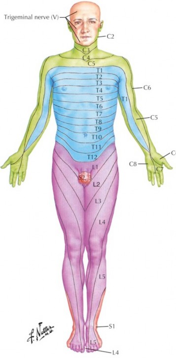
Note that visceral afferent neurons transmitting information from the heart enter the spinal cord through dorsal roots at levels T-1 to T-5. Once in the spinal cord, these neurons synapse on sensory nuclei whose axons ascend the spinal cord to the brain. Somatic afferent neurons from the skin also enter the same levels of the spinal cord through the same dorsal roots, synapse in the same sensory nuclei, and ascend to the brain through the same tract of neurons. This “sharing” of neuronal architecture by somatic and visceral organs may be the basis for “referred pain.” Indeed, pain emanating from stimuli in the heart may be interpreted by the brain as coming from T-1 to T-5 dermatomes, since these visceral sensations enter the spinal cord at those levels.By observing the dermatome map, one can see that the T-1 dermatome extends along the medial arm—this probably explains why patients experiencing heart attacks complain of pain in the chest and radiating down the inner arm.
Parasympathetic innervation of thoracic organs
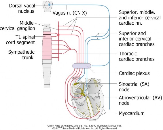
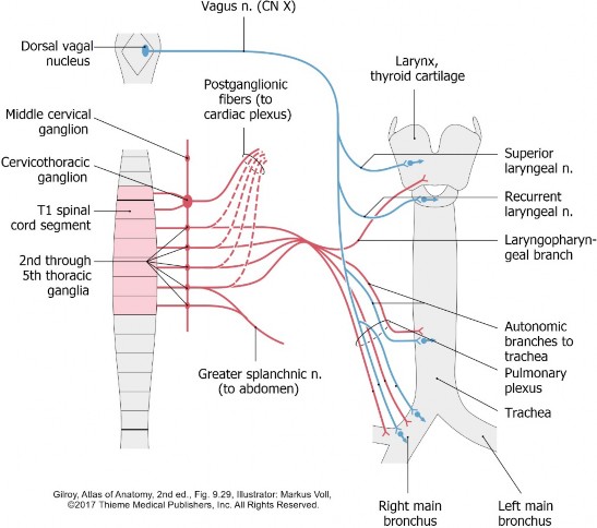
The vagus nerves supply parasympathetic fibers to thoracic organs and carry visceral afferent nerve fibers from thoracic organs.
The cell bodies of vagal preganglionic neurons are located in the dorsal nucleus of the vagus in the medulla of the brainstem. Preganglionic fibers leave the brainstem within the vagus, pass through an opening in the skull, descend through the neck, and enter thesuperior mediastinum via the superior thoracic aperture.
The vagi give off branches in the neck (cervical cardiac branches to the heart) and the thorax (thoracic cardiac and pulmonary branches). In the posterior mediastinum, fibers of the right and left vagi mix together onthe surface of the esophagus in the esophageal plexus.
Parasympathetic fibers destined for the heart (cervical and thoracic cardiac branches) and lungs (pulmonary branches in the thorax) enter the cardiac plexus, located at the tracheal bifurcation. From the cardiac plexus, fibers pass to the lungs via the pulmonary plexuses.
The cell bodies of postganglionic parasympathetic neurons are in intramural ganglia in the walls of thoracic organs: heart, bronchial tubes, and esophagus. Postganglionic parasympathetic nerve fibers are therefore very short (microscopic) = they terminate on the conducting system (SA and AV nodes) and cardiac muscle of the heart, smooth muscle of coronary arteries, bronchial tubes, and theesophagus, and on glands in the bronchial tubes and esophagus.
Parasympatheticinnervationofthoracic organs
1
Slows the heart rate and reduces strength of cardiac muscle contraction.
2
Constricts air tubes (stimulates smooth muscle in their walls).
3
Stimulates gland secretion (mucus) in the air tubes and esophagus.
4
Stimulates peristalsis in the esophagus.
Interactive 13.3
Draw parasympathetic pathways innervating (1) the heart and (2) a bronchus.
These 2-neuron pathways begin in a nucleus in the medulla and descend in the vagus. Most fibers to the heart leave the vagus in the neck; fibers to the bronchus leave the vagus in the thorax. Preganglionic fibers synapse on postganglionic cell bodies in intramural ganglia. Draw the pathway to the heart on the left side of the diagram; draw the route to the bronchus on the right side.
CORNELIUS ROSSE, M.D., D.SC., STUDY GUIDE FOR GROSS ANATOMY AND EMBRYOLOGY, UNIVERSITY OF WASHINGTON. USED WITH PERMISSION. (Tap to open; use your Apple Pencil to draw.)
Visceral afferents carrying other (non-pain) information from thoracic organs
Accompany parasympathetic pathways in reverse to the CNS
Vagal visceral afferents leave thoracic organs, pass through pulmonary, cardiac, or esophageal plexuses, and ascend in the vagi. The cell bodies of vagal visceral afferents are in vagal sensory ganglia—swellings of the vagus nerve just below the base of the skull. These sensory ganglia of the vagus are the equivalents of dorsal root ganglia of spinal nerves. The central processes of these visceral afferents synapse on sensory nuclei in the medulla of the brainstem.
Visceral afferents that follow the vagal pathway in reverse mediate pain from organs above the thoracic “pain line” (trachea, for example) and carry subconscious visceral reflex information from the heart and lungs.
examples
Blood pressure receptors in the great vessels associated with the heart Cough reflex and stretch in bronchial tubes Filling of air sacs in the lungs that signal the end of inspiration and the beginning of expiration.
Figure 13.16 Summary diagram: Autonomic innervation of the heart.
Figure 13.17 Summary diagram: Autonomic innervation of the esophagus.
Figure 13.18 Summary diagram: Autonomic innervation of the bronchi.