Gross Anatomy quick links
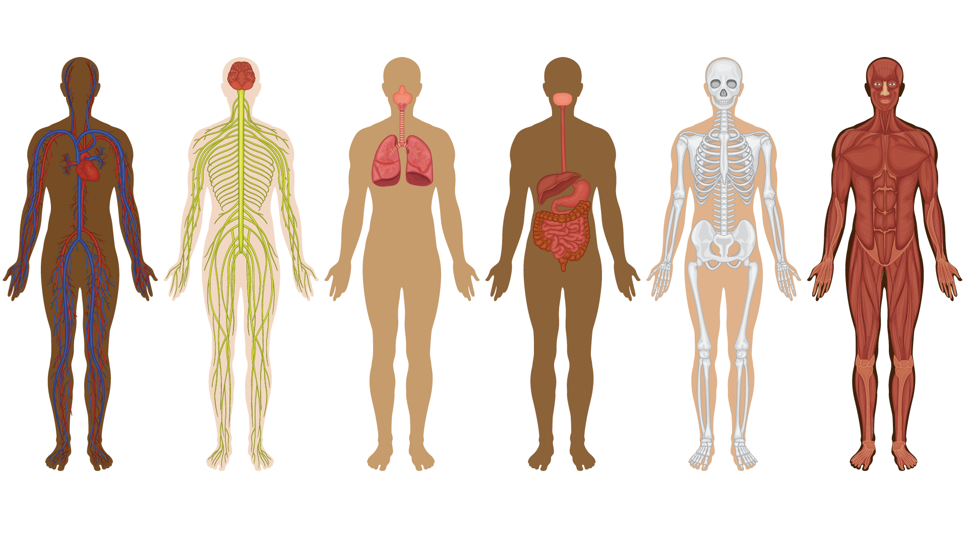
The Anatomy thread in the curriculum will provide students with a thorough introduction to the structure, development, and function of the human body. It is organized to encourage active participation in the learning process.
Students will gain the ability to recognize normal anatomic structures, appreciate their developmental histories, and understand relationships between structures and their clinical relevance. The major goal is to integrate embryonic development with study of the cadaver as a foundation for the examination of the living body.
An essential aspect of the thread will be learning and using the language of anatomy, since this vocabulary will allow students to communicate effectively with colleagues and patients throughout their professional lives. Anatomy is an excellent forum for practicing clear and precise description and orderly and structured thinking.
The thread will prepare students to apply knowledge of anatomy during their clinical training, when performing a physical exam, or when interpreting medical images. Since imaging is an essential tool for diagnosing and treating disease, anatomy provides the logical setting where it will be introduced to students and integrated with session topics.
Format
Gross anatomy begins with a session on embryogenesis and a lab orientation, and continues throughout the pre-clerkship curriculum.
Consult the schedule on E.Flo MD for the details.
- Large-group sessions
- Lab sessions
These sessions introduce concepts and principles and correlate gross anatomy with function and embryonic development. Although many details will be discussed, large-group sessions are not designed to cover all the material outlined under the objectives for the session, since most objectives overlap with the corresponding laboratory sessions. Large-group sessions provide a “big picture” so that details can be explored and discussed in the lab. Interactive activities to check comprehension and apply knowledge are included in most large-group sessions.
Most lab sessions involve dissection of the donor. Students will be assigned to a dissection team and a donor for the duration of the year. Instructions and checklists of structures to be identified and discussed will be provided in the Dissector.
A small number of lab sessions will be prosection (non-dissection) based. Students will be organized into groups that will rotate through interactive demonstrations with faculty using cadaveric specimens, draw-to-know exercises, and imaging.
expectations
Students are expected to come to anatomy sessions prepared, having reviewed materials in resources such as the Gross Anatomy textbook, anatomy atlases, and/or the Dissector beforehand.
Come to lab dressed in appropriate attire: scrubs or lab jacket, long pants, and closed-toe shoes.
Instructors


Local clinicians have graciously agreed to participate in anatomy lab sessions—taking time out of their busy practices to lend clinical perspective, experience, and expertise. This will be a real treat for students!
Resources for the component
Suggested primary resources
More anatomy resources
- Optional resources
Optional gross anatomy text
-
Clinically Oriented Anatomy, by K. L. Moore et al., 9th edition. Lippincott: LWW Health Library.
Optional embryology text
-
Langman’s Medical Embryology, by T.W. Sadler. 14th edition. Lippincott: LWW Health Library.
Optional anatomy atlases (owning one is highly recommended)
-
Atlas of Anatomy, by Gilroy et al., 4th edition. Thieme Medical Publishers.
-
Atlas of Human Anatomy, by Netter, 7th edition. Elsevier. Clinical Key. The optional resources listed above may be purchased as hard copies by students. Electronic versions of these resources (except the Gilroy atlas) can be accessed through the WSU Spokane Academic Library—listed under the Medicine lib guide.
Optional video and electronic resources
-
Acland’s Video Atlas of Human Anatomy: Access through the WSU Spokane Academic Library webpage.
-
Complete Anatomy : Purchased by the College of Medicine and available as an app on your iPad.
Donor CT scans
Full-body CT scan data is available for each donor. They are stored in a PACS (picture archive and communication system) hosted by a local imaging organization and can be accessed in lab or at home with a viewer program. Instructions for using the PACS system to view your donor’s scans will be given during the lab introduction and in a prerecorded video by Dr. Julie Kaczmark.
Assessment
Gross Anatomy is a thread in the larger FMS Courses. As such, anatomy items are included on the Mastery Knowledge Assessments (MKAs) in FMS courses. Consult the FMS Assessment Packages for more details.


