3. Functional neuroanatomy and vasculature, part 2

Long ascending and descending pathways The three major longitudinal pathways the corticospinal tract, posterior column-medial lemniscus, and anterolateral system can be followed systematically to and from the cerebral cortex, through brain stem and to and from spinal cord. A key feature of all these pathways is to note the location where their tracts cross to […]
3. Functional neuroanatomy and vasculature, part 1: The brain stem, cerebellum, cerebrum, and spinal cord
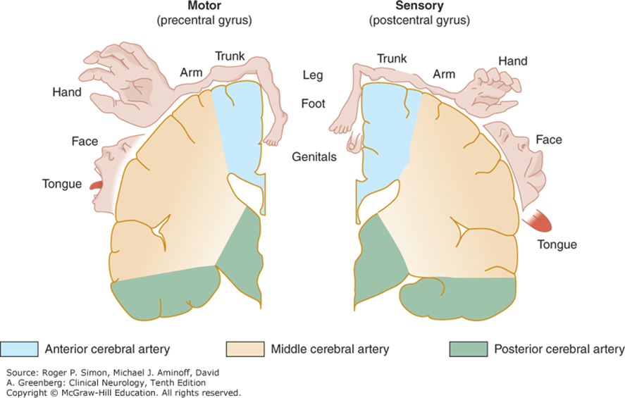
Blood supply to the Central Nervous System An understanding of the brain’s blood supply is crucial to understanding normal CNS function. Conversely, the consequences of cerebrovascular disease affecting the brainstem and cerebrum leading to a loss of function is also vital. Figure 3.1. Schematic of blood supply to brain stem regions (in transverse section). Simon, […]
Hematologic malignancy overview
Hematopoietic System

Overview of red blood cells More on RBC metabolism From Molecular Biology and Biochemistry: Red Blood Cell Metabolism
2. The meninges and ventricles

The meninges and meningeal spaces Figure 2.1. Meninges and meningeal spaces. By OpenStax, CC BY 4.0. Modified July 9, 2018, by Lampa. The meninges can be subdivided into cranial and spinal portions. The meninges function to protect, to act as a scaffold supporting blood vessels and venous sinuses, and finally to form a continuous sac, […]
Nervous System

Welcome to the Nervous System block of FMS 512! For the 2025–2026 school year, Shannon Brodersen, MD, and Kimberly Honn, PhD, have teamed up to co-direct and redesign the section. We have made changes based on student and faculty feedback from previous years, including beginning the block with a strong foundation of basic science—neuroanatomy including […]
Test Your Knowledge

Patient 1: Marie Tap the image to view the case. A 48-year-old female was brought to the emergency department via an emergency response vehicle following a high-speed motor vehicle collision. The air bags failed to deploy, and extrication was required. First responders report that she had to be resuscitated in the field and twice en […]
EKG self-assessments

Partially Occluded Artery Worksheets
These worksheets are for self-study only. Answers will not be evaluated. Instructions and examples Instructions for Chapter 13 Worksheets Complete basic measurements. Do all previous work. Note if inverted T waves or ST segment depression is present in two or more leads that represent a pattern in Table 13.2. Here the ST segments are diffusely […]
Partially Occluded Artery Case Studies
Case 1: Inferior ischemia or infarction Tap the arrow to view the case.EKG: The T wave is negative in leads II, III, and AVF, but positive in I and AVL (Figure 13.13).Visualization: The T is pointing to the left and superiorly, away from the inferior wall (Figure 13.14).Critical Thinking: The T wave is pointing away […]