Basic Fetal Anatomy Scan
Intro The fetal anatomy scan is the foundation for prenatal diagnosis and management. This scan is sometimes called a “Level II Scan,” although that nomenclature has been dropped by the AIUM. The elements of the basic fetal anatomy include evaluation of the uterus, fetal biometry and fetal anatomy. The sequence of ultrasound pictures shows how […]
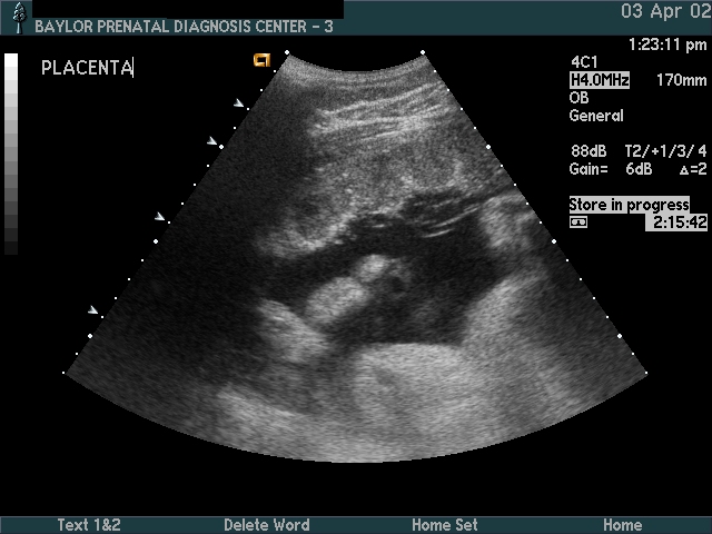
Clinical Images
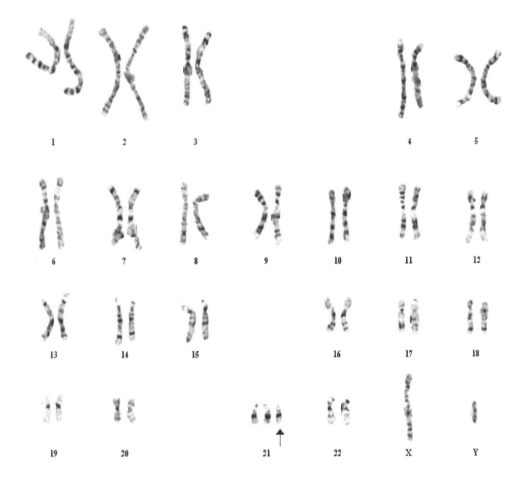
Table of Contents 7 days post c-section Abdominal Pregnancy Imaging MRI Ultrasound Abdomal Pregnancy (Surgery) Ambiguous Genitalia B-Lynch Suture Bicornate Uterus Caudal Regression Chromosome Transposition, Cleft Lip, Clenched Hands Cystic Teratoma Discordant Twins, Vel cord (1170g–1785g) Fetal surface. Fetal surface. Fetal surface. Maternal surface. Maternal surface. Early Twin Demise Exencephaly (Amniotic Band) Fetal Demise Umbilical […]
3-D Ultrasound
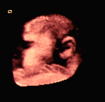
3-D Scans 3-D ultrasound is often used to create “pretty pictures” of fetal faces. It can also image other structures of interest (e.g., hand, foot, ear). The long term potential of 3-D ultrasound is to improve imaging of fine anatomical details. Face at 32 weeks Face at 20 weeks Face at 16 weeks Cleft Lip […]