Ob/Gyn Sub-Internship Expectations

Year 4
NCC electronic fetal monitoring game Can you recognize these strip elements? What are some downloadable resources that I can use? Ob-Gyn Notes Includes: Sample labor and delivery admit note Sample vaginal delivery note Sample operative note Sample post-partum note: Vaginal delivery Ob postpartum note: Cesarean delivery Sample gynecologic history and physical Sample new obstetrics presentation […]
Longitudinal Integrated Clerkship (LIC)
NCC electronic fetal monitoring game Can you recognize these strip elements?
What Ob/Gyn’s Wish All Other Physicians Knew

Benign and Malignant Breast Conditions

Reproductive Health: Preceptor Resources

Faculty Learning Portal The Faculty Development Unit’s eLearnings and microlearnings for faculty are now housed on Reach 360, a new platform that allows us to share our education and development opportunities while providing robust reporting for LCME Accreditation purposes. All faculty and staff are welcome to register and use the platform, and signing up is […]
Clerkship
In the clerkship
Fetal Anomalies

Listed In Order Of Level II Scan / Fetal Anatomy Survey Sequence Cervix Heart / Lung Placenta Face CNS / Spine Extremities Genitalia Other Bladder Aneuploidy Cord Insertion / Abdominal Wall Maternal Kidneys Multiple Pregnancy Stomach Cervix Outline Incompetent cervix Endovaginal ultrasound of the cervix Incompetent Cervix Patient had a “routine” ultrasound to evaluate fetus […]
Andersen
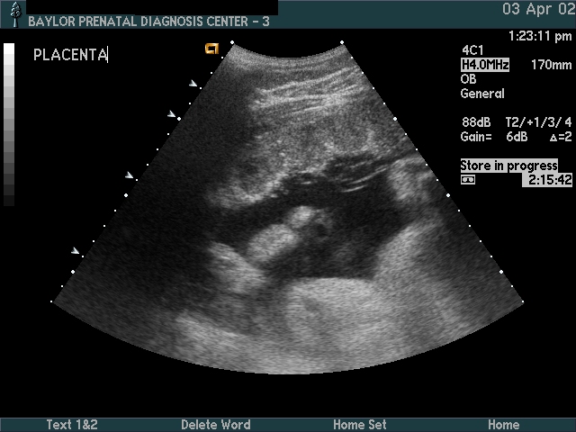
OB Ultrasound Primer Welcome to an introduction to OB Ultrasound This primer was developed at Baylor University Medical Center in Dallas from 1998 to 2006 to assist training OB/Gyn residents. Ultrasound technology has progressed steadily, and many “office” ultrasound machines now produce images as good, or better, than these images from what was then a […]
Basic Fetal Anatomy Scan

Intro The fetal anatomy scan is the foundation for prenatal diagnosis and management. This scan is sometimes called a “Level II Scan,” although that nomenclature has been dropped by the AIUM. The elements of the basic fetal anatomy include evaluation of the uterus, fetal biometry and fetal anatomy. The sequence of ultrasound pictures shows how […]