Protected: Lab 27: Dissection: Infratemporal Fossa and Floor of Mouth; Bony Anatomy of Pterygopalatine Fossa
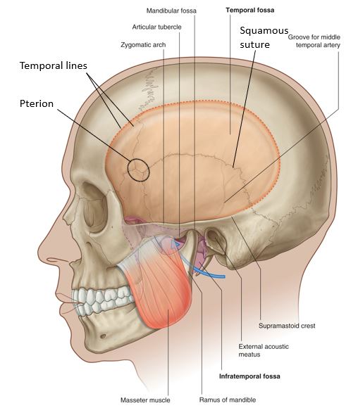
There is no excerpt because this is a protected post.
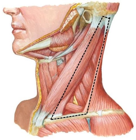
Protected: Lab 25: Neck: Anterior Triangle of Neck
There is no excerpt because this is a protected post.
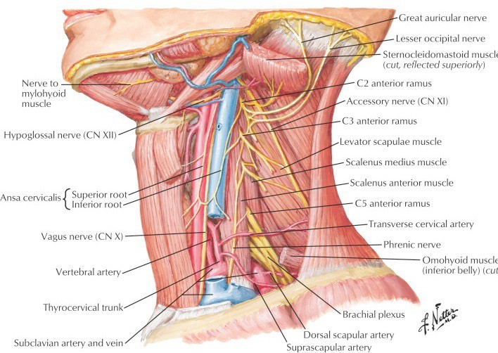
Protected: Lab 26: Dissection: Posterior Triangle of Neck, Deep Neck, and Root of Neck
There is no excerpt because this is a protected post.
Large vessel vasculitis
Optional resources American College of Rheumatology (ACR) Rheum2Learn: Musculoskeletal disorders Estimated time: 10–15 min.; case #1 Rheum2Learn 2.0 is a free, case-based online program from the ACR that uses interactive patient cases to build core skills in recognizing and managing rheumatic diseases. Flexible and self-paced, it’s designed for students, residents, and general internists. In this […]
Shockflix
Infectious arthritis
Optional resources Core EM Podcasts: Septic arthritis Estimated time: 20 min. This podcast reviews the essentials of septic arthritis, an orthopedic emergency that requires rapid recognition and treatment to prevent irreversible joint damage and systemic complications. It explains how bacteria reach the joint—most commonly through hematogenous spread—and how the resulting inflammatory cascade can destroy cartilage […]
How to succeed in Surgery: A peer-to-peer guide
Author Julia Todderud, Class of 2026 Welcome to Surgery! This module was developed in partnership by students and our surgical faculty to best prepare and support your surgical rotations. This includes a compilation of recommendations and tips to optimize your learning and the experience of your team members and patients. Key takeaway: You are a […]
Welcome to Wesford
Infections in the immunocompromised host
Learning goals Correlate types of opportunistic infections related to specific forms of immunodeficiency (cell-mediated, humoral, innate, adaptive, combined) Describe the epidemiology, clinical presentation, diagnosis, treatment approach, and preventative strategies for the opportunistic infections caused by Pneumocystis jirovecii, JC Virus, and Cytomegalovirus Compare and contrast the nature and degree of immunosuppression and the associated opportunistic infections […]
Degraded air quality
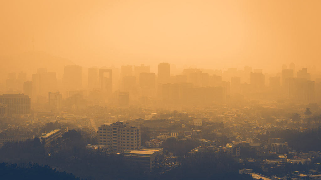
This module explores how climate change and pollution degrade air quality and the resulting health consequences, with attention to populations at greatest risk and strategies at multiple levels that create co-benefits for both climate and health. Sections Note Environmental and health policies evolve with changing political administrations. While federal approaches may shift, the scientific evidence […]