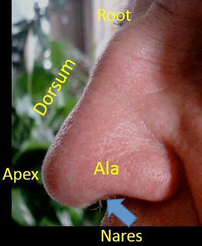
The external nose is on the face. It functions to direct air (and odors) into the nasal cavities. Identify these parts of the external nose on the face:
Apex (“tip”)
Root
Dorsum ("bridge")
Alae
Nares ("nostrils")
Figure 1. Parts of the external nose.
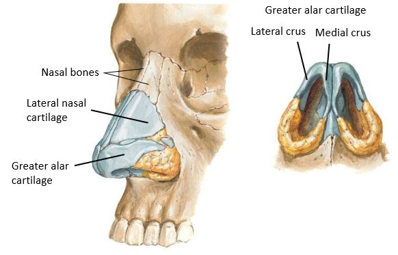
The “skeleton” of the external nose is composed of bones and cartilage.
Bones:
Nasal bones
Cartilages:
Major alar cartilages: Each is U-shaped, with a medial crus and a lateral crus. The two medial crura line up, side-by-side between the nostrils to form the mobile nasal septum (Columella)
Lateral nasal cartilages: Connect above to the nasal bones
Figure 2. Skeleton of the nose.
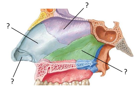
The internal nose is comprised of the left and right nasal cavities and the nasal septum that separates them.
The nasal septum is normally in the midline of the head. If not, it is called a “deviated septum.” The septum has three parts:
Bony nasal septum
Two bones contribute to the bony septum: the perpendicular plate of the ethmoid bone and the vomer.
Septal cartilage: Made of hyaline cartilage.
Mobile nasal septum (Columella): Discussed earlier with the external nose, it is composed of dense connective tissue and the medial crura of the two major alar cartilages.
In Figure 3, identify the parts of the nasal septum and the bones that contribute to the bony septum.
Figure 3. Nasal septum with mucosa removed.
Question
Vomer is Latin for "plow." Is it plow shaped?
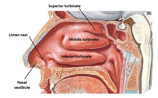
Nasal Cavities (Left and Right) Each nasal cavity has these boundaries:
Medial wall = smooth, formed by the nasal septum.
Lateral wall = complicated, formed by many bones and cartilage. The most prominent features are the three nasal conchae (also called the turbinates, because they produce turbulence in air flow). The turbinates are composed of thin bone covered by respiratory mucosa.
The turbinates are named superior, middle, and inferior. The superior and middle turbinates are parts of the ethmoid bone. The inferior turbinate is considered a separate bone.
The spaces deep (lateral) to the turbinates are the nasal meatuses: each nasal cavity has a superior, middle, and inferior meatus.
The highest part of each nasal cavity—above the superior turbinate—is called the spheno-ethmoidal recess.
The roof is formed by the cribriform plate of the ethmoid bone and the body of the sphenoid bone.
The floor is the hard palate.
The nasal cavities have openings at each end:
The anterior openings are the nares (nostrils). The anterior part of each nasal cavity just inside the nostril is called the vestibule. It is lined by skin and contains nose hairs (“vibrissae”).
The posterior openings are the choanae – they communicate with the nasopharynx.
Figure 4. Features of the lateral nasal wall.
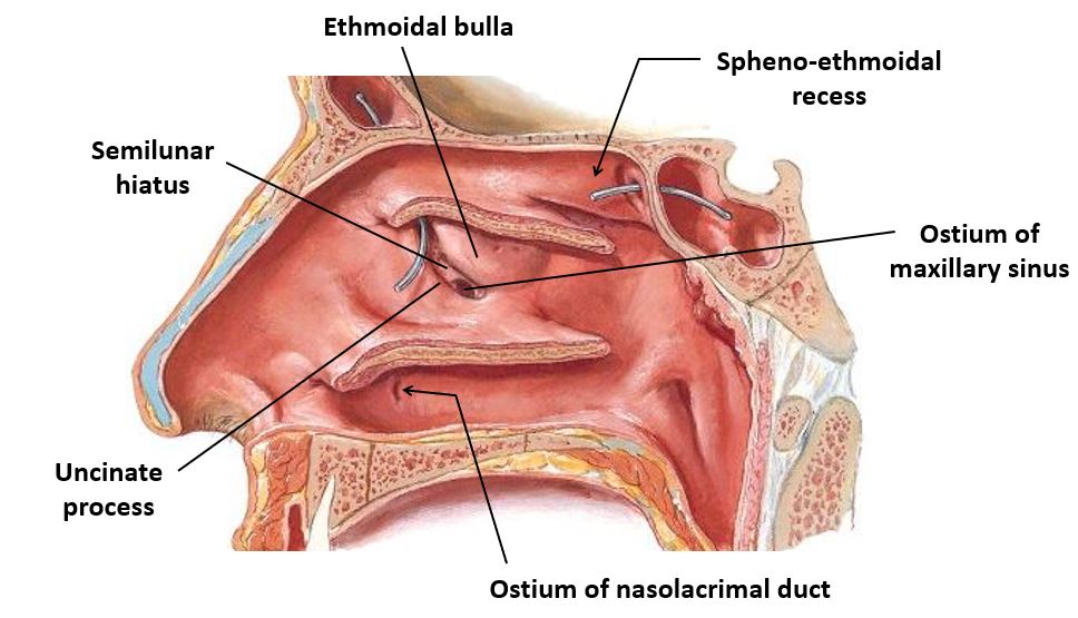
The anatomy of the middle nasal meatus is a bit intricate, since this is an area where most of the paranasal sinuses drain. Elevate the middle turbinate and identify these parts:
Ethmoidal bulla: A mound (“bubble”) in the lateral nasal wall produced by the underlying middle ethmoidal air cells.
Uncinate process: A curved, bony ridge anterior and inferior to the bulla.
Semilunar hiatus: The curved gap between the bulla and uncinate process.
Infundibulum: A small chamber lateral to the uncinate process. Slip a probe through the semilunar hiatus to demonstrate the infundibulum.