To access the pelvic cavity, you will need to mobilize the distal part gastrointestinal tract; namely freeing up the rectum.
Identify and protect and gonadal vessels.
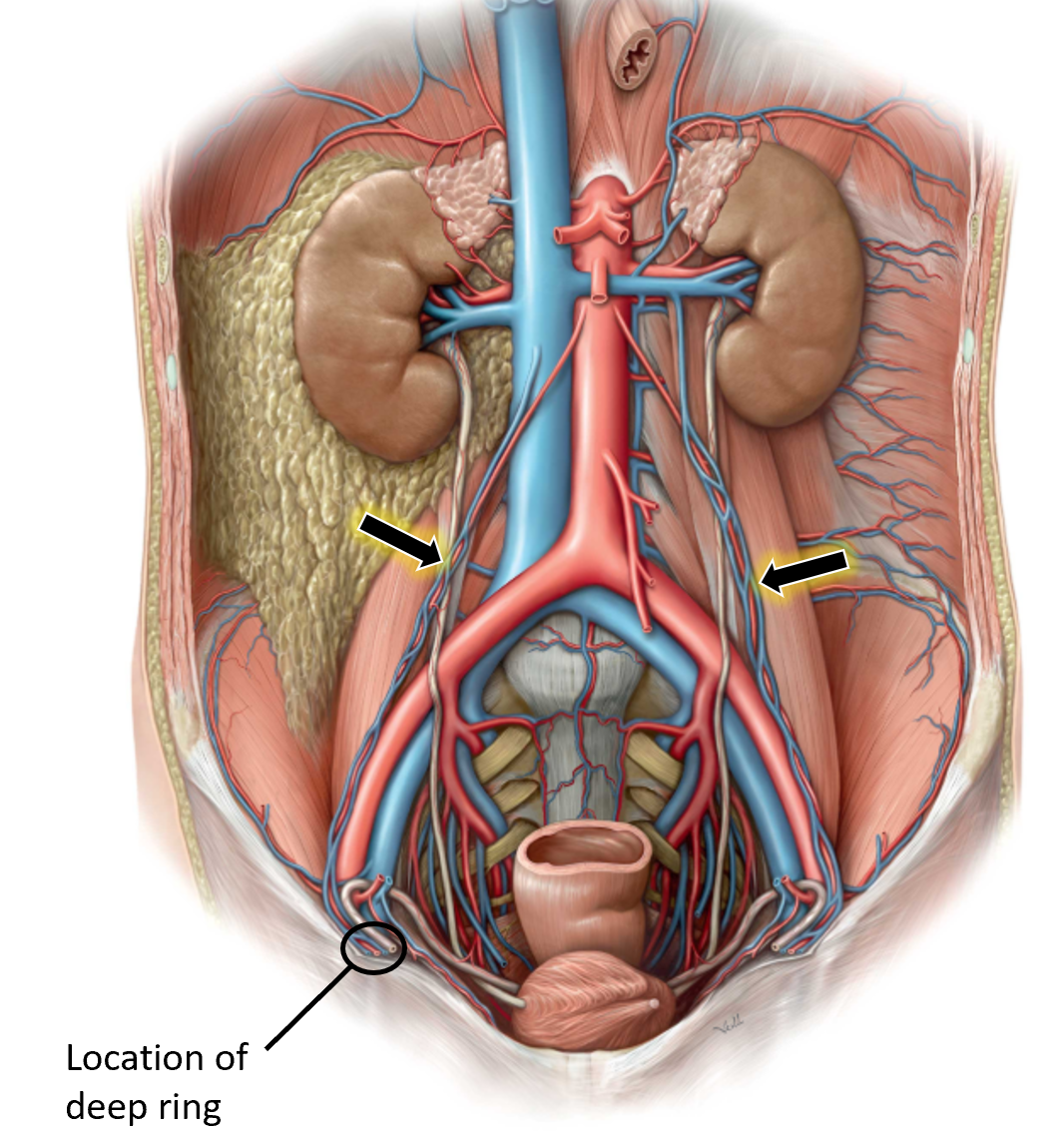
Locate the gonadal (testicular or ovarian) vessels. In the male, start at thedeep inguinal ring and trace back to the aorta and IVC (consult an atlas). In the female, trace back from the ovaries. Follow the left gonadal (testicular or ovarian) vein upwards and note that it flows into the left renal vein.
Figure 29.
They are very thin and vulnerable vessels, so having an idea where they are is important before you dissect!
Cut through the rectum in the transverse plane.
1Use plastic locking strips (cable ties) provided in lab to tie off the rectum.
■Apply two plastic ties to the rectum as far down in the pelvic cavity as possible.
■If the rectum is full of feces, move up to the sigmoid colon. The rectum has no mesentery, so you will have to pry it loose from the connective tissue anterior to the sacrum.
2Cut the rectum between the ties with scissors or a scalpel and lift the upper portion of the rectum out of the pelvic cavity.
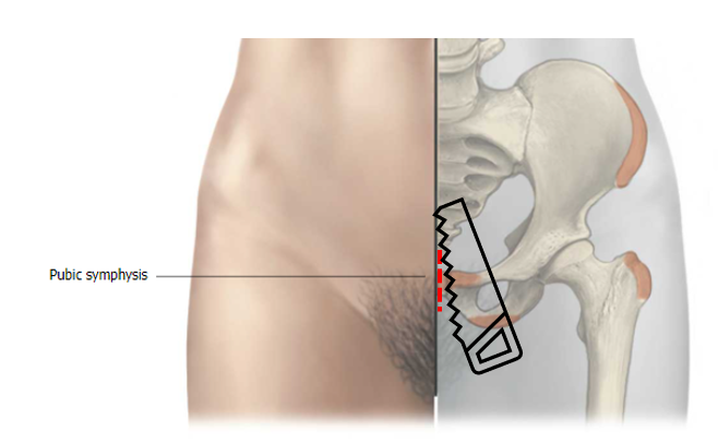
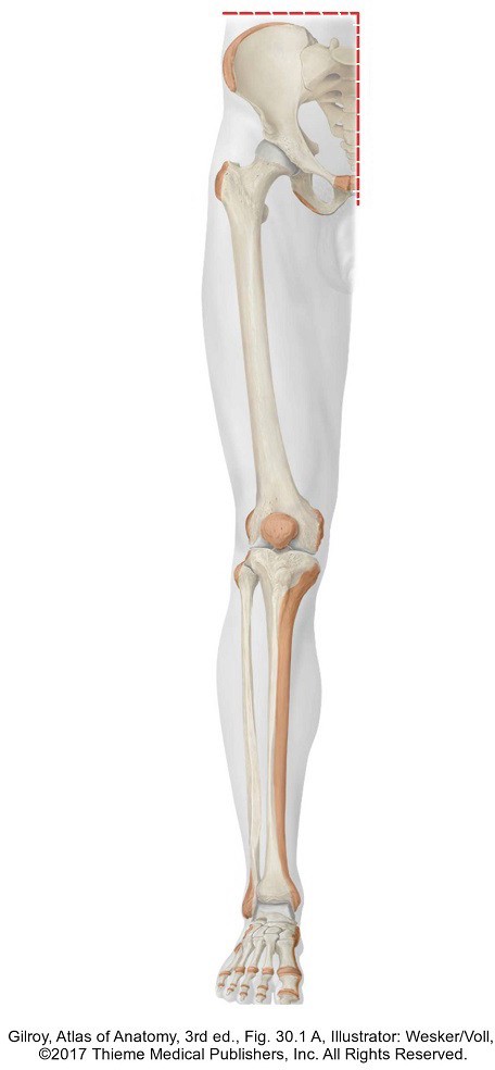
Section the pubic symphysis in the median plane.
1Lift the donor off the table and place a block under the lower back. With a scalpel and scissors clean tissue away from the pubic symphysis.
2Transect the pubic symphysis with a hand saw. As the cut proceeds through the interpubic disc, have several members of your team abduct the lower limbs as far as possible. Uncomfortable sounds may be heard as the sacroiliac joints tear.
Figure 30.
STOP HERE AND PAUSE
Don’t cut through the pelvic organs yet.
Section the perineum and pelvic organs in the median plane.
Note
The following parts of the dissection may elict some uncomfortable feelings. Brave teammates need to step up to do this work. The result will be worth the effort and the educational value of the view obtained will be high yield.
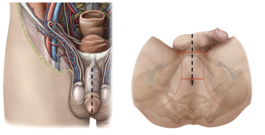
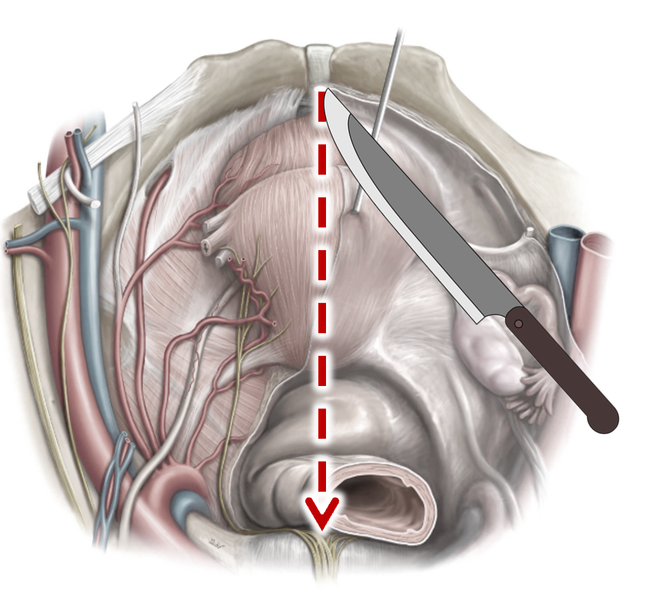
1Use a long knife to section the perineum in the midline. The perineum is the region between the thighs that contains the external genitalia.
■In the male, pass the knife through the midline of the penis, dividing it evenly in to left and right portions. Continue the cut down through the bulb of the penis and midline of the scrotum. See Figure 31.
Figure 31.
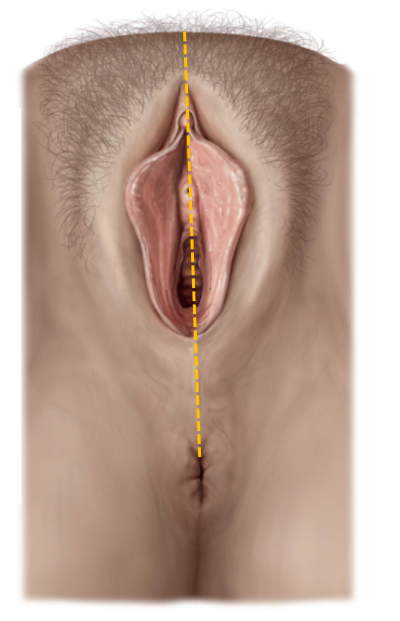
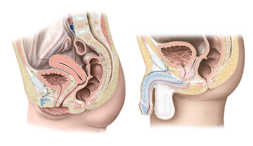
■In the female, the cut is made through the clitoris and down the center of the vestibule (the cavity that contains the openings of the urethra and vagina). See Figure 32.
Figure 32.
2Use the long knife to section the pelvic organs in the midline, working from anterior to posterior, while applying pressure to abduct the lower limbs.
■In the male, section the bladder and prostate.
■In the female, section the bladder and uterus.
3Remove the cable tie from the rectum and section the rectum and anal canal in the median plane. Anticipate that feces will leak out.
Figure 33.
Make sure you have a stack of paper towels at your table to clean up any feces in the rectum and anal canal.
Complete the hemisection.
4While two teammates abduct the lower limbs, use a hand saw to cut vertically through the sacrum and L-5 vertebra in the median plane. See Figure 33.
5On the right side of the body, use a long knife to make a deep horizontal incision, use a long knife to make a deep horizontal incision through the lateral abdominal wall just above the iliac crest. Carry the incision through the skin, body wall muscles, and the quadratus lumborum and psoas major muscles until you reach the vertebral column. See Figure 33.
6Now use a hand saw to cut horizontally through the vertebral column, until the horizontal cut meets the vertical cut you made earlier. The right side of the pelvis and right lower limb have now been separated from the rest of the body.
7Have several teammates help to remove the entire lower limb with the hemi pelvis. Rotate the specimen on its side in order to examine the pelvic organs in cross section. Now the structures in the lateral pelvic wall can be dissected and studied! See Figure 35.
Figure 34.
Figure 35.
Dissection of the Pelvic Cavity and Organs
The hemi-sectioned pelvis will allow your team to work on both sides of the pelvic cavity and have clear access to the pelvic floor and lateral pelvic walls.
Superficial side: Your team will identify organs and their parts on this side. Leave the peritoneum in place if possible—reflect it toward the midline in order to clean the subperitoneal space below it. The superficial side should be the side of the pelvis that is still attached to the body.
Deep side: Remove the pelvic peritoneum and reflect the organs toward the midline. Clean the pelvic walls, pelvic floor, and the branches/tributaries of the internal iliac vessels. The deep side should be the side of the pelvis that is detached with the lower limb.
Checklist, Lab #14
Checklist items at each of the five stations are indicated by checkboxes.