Picture Archival and Distribution System (PACS)
At the College of Medicine, you have access to a PACS (Picture Archival and Distribution System), where you can become familiar with how to work with PACS as this is how imaging studies are housed and distributed. Cases are continually added to the PACS. PACS login Open the PACS app on your iPad : WSU […]
6. Radiology Resources

Websites StartRadiology.com Radiology Café: Radiology Basics Radiopaedia.org Recommended radiology resources for medical students Essentials of Radiology, 4th edition. Fred A. Mettler, Jr. Digital version available for College of Medicine students. Picture Archival and Distribution System (PACS)
5. CT vs. MRI
When talking about CT vs. MRI, there is a lot of overlap with similar accuracies. The expertise and type of equipment your imaging partners have may determine the best test to use. However, for imaging of the central nervous system, spine, and spinal contents, MRI is superior in almost all applications. MRI is the best […]
4. Magnetic Resonance Imaging (MRI)

Magnets, computers, and ham radios MRI, like ultrasound and CT, uses sophisticated computer systems to collect and analyze data, i.e., reflected echos (think fish finder) in ultrasound and X-ray density for CT. MRI measures the mysterious values T1 and T2. T1 and T2 are values that relate to how molecules with a magnetic tendency (dipoles) […]
3. Computed Tomography (CT)
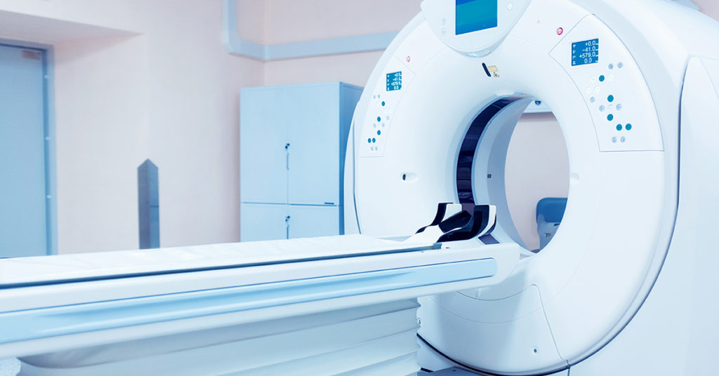
Computed Tomography (CT) is the marriage of computers and X-rays. In modern machines, a rotating gantry holds a specialized X-ray tube, which generates a highly contoured X-ray beam in opposition to a detector array surrounding the patient. The patient moves through the gantry. Gantries weigh around 6,000 kilograms and can achieve speeds of 300 milliseconds […]
2. Ultrasound
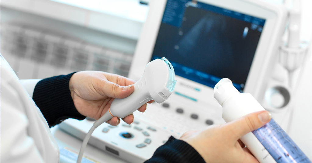
Ultrasound utilizes ultrasonic sound waves in the 2–10 MHz range generated by a handheld piezoelectric crystal, sonically coupled to the skin by gel. A computer controls the process, allowing complex modeling of the sound beam. Higher frequencies have better resolution but poorer depth penetration. Transducer selection is a compromise between the depth of the area […]
1. X-ray
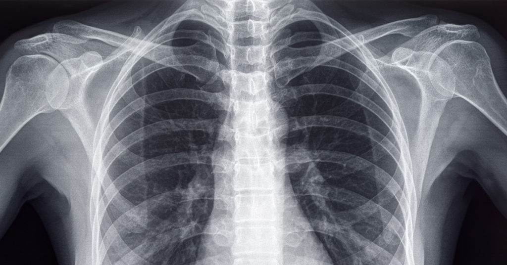
Craniocaudal magnification showing malignant breast calcifications. Case courtesy of Melbourne Uni Radiology Masters, Radiopaedia.org, rID: 43928 X-rays are produced by bombarding a dense target (the anode) with high-energy (5–110 Kv) electrons, which produces a beam that radiates outward from a small point source. The beam is shaped with lead shutters. This geometry can lead to […]
Radiology

In these learning materials Introduction to Imaging Please review this material prior to class and fill out the compare/contrast matrix as best you can. There is more information than can be covered in two 50-minute sessions, so the large-group session will attempt to cover concepts that tend to be confusing. Bring your questions. Remember, this […]