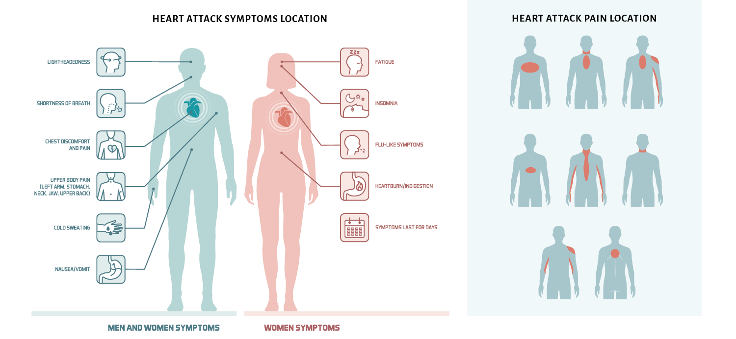
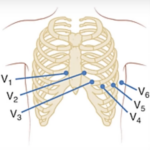
When there are changes in these leads (particularly ischemic changes):
-
- Consider right ventricular infarction.
- Beware of using agents that reduce preload (e.g., nitroglycerin).
- Hypotension is a risk—consider fluid resuscitation.
- Consider the risk of ventricular dysrhythmias.
- Consider right ventricular infarction.
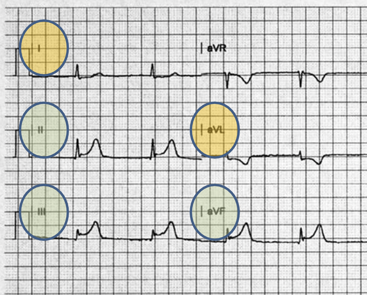
When there are changes in these leads (particularly ischemic changes):
-
- Consider ventricular dysrhythmias.
- Particularly ventricular fibrillation
- High probability this patient needs percutaneous coronary intervention (angioplasty or stent placement).
- Consider ventricular dysrhythmias.
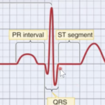

ST elevation
>1mm (one small box)
Two contiguous leads