Protected: Lab 19: Posterior Leg and Plantar Foot
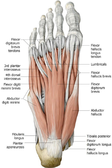
There is no excerpt because this is a protected post.
Protected: Lab 18: Anterior and Lateral Leg, Knee and Ankle Joints, and Dorsum of Foot
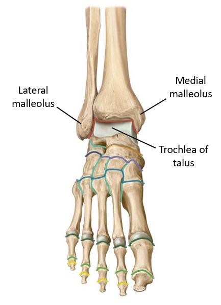
There is no excerpt because this is a protected post.
Protected: Lab 26: Scalp, Cranial Cavity, and Meninges
There is no excerpt because this is a protected post.
Protected: Lab 17: Gluteal Region, Posterior Thigh, and Popliteal Fossa
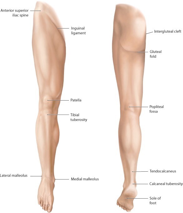
There is no excerpt because this is a protected post.
The ear
Optional reading Clinically Oriented Anatomy, 9th ed., Head chapter, Ear section through Auditory ossicles. The ear is the part of the head that contains the structures associated with the special sensations of hearing and balance. For descriptive and functional purposes, anatomists and clinicians organize the ear into three parts: external, middle, and internal. Figure 1. […]
Neck fascia and triangles
Optional reading Moore, Clinically Oriented Anatomy, 9th ed., Fascia of neck section through Surface anatomy of cervical regions and triangles of neck. Surface features and landmarks The anterior neck (“neck proper” or cervix): Extends from the inferior border of the mandible superiorly to the clavicles and sternum inferiorly. The posterior neck (nucha or “nape”): Extends […]
Face and parotid gland
Optional reading Clinically Oriented Anatomy, 9th ed., Head chapter, Face and scalp section through Surface anatomy of face; Parotid and temporal regions, infratemporal fossa, and temporomandibular joint section through Infratemporal fossa. The Developing Human: Clinically Oriented Embryology, 12th ed., Development of salivary glands section through Atresia of the nasolacrimal duct. The face is the anterior part […]
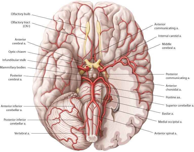
Scalp and cranial cavity
Optional reading Clinically Oriented Anatomy, 9th ed., Head chapter, Internal surface of cranial base section through Posterior cranial fossa; Face and scalp section through Lymphatic drainage of face and scalp; Cranial meninges section through Arachnoid mater and pia mater; Cerebral arterial circle section and Venous drainage of brain section. Scalp The scalp covers the skull and […]
Protected: Lab 16: Anterior and Medial Compartments of Thigh; Hip Joint
There is no excerpt because this is a protected post.
Protected: Lab 13: Dissection: Posterior Abdominal Wall (PAW) and Kidneys
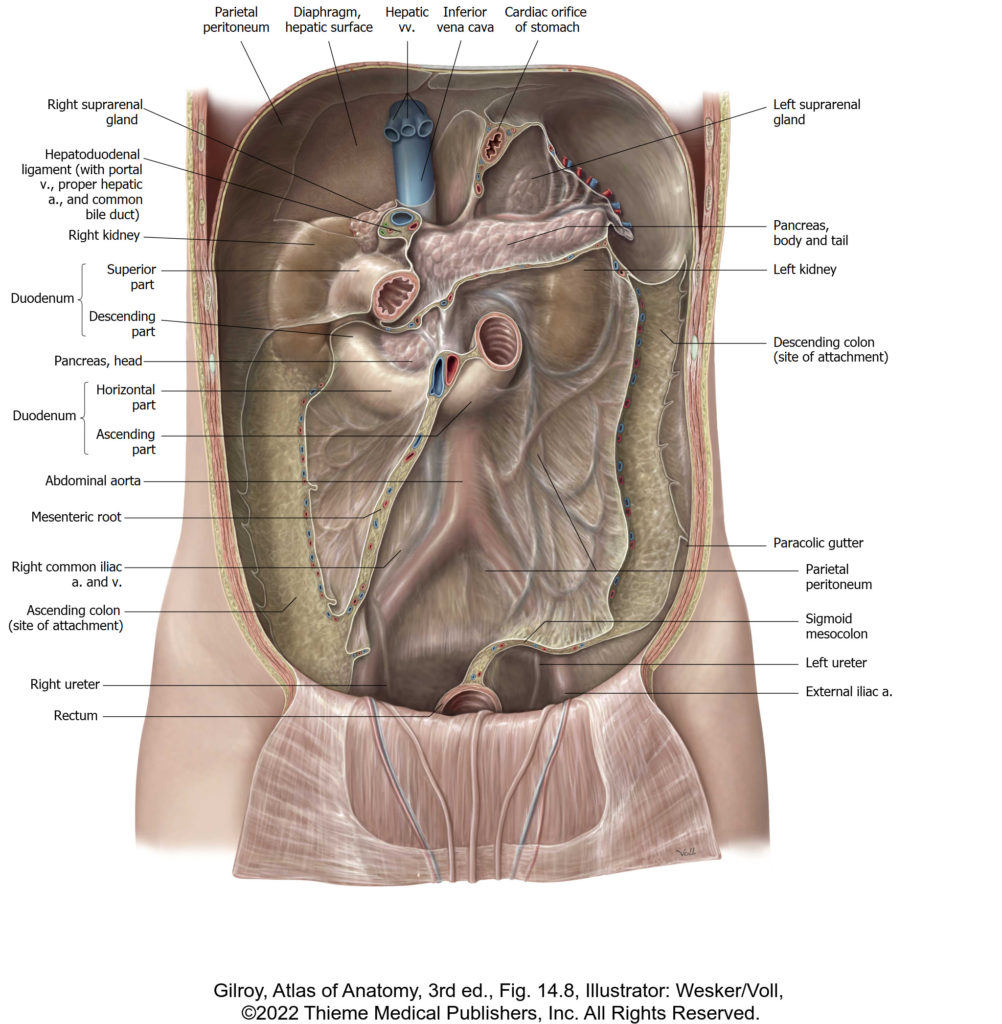
There is no excerpt because this is a protected post.