Scalp and cranial cavity
Optional reading Clinically Oriented Anatomy, 9th ed., Head chapter, Internal surface of cranial base section through Posterior cranial fossa; Face and scalp section through Lymphatic drainage of face and scalp; Cranial meninges section through Arachnoid mater and pia mater; Cerebral arterial circle section and Venous drainage of brain section. Scalp The scalp covers the skull and […]
Neuroanatomy lab knowledge checks

Station 1. Meninges and ventricles Ventricles Interactive 1 On a mid-sagittal image, label the parts of ventricular systems highlighted in blue: 3rd ventricle 4th ventricle cerebral aqueduct lateral ventricle (Tap to open; use your Apple Pencil to draw) Labeled image Interactive 2 What do each of the yellow lines indicate on this T2-grayscale inverted […]
Neuroanatomy lab introduction

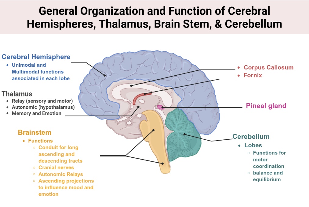
Neuroanatomy provides the essential framework for understanding the principles of neurology. This lab is designed to help you build that foundation through hands-on exploration and clinical application. Key focus areas Topographic anatomy Ventricular system, cerebrum, diencephalon, brain stem, and cerebellum Three-dimensional relationships Ventricular system and its connections to cerebral and brain stem structures Clinical imaging […]
Orientation required pre-reading
How to use this material First, please review this and the optional reading resources completely before your first day on rotation, as they provide important context for connecting with sites and communities. You are welcome to return to this resource throughout the rotation experience to deepen your knowledge and understanding, satisfy your curiosity, or dive […]
Community Engagement
Final Thoughts: Climate Change and Mental Health
Key takeaways The core message Climate change impacts mental health through multiple pathways—from acute trauma after disasters to chronic anxiety about the future. These impacts are not equally distributed; marginalized communities bear the greatest burden. As physicians, you have roles at multiple levels: Clinical care, community resilience building, and advocacy for climate action and health […]
Host-pathogen interactions
High-yield summary Core concept Host-pathogen interactions determine infection outcomes: clearance, persistence, latency, or disease. Immune responses are tailored to pathogen type Pathogens evolve mechanisms to evade immunity. Extracellular bacteria Innate Response Complement activation (Alternative and lectin pathways) Phagocytosis via PRRs, Fc receptors, complement receptors Inflammatory cytokines: IL-1, TNF, IL-6 Adaptive Response Antibodies: Opsonization, toxin neutralization CD4+ […]
Storms, Hurricanes, and Floods
An aerial view of extreme flooding: New Orleans inundated in the aftermath of Hurricane Katrina (2005). Such catastrophic floods illustrate how powerful storms can devastate entire regions, endanger lives, and incapacitate critical infrastructure. Heavy precipitation One of the clearest signals of climate change is a marked increase in heavy precipitation and flooding events. A warmer […]
Introduction to Extreme Weather Hazards
Even as far back as ancient times, people understood that weather could influence health—Hippocrates wrote 2,400 years ago that anyone practicing medicine must consider seasonal weather changes and their effects on disease. Today, our climate is changing at an unprecedented rate, and this is driving more extreme weather events that pose serious risks to human […]
Mini-literature review
Requirements Minimum 1,000 words. Minimum 10 citations Mini-reviews are essential when you Are writing a review paper. Draft the intro or discussion sections of research/scholarly work. Venture into a new research/scholarly field. Prepare a research/scholarly proposal. A mini-literature review should Summarize key findings in your topic area Evaluate the quality and impact of studies Show […]